Skip to content

Nails, Dry And Brittle (Chip, Peel, Crack, Break Easily)

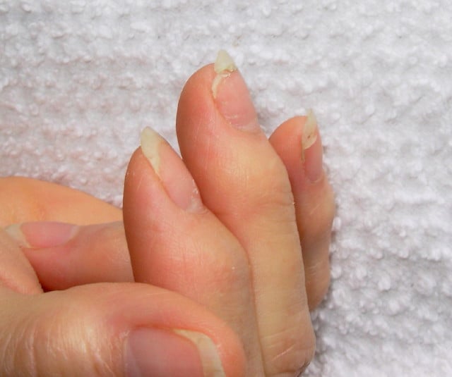
DSCN4758aWhat Are Brittle Nails?

[dropcap]B[/dropcap]rittle nails are abnormalities of the nail plate that are characterized by poor nail structure affecting all fingernails and toenails seen as thin, dry nails that easily chip, split, and are difficult to maintain a clean edge. Usually longitudinal ridging occurs from the nail base to the tips.

Q: What is the nail plate?

A: The nail plate is the hard keratin cover protecting the finger tip and toe tip. The nail plate (non-living tissue) is produced by the nail matrix (living tissue) at the base of the nail plate under the lunula (moon), which is the site of brittle nail development.

Nail Anatomy. A. Nail plate; B. lunula; C. root; D. sinus; E. matrix; F. nail bed; G. hyponychium; H. free margin. Courtesy Wikipedia.org

Poor nail structure affecting all nails may be a feature of nutritional deficiency in poor diet or malabsorption such as occurs in celiac disease.

Some other causes are: idiopathic (unknown cause), the result of aging, the effects of certain drugs, or an association with systemic autoimmune disorders such as vitiligo, alopecia areata (with pitting), psoriasis (with pitting), and lichen planus. 

External (non-nutritional or disease) causes of dry, brittle nails, such as detergents and cleaners, would only affect fingernails but not toenails.

Note: It has been shown that working with your hands in water or soaking them through activities like swimming does not cause dry, brittle nails but will worsen them.

What Are Brittle Nails In Celiac Disease and/or Gluten Sensitivity?

Dermatitis Herpetiformis or Duhring’s Disease

Dermatitis Herpetiformis. Blisters Opened Giving Relief From Pain and Itching.
Dermatitis Herpetiformis On Forearm. Skin Is Darkened Where Old Blisters Healed.

What Is Dermatitis Herpetiformis?

[dropcap]D[/dropcap]ermatitis herpetiformis (DH) is an autoimmune extremely itchy, painful bullous skin rash (blistering eruptions) arising from the underlying dermis layer of skin as a consequence of gluten sensitivity.

Dermatitis herpetiformis is characterized by multiple intensely itchy, red blisters appearing on the elbows which can extend down the forearm to the wrist and the knees. Less usual areas involve the back, buttocks, scalp, and abdomen.

Q: Do the blisters leave a mark when healed?

A: Crops of skin eruptions begin with itching or a burning sensation in reddened papules. There are grouped vesicles and tense blisters. The blister contents may be serous or bloody, with symmetrical distribution (eg, both knees or both elbows). Fluid filled elements rupture leaving denuded areas of sore skin and crust. Subsequently, there is residual hypopigmentation (a white area) or hyperpigmentation (dark area).1

Rupture of blisters begins relief from intense burning and itching.

Dermatitis Herpetiformis Eruptions On Knees.
Dermatitis Herpetiformis Eruptions On Knees. Notice White Areas Showing Loss of Pigmentation From Healed Blisters.

What Is Dermatitis Herpetiformis In Celiac Disease and/or Gluten Sensitivity?

Primary care providers should be aware of this skin condition, as they are more likely than a gastroenterologist to be confronted with this type of presentation of celiac disease.2

Sources:
  1. Mendes FB, Hissa-Elian A, de Abreu MA, Gonçalves VS. Review: dermatitis herpetiformis. An Bras Dermatol. 2013 Jul-Aug;88(4):594-9. []
  2. Robinson BL, Davis SC, Vess J, Lebel, J. Primary care management of celiac disease. Autoimmune Disorders. Nurse Practitioner. February 2015: Vol 40 – Issue 2; 28–34. []

DHA (Docosahexaenoic Acid) Deficiency

Smoked Salmon...Brain Food that's Good for the Eyes!
Salmon…Brain Food That’s Good for Your Eyes, Heart, Blood Vessels, Blood Sugar, Liver, Muscles, and Fights Inflammation!

What Is DHA?

[dropcap]D[/dropcap]ocosahexaenoic acid (DHA) is an essential omega-3 fatty acid that is abundant in the brain, being crucial in brain structure. As such DHA is a key component of neuronal membranes together with arachidonic acid (a major opposing omega-6 fatty acid), making up 15-20% of the brain’s dry mass.

This polyunsaturated fatty acid is obtained from fish sources of food.

In healthy human volunteers, positron emission tomography (PET) has shown that the normal human brain consumes 4.6 mg/day of DHA.1

DHA is particularly concentrated in highly active membranes such as nerve synapses (junctions) and photoreceptors in the eye (retina).

Q: How much DHA is in the retina?

A: DHA makes up more than 30% of the retina.2

In other roles, DHA is an  important building material for the eicosanoids, a large group of highly bioactive hormone-like substances including prostaglandins, leukotrienes, and thromboxanes that are involved in blood clotting, inflammation, and vasoconstriction.

DHA has been shown to increase insulin sensitivity as opposed to the opposite problem of insulin resistance, to improve muscle mass, and protect against non-alcoholic fatty liver disease.3

Egert et al. in a study of people aged 19 to 43 years with normal cholesterol showed that DHA intake significantly increased serum HDL (good) cholesterol. Also, DHA significantly decreased fasting serum triglycerides.4

What Is DHA Deficiency In Celiac Disease and/or Gluten Sensitivity?

Sources:
  1. Rapoport SI. Brain arachidonic and docosahexaenoic acid cascades are selectively altered by drugs, diet and disease. Prostaglandins Leukot Essent Fatty Acids. 2008 Sep-Nov;79(3-5):153-6. Epub 2008 Oct 29. []
  2. Richardson AJ. The importance of omega-3 fatty acids for behavior, cognition, and mood. Scandinavian Journal of Nutrition. 2003;47(2):92-8 []
  3. Espinosa A, Valenzuela R, González-Mañán D, D’Espessailles A, Guillermo Gormaz J, Barrera C, Tapia G. Prevention of liver steatosis through fish oil supplementation: correlation of oxidative stress with insulin resistance and liver fatty acid content. Arch Latinoam Nutr. 2013 Mar;63(1):29-36. []
  4. Egert S, Kannenberg F, Somoza V, et al. Dietary alpha-linolenic acid, EPA, and DHA have differential effects on LDL fatty acid composition but similar effects on serum lipid profiles in normolipidemic humans. J Nutr. 2009;139:861–868. doi: 10.3945/jn.108.103861 []

Dermatomyositis

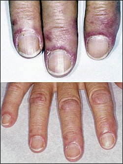
dermatomyositisWhat Is Dermatomyositis?

[dropcap]D[/dropcap]ermatomyositis is a rare autoimmune systemic disease of the connective tissue that is characterized by inflammatory and debilitating degenerative changes in the muscles and in the skin. 

Dermatomyositis results in symmetric, proximal muscle weakness of limbs (upper arms and legs), and skin manifestations. 50-70% of patients have circulating myositis-specific auto-antibodies.

The course of dermatomyositis is unpredictable being marked by spontaneous flare-ups and remissions. It can begin slowly or abruptly according to the factor that is triggering the onset such as infection, medications like phenytoin, and autoimmune disease.

Q: What are the skin manifestations of dermatomyositis?

A: Classic skin manifestations of dermatomyositis include these features:

  • The heliotrope rash (lilac color) on upper eyelids.
  • Rash on face, neck, shoulders, upper chest, elbows, knees, knuckles, and back.
  • Gottron’s papules (scaly, red eruptions or  patches over the knuckles, elbows, and knees).
  • The V-sign (rash front of neck and chest).
  • The shawl sign (rash distribution on shoulders and back).1

Additional cutaneous manifestations are described below under symptoms.

Dermatomyositis is associated with an increased risk of  cancer, other autoimmune diseases, such as lupus and psoriasis, and it can be a complication of interferon-α therapy. About 1 person in 100,000 are affected according to various studies. While it affects all ages, women have twice the occurence of men.

There is no cure for dermatomyositis, but the symptoms can be treated.  Options include medication, physical therapy, exercise, heat therapy (including microwave and ultrasound), orthotics and assistive devices, and rest.  The standard treatment for dermatomyositis is a corticosteroid drug, given either in pill form or intravenously.  Immunosuppressant drugs, such as azathioprine and methotrexate, may reduce inflammation in people who do not respond well to prednisone.2

What Is Dermatomyositis In Celiac Disease and/or Gluten Sensitivity?

Sources:
  1. Marvi U, Chung L, Fiorentino DF. Clinical presentation and evaluation of dermatomyositis. Indian J Dermatol. 2012 Sep;57(5):375-81. doi: 10.4103/0019-5154.100486. []
  2. National Institute of Neurological Disorders and Stroke. []

Eczema (Atopic Dermatitis)

eczemaWhat Is Eczema?

[dropcap]E[/dropcap]czema, or atopic dermatitis, is a chronic recurring inflammatory skin disorder characterized by itching, eczematous plaques and a defective epidermal barrier (surface layer of skin cells) that fails to hold moisture, with affected areas becoming dry then inflamed.

Q: Why does eczema develop?

A: Patients with atopic dermatitis have genetically determined risk factors that affect the barrier function of the skin and immune responses that interact with environmental factors.1

Atopic eczema is associated both with an abnormal skin matrix and impaired systemic immune response. Therefore, isolated topical treatment may have suboptimal effect.2 

Atopic eczema is a common presentation of  immune response towards food proteins in infancy.3

Global evidences reflect a marked increase in prevalence, which has tripled since 1960. In the United States, the current prevalence rates range from 10% to 20% in children and 1 to 3% in adults.4

What Is Eczema In Celiac Disease and/or Gluten Sensitivity?

Sources:
  1. Mesquita Kde C, Igreja AC, Costa IM. Atopic dermatitis and vitamin D: facts and controversies. An Bras Dermatol. 2013 Nov-Dec;88(6):945-53. doi: 10.1590/abd1806-4841.20132660. []
  2. Caffarelli C, Santamaria F, Vottero A, Bernasconi S. Progress in Pediatrics in 2012: choices in allergy, endocrinology, gastroenterology, hematology, infectious diseases, neurology, nutrition and respiratory tract illnesses. Ital J Pediatr. 2013 May 8;39(1):26. []
  3. Ho MH, Wong WH, Chang C. Clinical Spectrum of Food Allergies: a Comprehensive Review. Clin Rev Allergy Immunol. 2012 Nov 16. []
  4. Mesquita Kde C, Igreja AC, Costa IM. Atopic dermatitis and vitamin D: facts and controversies. An Bras Dermatol. 2013 Nov-Dec;88(6):945-53. doi: 10.1590/abd1806-4841.20132660. []

Vitamin B12 Deficiency

Clams Are Chock Full of Vitamin B12.
Lovely Clams For Vitamin B12.

What Is Vitamin B12?

[dropcap]V[/dropcap]itamin B12, also called cobalamin, is a highly complex vitamin that functions in two coenzyme forms: adenosylcobalamin and methylcobalamin.

These forms of the vitamin play important roles in the physical and chemical processes by which amino acids become proprionate, proprionate that becomes acetate,  and single carbons.

Q: Why are these steps important?

A: These steps are essential for normal function in the workings of all cells, especially for those of the digestive tract, bone marrow and nervous tissue.

Vitamin B12 is mainly excreted through bile into the duodenum (first part of the small intestine) for excretion in stool.1 However, if vitamin B12 is needed, it is reabsorbed in the ileum (end of the small intestine) while excess is excreted in stool and very little in urine.2

The blood level of vitamin B12 in healthy people ranges between 140 and 750 pg/ml.

What Is Vitamin B12 Deficiency In Celiac Disease and/or Gluten Sensitivity?

Sources:
  1. Shibata K, Hirose J, Fukuwatari T. Relationship Between Urinary Concentrations of Nine Water-soluble Vitamins and their Vitamin Intakes in Japanese Adult Males. Nutr Metab Insights. 2014 Aug 5;7:61-75. doi: 10.4137/NMI.S17245. []
  2. Shinton N K. Vitamin B 12 and folate metabolism. Br Med J. Feb 26, 1972; 1(5799): 556–559. []

Nails, Horizontal Ridges (Beau’s Lines), Fragile

Beaus lines in thin nails (The tiny brown streak is a splinter hemorrhage.)
Beau”s lines in a thin nail. (The tiny brown streaks are splinter hemorrhages due to vitamin C deficiency.)

What Are Horizontal Ridges In Fragile Nails?

Horizontal ridges, also called “beau’s lines,” are abnormalities of the nail plate that appear as rumpling from the base to the tips of nails and are characterized by poor nail structure of both fingernails and toenails.

The nail plate is the hard keratin cover of the finger tip and toe tip which we ordinarily call “nails.” The nail plate is produced by the nail matrix. 

Q: Why do Beau’s lines develop in nails?

A: Beau’s lines occur due to temporary cessation of proliferation (growth) of proximal nail matrix at the nail base. As the finger nail grows at the rate of 0.1 mm/day, the time course of the illness can be estimated from the position of the Beau’s line from proximal nail fold.1

Nail Anatomy. Nail Anatomy. A. Nail plate; B. lunula; C. root; D. sinus; E. matrix; F. nail bed; G. hyponychium; H. free margin. Courtesy Wikipedia.org.

A. Nail plate; B. lunula; C. root; D. sinus; E. matrix; F. nail bed; G. hyponychium; H. free margin. Courtesy Wikipedia.

Beau’s lines are frequently seen in nutritional deficiency states, bacterial illness, acute stress, and systemic disease. The conditions where Beau’s lines have been described include severe systemic illness, chemotherapy, malnutrition, zinc deficiency, trauma, paronychia, pemphigus, and Kawasaki disease.2 Beau’s lines are commonly seen in patients undergoing chemotherapy.3

 This condition of nails was named after Joseph Honoré Simon Beau (1806-1865).

What Are Horizontal Nail Ridges In Celiac Disease and/or Gluten Sensitivity?

Sources:
  1. Naik GS1, Harikrishna J. Beau’s lines. Indian J Med Res. 2013 Jan;137(1):220. []
  2. Naik GS1, Harikrishna J. Beau’s lines. Indian J Med Res. 2013 Jan;137(1):220. []
  3. Patel LM, Lambert PJ, Gagna CE, Maghari A, Lambert WC. Cutaneous signs of systemic disease. Clin Dermatol. 2011 Sep-Oct;29(5):511-22. doi: 10.1016/j.clindermatol.2011.01.019. []

Koilonychia (Washboard Nails)

Koilonychia (Hordinsky, Sawaya, and Scher, 2000)

What Is Koilonychia?

[dropcap]K[/dropcap]oilonychia is a feature of an iron deficiency state characterized by abnormally shaped nails, called spoon shaped nails, meaning that the normal downward curve is reversed.

Depending on the inadequacy of iron, nails may simply remain flat or with a slight dip without progressing to a reversed curve.

These nails develop vertical ridges from the base to the nail tip that can become deep, called washboard nails.

Q: Are all nails affected?

A: All nails are affected in varying degrees. The small finger shows the least effect while the thumb shows the greatest change which begins as flattening before scooping upward.

Nail Anatomy. Nail Anatomy. A. Nail plate; B. lunula; C. root; D. sinus; E. matrix; F. nail bed; G. hyponychium; H. free margin. Courtesy Wikipedia.org.

A. Nail plate; B. lunula; C. root; D. sinus; E. matrix; F. nail bed; G. hyponychium; H. free margin. Courtesy Wikipedia.

What Is Koilonychia In Celiac Disease and/or Gluten Sensitivity?

Nails, Splinter Hemorrhages In

The tiny brown streak is a splinter hemorrhage.

What Are Splinter Hemorrhages In Nails?

[dropcap]S[/dropcap]ubungual (under the nail) splinter hemorrhage is characterized by fragility of the underlying capillaries resulting in bleeding under the nail . Bleeding that results looks like a brown streak or splinter.1

Because the streak of blood is not in the nail but under the nail, it does not grow out with the nail. It can be a feature of vitamin C deficiency which resolves with adequate nutrition.

Nail Anatomy. Nail Anatomy. A. Nail plate; B. lunula; C. root; D. sinus; E. matrix; F. nail bed; G. hyponychium; H. free margin. Courtesy Wikipedia.org.

A. Nail plate; B. lunula; C. root; D. sinus; E. matrix; F. nail bed; G. hyponychium; H. free margin. Courtesy Wikipedia.

Splinter hemorrhage can be seen in a diet poor in vitamin C, disorders of food absorption, systemic disease such as psoriasis, nail lichen planus, an aggressive disease that may destroy the nail plate, and endocarditis. Some drugs, such as oral contraceptives, cause vitamin C deficiency.

Who is Affected in the General Population? Both sexes are commonly affected, though it occurs significantly more frequently in males. Where present in females, they were confined to a single digit.2

What Are Splinter Hemorrhages In Nails In Celiac Disease and/or Gluten Sensitivity?

Sources:
  1. Krause’s Food, Nutrition, & Diet Therapy. 10th Edition. Kathleen Mahan, Sylvia Escott-Stump. 2000. W.B. Saunders Company. []
  2. Monk BE. The prevalence of splinter haemorrhages. Br J Dermatol. 1980 Aug;103(2):183-5. []

Nails, White Spots And White Bands

This White Spot Is A Feature of Zinc Deficiency.

What Are White Spots And White Bands In Nails?

[dropcap]W[/dropcap]hite spots in the nails is a feature of zinc deficiency and white bands signify protein deficiency characterized by abnormal appearance anywhere in one or more nails.

White spots and bands can appear separately or together in nails. There may be just one mark or many marks depending on the level of deficiency.

Q: What part of the nail is affected by zinc and/or protein deficiency?

A: Zinc and protein deficiencies affect the nail plate.

The nail plate is the hard keratin cover of the finger tip and toe tip, simply called “the nail.” The nail plate is produced by the living nail tissue at its base. Here is an illustration of the parts that make up a fingernail:

Nail Anatomy. Nail Anatomy. A. Nail plate; B. lunula; C. root; D. sinus; E. matrix; F. nail bed; G. hyponychium; H. free margin. Courtesy Wikipedia.org.

A. Nail plate; B. lunula; C. root; D. sinus; E. matrix; F. nail bed; G. hyponychium; H. free margin. Courtesy Wikipedia.

What Are White Spots And White Bands In Nails In Celiac Disease and/or Gluten Sensitivity?