Skip to content

Nails, Rounded With Curved Ends

What Is Angina Pectoris?Coronary Artery Lesion
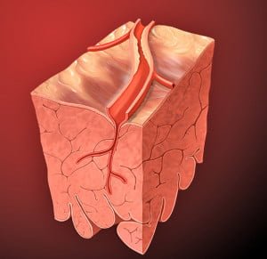
[dropcap]A ngina pectoris, or simply angina, is a coronary syndrome characterized by an oppressive substernal pain (pain under breastbone) or pressure brought on by exertion and relieved by rest that results from failure of coronary arteries to deliver adequate oxygen to heart tissue due to ischemic heart disease.

Q: Why do coronary arteries fail to deliver adequate oxygen to heart tissue?

A: Coronary arteries are the blood vessels that serve the heart. In angina, these vessels fail to deliver adequate oxygen to heart tissue because they are narrowed or blocked by fatty buildups, called atherosclerotic plaques or by a blood clot which impair their ability to carry adequate blood that carries the oxygen. Diseased coronary arteries cannot deliver adequate oxygenated blood pumped by the heart to its own muscle cells.

The heart is a muscular organ that is working all the time without rest, so it needs a constant supply of oxygen. When heart muscle has to work harder, it needs more oxygen. Lack of oxygen causes pain which makes the affected person stop activity and rest.

Angina can be stable or unstable. Unstable angina is much more serious and can be life-threatening.

  • Stable angina produces predictable pain and responds to rest and/or medication. It is less serious than unstable angina but can be very painful or uncomfortable. Anything that makes the heart muscle need more oxygen can cause an angina attack in someone with heart disease, including: smoking, cold weather, exercise, emotional stress, obesity, and large meals. Other causes of angina include: abnormal heart rhythms (usually ones that cause the heart to beat quickly), anemia, coronary artery spasm, heart failure, heart valve disease, and hyperthyroidism (overactive thyroid).1
  • Unstable angina produces unpredictable pain that may occur at rest, lasting more than 20 minutes. It is more severe than stable angina and less responsive to medication. Atherosclerosis is by far the most common cause of unstable angina. Oxidized low-density lipoprotein, so-called bad cholesterol, and oxysterols play an important role in atherogenesis, the development of atherosclerosis. Coronary arteries that are narrowed by atherosclerotic plaques can rupture causing injury to the coronary blood vessel resulting in blood clotting which blocks the flow of blood to the heart muscle. Blood clots may form, partially dissolve, and later form again and angina can occur each time a clot blocks blood flow in an artery. People with unstable angina are at increased risk of having a heart attack.2

What Is Angina In Celiac Disease and/or Gluten Sensitivity?

Sources:
  1. http://www.ncbi.nlm.nih.gov/pubmedhealth/PMH0001247/ []
  2. http://www.heart.org/HEARTORG/Conditions/HeartAttack/SymptomsDiagnosisofHeartAttack/Unstable-Angina_UCM_437513_Article.jsp# []

Itchy (Pruritic) Skin  

tongue in B12 deficiencyWhat Is Beefy Red, Smooth, Burning Tongue?

[dropcap]B eefy, red, smooth, burning tongue is an inflammatory alteration in tongue tissue characteristic of vitamin B12 deficiency.

Inflammation makes the tongue red and sore which, when untreated, progresses to atrophied papillae (shrunken taste buds) that makes the tongue smooth.

Q: Why does vitamin B12 alter the tongue?

A: Vitamin B12 is essential for the metabolism of all cells, especially for those of the digestive tract of which the tongue is an organ.1

These easily seen changes to tongue tissue are an important diagnostic feature of deficient vitamin B12, which causes other devastating unseen damage, because the discomfort generally causes the individual to seek medical care.

What Is Beefy Red, Smooth, Burning Tongue In Celiac Disease and/or Gluten Sensitivity?

Sources:
  1. Krause’s Food, Nutrition, & Diet Therapy. 10th Edition. Kathleen Mahan, Sylvia Escott-Stump. 2000. W.B. Saunders Company. []

Cutaneous Vasculitis 

Heart showing dilated cardiomyopathy at autopsy. Courtesy
Enlarged heart showing dilated cardiomyopathy at autopsy. 

What Is Idiopathic Dilated Cardiomyopathy?

[dropcap]I diopathic dilated cardiomyopathy (IDCM) is a disorder of myocardial (heart muscle) function characterized by dilation or enlargement of the cardiac chambers and reduction in the ability of the ventricles (lower chambers) to contract with the required force needed to pump blood out of the heart to the body.

Idiopathic means the cause is not known.

Q: What is the prognosis for idiopathic dilated cardiomyopathy?

A: This disorder often results in symptomatic heart failure due to the inability of the heart to pump blood as required to supply the body with oxygen and meet metabolic needs.

What Is Idiopathic Dilated Cardiomyopathy In Celiac Disease and/or Gluten Sensitivity?

Cutis Laxa, Acquired

Image on left shows how atherosclerosis impedes blood flow through coronary arteries while blood clots block blood flow. Courtesy Google.
Figure on right shows how atherosclerosis impedes blood flow through coronary arteries while blood clots block blood flow. Courtesy Google.

What Is Coronary Artery Disease (CAD)?

[dropcap]C oronary artery disease (CAD), also called ischemic heart disease, is a gradual narrowing of medium and large arteries of the heart by fatty buildups, called atherosclerotic plaques.

It is characterized by slowly developing interference with blood flow to heart tissue itself, resulting in oppressive chest pain called angina and, ultimately, thrombosis (clot) causing heart attack.  

The heart is a muscular organ that is working all the time, so it needs a constant supply of oxygen. Oxygen is brought to the working heart tissue by the coronary arteries with each beat of the heart. When heart muscle has to work harder, it needs more oxygen delivered to itself. Lack of oxygen causes pain.

In fact, failure of diseased coronary arteries to deliver adequate oxygen to heart tissue is the most common cause of angina pectoris – substernal pain (under breastbone) or pressure brought on by exertion and relieved by rest. 

Thrombosis, or clot formation, occurs when blood cells within a narrowed artery can no longer get through. Trapped, blood cells pile up and block the artery thus triggering a cascade of events called heart attack. Coronary arteries that are narrowed by atherosclerotic plaques can rupture causing injury to the coronary blood vessel resulting in blood clotting which blocks the flow of blood to the heart muscle. Blood clots may form, partially dissolve, and later form again and angina can occur each time a clot blocks blood flow in an artery.1

Q: How does coronary artery disease develop?

A: Coronary artery disease slowly develops from this combination of events:

  • Dysfunction of epithelial cells that line the inside of arteries cause the vessels to stiffen, and subsequently

  • Accumulation of lipid (fat) in smooth muscle cells beneath the inside lining of arteries and in foam cells cause buildup of fatty deposits on the inside walls progressing to fibrous plaque formation.

Oxidized low-density lipoprotein (oxLDL), so-called bad cholesterol, and oxysterols play important roles in the development of  atherosclerosis. OxLDL triggers the immune system to produce autoantibodies against oxLDL that are detectable in serum. These antibodies are called anti-oxLDL. Anti-oxLDL antibody and oxysterol concentrations are associated with coronary artery stenosis. Oxidative stress may be greatly increased in unstable angina.2 and Chronic inflammation in the general population is a major risk factor for ischemic heart disease.

The pathophysiology of atherosclerosis is, clearly, different in women when compared to the men. The women have a higher risk of blood coagulability making them at high risk for the blood clot formation. In a large number of women endothelial dysfunction, small vessel size and diffuse atherosclerosis have been identified as causes of ischemia without evidence of blockade in the coronary arteries.3

Also, atherosclerotic plaque in women is less fibrotic and contains more lipid filled foam cells, implying greater potential for reversibility but also potentially greater vulnerability for plaque rupture and thrombosis.4

Who is Affected in the General Population?

  • Coronary artery disease remains the leading cause of death in developed countries despite significant progress in primary prevention and treatment strategies.

  • It is the leading cause of death in women, as well as an important cause of disability.

  • Older patients are at particularly high risk of poor outcomes following acute coronary syndrome.5

What Is Coronary Artery Disease In Celiac Disease and/or Gluten Sensitivity?

Ischemic heart disease is the leading cause of death in the United States, making cardiovascular risk assessments and potential interventions or treatments imperative for patients with celiac disease.6

Sources:
  1. http://www.heart.org/HEARTORG/Conditions/HeartAttack/SymptomsDiagnosisofHeartAttack/Unstable-Angina_UCM_437513_Article.jsp# []
  2. Yasunobu Y, Hayashi K, Shingu T, Yamagata T, Kajiyama G, Kambe M. Coronary atherosclerosis and oxidative stress as reflected by autoantibodies against oxidized low-density lipoprotein and oxysterosis. Atherosclerosis. Apr 2001;155(2):445-53. []
  3. Kunadian V, Ford GA, Bawamia B, Qiu W, Manson JE. Vitamin D deficiency and coronary artery disease: A review of the evidence. Am Heart J. 2014 Mar;167(3):283-291. doi: 10.1016/j.ahj.2013.11.012. Epub 2013 Dec 19. []
  4. Kunadian V, Ford GA, Bawamia B, Qiu W, Manson JE. Vitamin D deficiency and coronary artery disease: A review of the evidence. Am Heart J. 2014 Mar;167(3):283-291. doi: 10.1016/j.ahj.2013.11.012. Epub 2013 Dec 19. []
  5. Kunadian V, Ford GA, Bawamia B, Qiu W, Manson JE. Vitamin D deficiency and coronary artery disease: A review of the evidence. Am Heart J. 2014 Mar;167(3):283-291. doi: 10.1016/j.ahj.2013.11.012. []
  6. Robinson BL, Davis SC, Vess J, Lebel, J. Primary care management of celiac disease. Nurse Practitioner. February 2015: Vol 40 – Issue 2; 28–34. []

Edema

Cachexia with Wasted Muscles, Weakness, and That Developed over 3 Years Time.
Cachexia with Wasted Muscles, Weakness, Weight Loss, Anemia, Skin Hemorrhages, Anorexia. GFW

What Is Cachexia?

[dropcap]C achexia is a state of ill health involving deteriorating body composition that is characterized by general malnutrition and loss of lean tissue such as muscle.

Q: What are typical findings in cachexia?

A: Arm muscle triceps (the muscles at the back of the upper arm), skin folds, subscapular skin folds, fat area index, and bone mineral content are significantly lower than normal.

Cachexia may develop in protein-losing enteropathy such as celiac disease, chronic or severe infection such as pneumonia, tuberculosis, malaria, or many chronic diseases such as heart failure and cancer.

Cachexia can develop in persons of any age.

What Is Cachexia In Celiac Disease and/or Gluten Sensitivity?

Erythema Elevatum Diutinum (EED)

unexpected weight loss celiac disease symptomWhat Is Unexpected Weight Loss?

[dropcap]U nexpected weight loss is unintentional loss of body mass composition or inability to gain weight marked by decreased serum proteins and increased stool fat.1

What Is Unexpected Weight Loss In Celiac Disease and/or Gluten Sensitivity?

Sources:
  1. Krause’s Food, Nutrition, & Diet Therapy. 10th Edition. Kathleen Mahan, Sylvia Escott-Stump. 2000. W.B. Saunders Company. []

Erythema Nodosum 

anorexia nervosa celiac disease gluten symptomWhat Is Anorexia?

[dropcap]A norexia or loss of appetite is a reduced desire to eat which can cause unintentional weight loss.

Anorexia is a feature of many disorders caused by malutrition such as celiac disease and aging or gastrointestinal problems such as chronic gastroenteritis, and dysgeusia (loss of taste).

Q: Are there other conditions that cause anorexia?

A: Other underlying problem include medication side effects, infection, chronic pain, depression, grief, dehydration, hormonal dysfunction such as thyroid disease, primary hyperparathyroidism, or Addison’s disease, autoimmune mechanisms such as sarcoidosis,  and cancer growth.

What Is Anorexia In Celiac Disease and/or Gluten Sensitivity?

Follicular Hyperkeratosis (goose flesh or corkscrew hair)

increased appetite gluten celiac symptomWhat Is Increased Appetite?

[dropcap]I ncreased appetite is the increased desire for food.

Q: What causes appetite.

A: Appetite is caused by hunger.

The brain monitors nutritional needs and employs a hunger mechanism to alert us when fresh food is needed. Hunger is mainly caused by the need for protein. Low blood sugar also triggers hunger.

The tempting sight, smell, taste and expectation of food stimulates involuntary sensory nerves. By reflex action, these sensory nerves cause muscle and sensory activity in various digestive organs. Salivary glands in the mouth begin to secrete saliva as stomach glands and muscles become active.1

What Is Increased Appetite In Celiac Disease and/or Gluten Sensitivity?

Sources:
  1. Cleo Libonati. Recognizing Celiac Disease. Gluten Free Works Publishing, Fort Washington, PA 19034 USA []

Hair, Fine With Rough Texture

Inflamed Aorta. Courtesy Quizlet.com

What Is Aortic Vasculitis?

[dropcap]A ortic vasculitis is an inflammatory disease of the aorta that causes dilation of the aorta wall with narrowing of the inside passageway and results in widespread impairment of blood flow to tissues served by the aorta.

Q: What is the result of inflammatory disease of the aorta?

A: The aorta is the largest artery in the body. It extends from the left ventricle (lower chamber) of the heart and carries blood out of the heart with each beat to rest of the body. Narrowing of the aorta’s inside diameter due to swelling from inflammation causes elevated blood pressure and enlarged heart because of back pressure of blood unable to be fully pumped out of the heart with each beat. The heart enlarges because the difficulty of pumping blood out makes it work harder than normal.

Vasculitis that causes midaortic syndrome is a variety of aortic coarctation (narrowing) located in the lower thoracic aorta, the abdominal aorta or both, involving the intestinal and renal vessels (kidney). It usually presents with arterial hypertension.1

What Is Aortic Vasculitis  In Celiac Disease and/or Gluten Sensitivity?

Sources:
  1. Massel D. n-3 polyunsaturated fatty acids reduced mortality and morbidity after recent myocardial infarction. Therapeutics. Jan-Feb 2000:6 []

Hangnail

Aphthous_ulcer[1]What Are Aphthous Ulcers?

[dropcap]A phthous ulcers, also called canker sores, are a chronic disorder of soft mouth tissue characterized by small, painful purpuric, papular, or erosive lesions that are often surrounded by erythematous (red) margins.1

Q: What soft mouth tissues develop canker sores?

A: Canker sores can erupt on the mucosal surface of the inside lip, sides of mouth, under the tongue and along the side of the tongue. When they are forming, the area swells into a papule, or small bump. Later, the surface erodes and a crater forms with firm pus.

What Are Aphthous Ulcers In Celiac Disease and/or Gluten Sensitivity?

Sources:
  1. Lahteenoja H, Toivanen A, Viander M, Maki M, Irjala K, Raiha I, Syrjanen S. Oral mucosal changes in coeliac patients on a gluten-free diet. European Journal of Oral Sciences. Oct 1998;106(5):899,8p. []