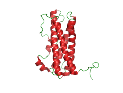
This is a model of Immunoglobulin-G Antibody (IgG)
What Are Anti-Gliadin Antibodies?
[dropcap]A nti-gliadin antibodies (AGA) are produced by the body as an immune response against partially digested gliadin peptides (fragments) that have abnormally entered the body from the intestinal tract. Gliadin is a protein in wheat grain.
Anti-gliadin antibodies circulating in blood are measured by laboratory testing on blood that is drawn. The antibodies are both immunoglobulin A (IgA) and immunoglobulin G (IgG) types, which are termed AGA-IgA and AGA-IgG.
The presence of anti-gliadin antibodies in blood is an abnormal laboratory finding. That is, when the intestinal lining is healthy and able to keep out undigested matter from the body, there are no molecules of gliadin present to trigger the immune system for defense.
Q: Who produces anti-gliadin antibodies?
A: Persons, with or without celiac disease, can produce anti-gliadin antibodies if and when leaky gut (increased intestinal permeability) develops that then allow gliadin peptides to get into the bloodstream.
Gliadin itself causes leaky gut, but there are other causes such as various foods, intestinal infections, medications, and other diseases of the gastrointestinal tract. Whatever the cause of leaky gut, if poorly digested gliadin is present, it can slip through the leaky lining and enter the bloodstream where it can trigger an antibody response by anti-gliadin antibodies.
What Are Anti-Gliadin Antibodies In Celiac Disease and/or Gluten Sensitivity Or Non-Celiac Gluten Sensitivity?
[dropcap]A nti-tissue transglutaminase antibodies (anti-tTG) are connective tissue autoantibodies and can be detected in blood samples from affected persons who are reacting to gluten in the diet.
Autoantibodies are abnormal because they attack the body’s own tissue, which in the case of these antibodies is tissue transglutaminase 2 (TG2).
Q: What is tissue transglutaminase 2 (TG2)?
A: Tissue transglutaminase 2 (TG2) is an enzyme that appears in many cell locations and is particularly abundant in endothelial cells that line the small intestine. It has been implicated in a variety of cellular processes, such as differentiation, cell death, inflammation, cell migration and wound healing.
The cell appears to adapt the dynamics of this enzyme to meet specific sub-cellular needs or to respond to stress or other stimuli. Substantial evidence indicates that the location of TG2 within cells is critical for the regulation of its various biochemical activities, which subsequently trigger diverse downstream events,1
Although initially studied as an enzyme within cells, TG2 is now known to be secreted also into the extracellular space (between cells) or onto the cell surface.1
Abnormal activation of TG2 or deregulation of its function(s) is involved in a variety of human diseases, such as celiac disease, diabetes, neurodegenerative diseases, multiple sclerosis and rheumatoid arthritis. A role in inflammatory disorders and septic shock has also been shown. Moreover, multiple studies have revealed elevated TG2 expression in many types of cancer cells.1
What Are Anti-tissue Transglutaminase Antibodies In Celiac Disease and/or Gluten Sensitivity?
Sources:
Piacentini M, D’Eletto M, Farrace MG, Rodolfo C, Del Nonno F, Ippolito G, Falasca L. Characterization of distinct sub-cellular location of transglutaminase type II: changes in intracellular distribution in physiological and pathological states. Cell Tissue Res. 2014 Dec;358(3):793-805. doi: 10.1007/s00441-014-1990-x. [↩] [↩] [↩]
[dropcap]E levated bone alkaline phosphatase (BALP) is a laboratory result that indicates an abnormal blood level of this bone enzyme.
A bone alkaline phosphatase blood level is one of the most frequently used biochemical markers of bone formation.
Q: Why is the purpose of bone alkaline phosphatase?
A: Bone alkaline phosphatase is produced by bone cells called osteoclasts in normal bone maintenance for the purpose of breaking down old or damaged bone so that other bone cells called osteoblasts can fill in the excavated areas with new bone. This process keeps bone stong and healthy.
Elevated bone alkaline phosphatase shows that more bone is being broken down than is being replaced. It can be caused by hyperparathyroidism, bone tumors from cancer, and malnutrition.
What Is Elevated Bone Alkaline Phosphatase (BALP) In Celiac Disease and/or Gluten Sensitivity?
This photo taken during laparoscopy shows the gall bladder (small white organ in middle) surrounded by yellow fat. Liver (dark red organ) is overlying.
What Is Impaired Gall Bladder Motility?
[dropcap]I mpaired gall bladder motility means the gall bladder is slow to empty or is dysfunctional.
The functional disorder of the gallbladder is caused initially either by metabolic abnormalities or by an alteration in its muscular ability to contract (motility dysfunction).
The diagnostic criteria based on symptoms of motility dysfunction of the gallbladder are episodes of moderate to severe steady pain located in the epigastrium and right upper abdominal quadrant that last at least 30 minutes.
Gallbladder motility disorder is suspected after gallstones and other structural abnormalities have been excluded.1
Q: What does the gallbladder do?
A: The gallbladder is a small pouch-like organ about the size of a pear that receives bile produced by the liver and stores it until needed during digestion. It lies just under the liver.
Bile is a complex fluid containing water, electrolytes and many organic molecules including bile acids, cholesterol, phospholipids and bilirubin. Bile acids are critical for digestion and absorption of fats and fat-soluble vitamins in the small intestine. Many waste products, including bilirubin, are eliminated from the body by secretion into bile and elimination in feces.2
Before a meal, the gallbladder is usually full of bile. In response to fat in the diet, the gallbladder squeezes stored bile into the small intestine through a series of ducts. When emptied after meals, the gallbladder is flat.
What Is Impaired Gall Bladder Motility In Celiac Disease and/or Gluten Sensitivity?
Sources:
Behar J, Corazziari E, Guelrud M, Hogan W, Sherman S, Toouli J. Functional gallbladder and sphincter of oddi disorders. Gastroenterology. 2006 Apr;130(5):1498-509. [↩]
[dropcap]L ow cholesterol found in blood indicates an abnormal blood level of this essential lipid (fat) that is characterized by decreased production of steroid hormones and bile.
Cholesterol is a soft, fat-like substance found in the bloodstream and is an essential structural component in all body cells. It is required for membrane fluidity and permeability of cells. Within the cell membrane, cholesterol also functions in intracellular transport, cell signaling and nerve conduction.
Cholesterol is required for these essential body processes:
Cholesterol is converted to bile by the liver and stored in the gallbladder until needed for digestion of fats in the small intestine.
Cholesterol is used by the adrenal glands to produce the steroid hormones cortisol and aldosterone.
Cholesterol is used by the sex glands to make progesterone, estrogen, and testosterone and their derivatives.
Cholesterol is needed for the production of vitamin D.
Q: What makes cholesterol bad for you?
A: There are different forms of cholesterol that can be bad or good depending on the level. In blood vessels, low-density lipoprotein (LDL or ‘bad’) cholesterol can abnormally join with fats and other substances to build up in the inner walls of arteries. The arteries can become clogged and narrow, and blood flow is reduced. This is atherosclerosis which can lead to stroke, heart attack and thrombosis.
High-density lipoprotein (HDL or ‘good’) carries harmful cholesterol away from the arteries and helps protect from heart attack and stroke.1
What Is Low Cholesterol In Celiac Disease and/or Gluten Sensitivity?
[dropcap]C oagulation factors II, VII, IX, X found in blood are essential for normal blood clotting. Low coagulation factors on blood assay indicate an altered secondary coagulation disorder that is characterized by impaired clot formation.
Each coagulation factor must be present in sufficient quantity in order for normal clotting to occur, but the level required is different for each factor. Results are frequently reported as a percentage with 100% being normal. For example, a factor VIII that is 30% would be considered abnormally low.1
The production of the coagulation factors II, VII, IX, and X requires vitamin K without which the factors will be low.
Q: What happens when coagulation factors II, VII, IX, and X are low?
A: When any of the blood clotting factors are lacking or not working properly, the blood tests prothrombin (PT) and partial thromboplastin time (PTT) will be abnormally prolonged. Prothrombin and partial thromboplastin time measure the time it takes for blood to clot. When you bleed, the body launches a series of activities that help the blood clot. This is called the coagulation cascade. There are three pathways to this event. These tests looks at coagulation factors, found in two of these pathways.2
What Are Low Coagulation Factors In Celiac Disease and/or Gluten Sensitivity?
A 3D Image From Magnetic Resonace Cholangiography showing biliary tree. NIHMS
What Is Primary Sclerosing Cholangitis?
[dropcap]P rimary sclerosing cholangitis (PSC) is an uncommon, slowly progressive bile duct disease that results in stagnation or build-up of bile in the liver, called cholestasis.
Primary sclerosing cholangitis is characterized by sclerosis, or scarring inflammation in bile ducts both within the liver (intra-hepatic ducts), and outside the liver (extra-hepatic ducts), causing progressive narrowing and, eventually, obliteration of the bile ducts.
Primary sclerosing cholangitis comes under the umbrella term autoimune liver disease in which the end result is immune-mediated hepatocellular (liver cell) or hepatobiliary (bile duct) injury.1
Q:What happens when scarred bile ducts can no longer transport bile out of the liver?
A:Bile that cannot be removed from the liver by the biliary duct system backs up and damages the liver, causing cirrhosis.
Bile is continually made by the liver from phospholipids salt, cholesterol, and aging blood cells that it removes from circulation to be carried out of the liver. Bile also carries away waste products produced by normal metabolism and toxic substances that are removed by the liver for eventual elimination in stool. As such, bile must continually flow out of the liver to prevent build-up in the liver.
Bile is a greenish brown liquid made by the liver. Bile ducts carry it out of the liver to the gall bladder for storage until needed to aid in the digestion and absorption of fat from the small intestine. Bile emulsifies fat eaten in the diet so that the pancreatic enzyme called lypase can break it down into its fatty acid and glycerol components.
The liver is the largest organ within the body. It lies mostly in the upper part of the abdomen on the right side just under the diaphragm. About 70% of liver tissue is made up of cube shaped cells called hepatocytes that do the main work of the liver. Other cells (epithelial) form structure and are arranged in single layers around blood vessels, sinusoids, and bile ducts.
Build-up of bile in the liver is the end result of the inflammatory process in primary sclerosing cholangitis, that by swelling and scarring of bile ducts impedes and eventually prevents bile flow out of the liver, leading to liver failure. There is no curative treatment available for primary sclerosing cholangitis, besides liver transplantation.2
The appearance of the intrahepatic and extrahepatic biliary ducts can be assessed by use of cholangiography, and magnetic resonance (MR) imaging is the best way to identify patients. See image above.3
MR cholangiography offers a noninvasive method of obtaining images of the biliary system without the use of a contrast agent. There is no radiation exposure. Pulse sequences can be chosen to obtain bright bile or black bile cholangiograms. Image processing algorithms can be selected to obtain a three-dimensional representation of biliary anatomy and pathology, and those images can be rotated in any plane so that ductal anatomy and pathology can be seen to best advantage.4
There is no cure for primary sclerosing cholangitis but there are symptom treatments one of which is supplementation for low levels of vitamins A,D,E, and K. Liver transplant is the only effective option.
What Is Primary Sclerosing Cholangitis In Celiac Disease and/or Gluten Sensitivity?
Sources:
Trivedi PJ, Adams DH. Mucosal immunity in liver autoimmunity: a comprehensive review. J Autoimmun. 2013 Oct;46:97-111. doi: 10.1016/j.jaut.2013.06.013. [↩]
Kummen M, Schrumpf E, Boberg KM. Liver abnormalities in bowel diseases. Best Pract Res Clin Gastroenterol. 2013 Aug;27(4):531-42. doi: 10.1016/j.bpg.2013.06.013. [↩]
Eaton JE, Talwalkar JA, Lazaridis KN, Gores GJ, Lindor KD. Pathogenesis of primary sclerosing cholangitis and advances in diagnosis and management. Gastroenterology. 2013 Sep;145(3):521-36. doi: 10.1053/j.gastro.2013.06.052. Epub 2013 Jul 1. [↩]
Meakem TJ 3rd, Schnall MD. Magnetic resonance cholangiography. Gastroenterol Clin North Am. 1995 Jun;24(2):221-38. [↩]
Prolactin Structure. Courtesy Boris TM at Wikipedia.
What Is Hyperprolactinemia?
[dropcap]H yperprolactinemia is an excess secretion of prolactin hormone causing an elevated blood level that is characterized by its effects on reproduction: in females causing amenorrhea and in males causing reduced sexual potency.
Q: What is prolactin?
A: Prolactin is a hormone produced by the the anterior pituitary gland that is transported by the bloodstream to stimulate androgen hormone production in male and estrogen hormone production in female sex glands. However, too much prolactin inhibits hormone production of estrogen in women and testosterone in men.
In pregnant females upon delivery of a baby, prolactin stimulates breast development and milk production for the purpose of feeding the infant. Sucking by the infant stimulates the release of prolactin which keeps the milk supply adequate.
Prolactin also promotes autoimmune mechanisms. Prolactin interferes specifically with B cell tolerance induction, enhances proliferative response to antigens and mitogens and increases the production of immune globulins, cytokines and autoantibodies.1
Other conditions that can stimulate prolactin release include nutritional deficiencies, thyroid stimulating hormone (TSH) produced by the pituitary gland, stress, exercise, and anterior pituitary tumors.
What Is An Elevated Prolactin Level In Celiac Disease and/or Gluten Sensitivity?
Sources:
Shelly S, Boaz M, Orbach H. Prolactin and autoimmunity. Autoimmun Rev. 2012 May;11(6-7):A465-70. doi: 10.1016/j.autrev.2011.11.009. [↩]
Enlarged heart showing dilated cardiomyopathy at autopsy.
What Is Idiopathic Dilated Cardiomyopathy?
[dropcap]I diopathic dilated cardiomyopathy (IDCM) is a disorder of myocardial (heart muscle) function characterized by dilation or enlargement of the cardiac chambers and reduction in the ability of the ventricles (lower chambers) to contract with the required force needed to pump blood out of the heart to the body.
Idiopathic means the cause is not known.
Q: What is the prognosis for idiopathic dilated cardiomyopathy?
A: This disorder often results in symptomatic heart failure due to the inability of the heart to pump blood as required to supply the body with oxygen and meet metabolic needs.
What Is Idiopathic Dilated Cardiomyopathy In Celiac Disease and/or Gluten Sensitivity?
What Is Carbohydrate Malabsorption? [dropcap]C arbohydrate malabsorption is a digestive disorder characterized by the inability to properly digest and absorb carbohydrates within the small intestine to supply needed energy to the body. Q: What carbohydrates should be…