[dropcap]M uscle pain and tenderness, also called myalgia, is a disorder of muscle tissue.
Muscle pain that is ongoing or chronic can result from a variety of disorders, infections, overuse, injury, as an unwanted effect of certain medications, toxic substances, celiac disease, and certain nutritional deficiencies.
What Is Muscle Pain and Tenderness In Celiac Disease and/or Gluten Sensitivity?
Testing the Eye for Tear Production (L) and Damage to Conjunctiva from Dryness (R).
What Is Sjögren’s Syndrome?
[dropcap]S jögren’s syndrome is a systemic inflammatory autoimmune disease with a chronic, progressive course that primarily attacks the lacrimal glands of the eye and the salivary glands of the mouth, which are exocrine glands. Exocrine glands secrete the substances they produce through a duct.
Sjögren’s syndrome is ordinarily characterized by dysfunction of the lacrimal glands to produce tears causing dry eye and the salivary glands to produce saliva causing dry mouth, but is not limited by or to these features.
Besides involvement of these exocrine glands, there may be involvement of other parts of the body, termed extraglandular, which may be more severe than eye or mouth features.
There is not yet agreement on classifying Sjögren’s syndrome. Primary and secondary are the two forms generally accepted.1 Both forms can cause mild to severe disease, called the spectrum:
Primary Sjögren syndrome. Disease occurs without involvement of other linked autoimmune disorders. In addition to the eyes and mouth, the nose, throat and skin may also be affected and joints, lungs, kidneys, blood vessels, digestive organs and nerves as well.2 Systemic manifestations (other than eyes and mouth) concern a third of patients, including lymphoma in 5% of the patients.3
Secondary Sjögren’s syndrome. Disease complicates other autoimmune disease such as systemic lupus erythematosus, rheumatoid arthritis, primary biliary cirrhosis, and celiac disease.
Diagnosis of Sjögren’s syndrome is made by most doctors based on Schimer’s test for tears and unstimulated whole salivary flow to assess objective eye and oral involvement, since these are the tests most physicians use in clinical practice.4 Specific antibody tests would be positive for anti-Ro (SSA)/anti-La (SSB) autoantibodies. Sjögren’s syndrome should also be considered when extraglandular manifestations such as vasculitis, polyneuropathy or arthritis occur, even when the patients do not complain of dry eyes and mouth.5
There is no cure for Sjögren’s syndrome. Treatment is aimed to diminish symptoms. For example, steroids and Ibupropen are used to decrease inflammation and pain in joints. Artificial tears and ointments are used for dry eye.
Most people who develop Sjogren’s syndrome are older than 40 years. Nine of ten people with Sjögren’s syndrome are women.2
What Is Sjögren’s Syndrome In Celiac Disease and/or Gluten Sensitivity?
Sources:
Huang YF, Cheng Q, Jiang CM, An S, Xiao L, Gou YC, Yu WJ, Lei L, Chen QM, Wang Y, Wang J. The immune factors involved in the pathogenesis, diagnosis, and treatment of Sjogren’s syndrome. Clin Dev Immunol. 2013;2013:160491. doi: 10.1155/2013/160491. Epub 2013 Jul 9. [↩]
Fazaa A, Bourcier T, Chatelus E, Sordet C, Theulin A, Sibilia J, Gottenberg JE. Classification criteria and treatment modalities in primary Sjögren’s syndrome. Expert Rev Clin Immunol. 2014 Apr;10(4):543-51. doi: 10.1586/1744666X.2014.897230. [↩]
Cornec D, Saraux A, Cochener B, Pers JO, Jousse-Joulin S, Renaudineau Y, Marhadour T, Devauchelle-Pensec V. Level of agreement between 2002 American-European Consensus Group and 2012 American College of Rheumatology classification criteria for Sjogren’s syndrome and reasons for discrepancies. Arthritis Res Ther. 2014 Mar 19;16(2):R74. [↩]
Witte T. Pathogenesis and diagnosis of Sjögren’s syndrome. Z Rheumatol. 2010 Feb;69(1):50-6. doi: 10.1007/s00393-009-0519-2. [↩]
Small Bowel Volvulus Caused the Enlarged Dark Loops. Courtesy African Journal of Pediatric Surgery.
What Is Volvulus?
[dropcap]V olvulus is twisting of a loop(s) of bowel onto itself which effectively closes it thus preventing digested matter from passing and causing engorgement of the closed loop with gas and fluid.
Q: How does the bowel get untwisted?
A: This condition usually necessitates surgical correction although some do return to their proper position.
The danger is that there may develop necrosis of the twisted loop(s) in which case, if left untreated,death will ensue.
What Is Volvulus In Celiac Disease and/or Gluten Sensitivity?
Nail Anatomy. A. Nail plate; B. lunula; C. root; D. sinus; E. matrix; F. nail bed; G. hyponychium; H. free margin. Courtesy Wikimedia.org
What Are Rounded Nails With Curved Ends?
[dropcap]R ounded, curved, dark and dry nails are a well known feature of vitamin B12 deficiency.
Q: How does vitamin B12 deficiency cause the nails to grow this way?
A: Vitamin B12 deficiency affects the nail plate. The nail plate is the hard keratin cover of the finger tip and toe tip, simply called “the nail.” The nail plate is produced by the living nail matrix at its base.
What Are Rounded Nails With Curved Ends In Celiac Disease and/or Gluten Sensitivity?
[dropcap]W hite spots in the nails is a feature of zinc deficiency and white bands signify protein deficiency characterized by abnormal appearance anywhere in one or more nails.
White spots and bands can appear separately or together in nails. There may be just one mark or many marks depending on the level of deficiency.
Q: What part of the nail is affected by zinc and/or protein deficiency?
A: Zinc and protein deficiencies affect the nail plate.
The nail plate is the hard keratin cover of the finger tip and toe tip, simply called “the nail.” The nail plate is produced by the living nail tissue at its base. Here is an illustration of the parts that make up a fingernail:
Nail Anatomy. .
A. Nail plate; B. lunula; C. root; D. sinus; E. matrix; F. nail bed; G. hyponychium; H. free margin. Courtesy Wikipedia.
What Are White Spots And White Bands In Nails In Celiac Disease and/or Gluten Sensitivity?
What Are Gastro-Intestinal Complications Of Type 1 Diabetes?
[dropcap]G astrointestinal complications of type I diabetes mellitus are functional or organic changes that result from diabetes affecting every organ of the gastrointestinal tract.
Q: How do gastrointestinal tract complications affect diabetes?
A: Impaired function of individual digestive organs in diabetes can significantly influence level of diabetes compensation and vice versa. On the other side, unsatisfactory diabetes compensation can result in manifestation of digestive problems.
The most frequent (55 to 75%) and the most serious clinical complication is diabetic gastroparesis characterized by impaired evacuation and motility of the stomach and small intestine. Gastroparesis results in nausea, vomiting, early fullness after eating and constipation.
Treatment is with prokinetics. These oral medicines help motility and transport of food in the esophagus, stomach and intestines and improve sphincter function to keep food from going backwards.
Gastroesophageal reflux and heartburn are other complications in diabetes. These are treated with antacids and acid reducing medications.
Various autoimmune disease interfere with digestion. One connection between diabetes mellitus and the gastrointestinal tract can be celiac disease.1
What Are Gastro-Intestinal Complications Of Diabetes In Celiac Disease and/or Gluten Sensitivity?
Sources:
Perusicova J. Gastrointestinal complications in diabetes mellitus. Vnitri Lekarstvi. May 2004;50(5):338-43. [↩]
Image of Pancreas From Gray’s Anatomy. Courtesy Wikipedia.org
What Is Pancreatic Insufficiency?
[dropcap]P ancreatic insufficiency is a disorder characterized by insufficient exocrine production of pancreatic enzymes for normal digestion of fats, proteins, and carbohydrates that results in maldigestion of these foodstuffs.
Pancreatic insufficiency also causes malabsorption of the fat-soluble vitamins: vitamin A, vitamin D, vitamin E, and vitamin K.
Q: What are the exocrine enzymes produced by the pancreas?
A: Exocrine enzymes produced by the pancreas include amylase for the digestion of carbohydrates, lipase for the digestion of fats, and protease for the digestion of proteins.
While lipases and amylase are secreted in the active form, proteases are secreted as pro-enzymes (need to be activated). Trypsinogen is converted to its active form trypsin in the duodenum by enterokinase, a protease secreted by the enterocytes (surface cells that line the duodenum), and trypsin in turn activates the other pancreatic proteases.1
Pancreatic enzymes are released by the pancreas into the internal pancreatic duct that empties into the common bile duct from which they are pumped through the Sphincter of Odi directly into the duodenum as needed. Tthe Sphincter of Odi is a strong circular muscle that controls the entrance of pancreatic enzymes into the duodenum.
After a meal, enzyme secretion into the duodenum increases quickly reaching peak output within the first 20 to 60 minutes, then decreasing to a stable level before reaching an interdigestive level at the end of the digestive period, that is, about 4 hours after meal intake.2
Medical treatment is with medication that contains enzymes to break down carbohydrates, fats, and protein in food at mealtime. This treatment is safe, effective, and has few side effects.
What Is Pancreatic Insufficiency In Celiac Disease and/or Gluten Sensitivity?
Sources:
Fieker A, Philpott J, Armand M. Enzyme replacement therapy for pancreatic insufficiency: present and future. Clin Exp Gastroenterol. 2011;4:55-73. doi: 10.2147/CEG.S17634. [↩]
Fieker A, Philpott J, Armand M. Enzyme replacement therapy for pancreatic insufficiency: present and future. Clin Exp Gastroenterol. 2011;4:55-73. doi: 10.2147/CEG.S17634. [↩]
Tissue Changes in Hyperparathyroidism Causing Brown Tumor. Courtesy Quizlet.com
[dropcap]P rimary hyperparathyroidism is a parathyroid disorder characterized by excessive secretion of parathyroid hormone by one or more parathyroid glands for more than 6 months.
In primary hyperparathyroidism, blood calcium levels are high while phosphorus levels are decreased due to the action of parathyroid hormone.
Parathyroid hormone is produced by the four pea sized parathyroid glands that are located on the thyroid gland in the front of the neck. Partly because the thyroid and parathyroid glands share the same anatomic place in the body and partly because they have similar names, they are often confused although they have completely different actions.
Parathyroid hormone normally keeps calcium and the opposing mineral phosphorus levels in balance by drawing calcium as needed from bones to increase it in blood and releasing excess phosphorus through the kidneys to decrease blood levels.
Primary hyperparathyroidism is commonly caused by an adenoma (tumor) in a parathyroid gland (80%) or 15% due to hyperplasia of gland tissue (overgrowth). It is seldom associated with autoimmune disorders. However, cancer is a possibility.
Q: What is a parathyroid adenoma?
A: A parathyroid adenoma is usually a solitary, well circumscribed, soft, tan reddish-brown nodule with a capsule. Gland tissues outside of the adenoma are normal or slightly shrunken (not needed anymore).1
Untreated, primary hyperparathyroidism results in cyst formations in bone marrow (osteitis fibrosa cystica) and brown tumors in bone tissue. Cysts contain large amounts of fibrous tissue with areas of hemorrhage. Brown tumors contain aggregates of osteoclasts (bone cells), hemorrhage and giant cells resembling neoplasms.2
Here is the symptomatolgy: “Painful Bones, Renal Stones, Abdominal Groans, and Mental Moans.”
What Is Primary Hyperparathyroidism In Celiac Disease and/or Gluten Sensitivity?
Endocrine glands targeted in polyglandular autoimmune syndrome.
What Are Autoimmune Polyglandular Syndromes?
[dropcap]A utoimmune polyglandular syndromes (APS) are rare clusterings of two or more endocrine and non-endocrine autoimmune disorders in the same affected person.
Polyglandular is somewhat of a misnomer since many of the manifestations of the diseases do not concern endocrine glands.1
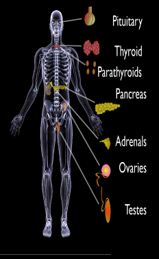
Endocrine autoimmune disorders involve the abnormal production of autoantibodies that target and destroy the body’s own endocrine tissues, causing loss of essential hormone production by the targeted glands. Endocrine glands include the pituitary, thyroid, adrenal, parathyroid, islets of Langerhans (pancreas), testes in males, and ovaries in females.
First degree relatives (siblings of same parents, parents, children) have an increased incidence of latent, meaning not apparent, autoimmune pathology.2
Q: How many autoimmune polyglandular syndromes are described?
A:Three syndromes have been identified and they are all inherited: APS type-1, APS type-2, and APS type-3.
APS type-1 is a genetic mutation inherited in an autosomal recessive manner. A child with APS type-1 has inherited two mutated copies of a gene called the AIRE (autoimmune regulator) gene, which is on the long arm of 21st chromosome present in each cell.3 The parents, called carriers, are unaffected since they each have only a single copy of the AIRE mutated gene. Humans have a total of 23 pairs of chromosomes that contain genes inherited from each parent. Mutations in the genes cause disease.
Diagnosis criteria for autoimmune polyglandularsyndrome type-1 includes these three disorders:
Chronic candida infection (CMC), which usually develops first, typically attacks skin, but very commonly also nails, mouth, vagina, esophagus and intestine. CMC in APS type-1 patients is usually mild, and in most cases, it is chronic. It is found in 73–100 % of APS type-1 patients.
Hypoparathyroidism, causing loss of parathyroid function (hypoparathyreosis) is found in 76–93 % of APS type-1 patients.
Autommune Addison’s disease, also called autoimmune adrenalitis, is found in 72-100 % of APS type-1 patients. Still many of them die for unrecognized or late diagnosed autoimmune Addison’s disease, so regular follow-up for children in suspicion of APS type-1 (with CMC or/and hypoparathyroidism) is necessary.4
Other assocated disorders that may develop, but are not required for diagnosis, include: vitiligo, premature menopause, pernicious anemia, parathyroid gland failure, alopecia, and celiac disease. Thyroid disease rarely occurs.5
APS type-2 is linked to the inheritance of HLA antigens on chromosome 6 and appears to be autosomal dominant with incomplete penetrance. This suggests the contribution of environmental factors, such as bacterial and viral infections, medications, psychological factors, etc.6It does not have an identified mutation of the AIRE gene.
Diagnosis criteria for autoimmune polyglandular syndrome type-2 includes these two disorders:
Autommune Addison’s disease combined with
Autoimmune thyroid disease (thyroid atrophy, hypertrophic goiter related to Hashimoto’s thyroiditis, Graves’ disease, asymptomatic autoimmune thyroiditis)6 and/or type I diabetes mellitus. The conditions may occur in any order.
Polyglandular autoimmune syndrome type-2 is also known as Schmidt’s syndrome when adrenalitis (adrenal insufficiency) is associated with thyroiditis and Carpenter’s syndrome for adrenal insufficiency with hypoparathyreosis (impaired function of parathyroid glands).
Other disorders that may develop, but are not required for diagnosis, include: type 1 diabetes (50%), frequently gonadal failure or vitiligos, also celiac disease, autoimmune hepatitis, alopecia, pernicious anemia, and myasthenia gravis.7 Decades may arise between the onset of one disease and the onset of the second in the same patient.8
Therapy of APS type-2 consists of hormone replacement therapy for each separate condition, except that treatment for adrenal insufficiency must be given before thyroid therapy is started when the conditions occur together.9 Thyroxin replacement may induce life-threatening adrenal failure in a patient with untreated Addison’s disease. Thus, in case of doubt hydrocortisone should be given before the thyroxine administration is started.10
APS type-3 has a strong genetic background. Diagnosis criteria for autoimmune polyglandular syndrome type-3 involves these conditions:
Autoimmune thyroiditis that occurs with another organ-specific autoimmune disease, but not with autoimmune Addison’s disease, and
Other autoimmune diseases can include diabetes mellitus, pernicious anemia, vitiligo, alopecia, myasthenia gravis, celiac disease, and Sjögren’s syndrome. The most common APS type-3 combination is autoimmune disease of thyroid gland and pernicious anemia.11
Who is Affected in the General Population?
APS type-1 is usually apparent in childhood with the incidence of 1 in100,000 persons. It is more common among Finns (1 in 25,000), Sardinians (1 in 14,000), and Iranian Jews (1 in 6,500 to 1 in 9,000). The age of onset is usually early childhood, but new symptoms can develop throughout life. It affects both sexes equally.
APS type-2 has a peak onset in middle age, although the first signs usually develop between 20–30 years of age. Its prevalence is 1 in 20,000 persons. It is three times more frequent among women than men.12
APS type-3 is most frequent among middle-aged women.7[/box]
What Is Autoimmune Polyglandular Syndrome In Celiac Disease and/or Gluten Sensitivity?
Sources:
Wémeau JL, Proust-Lemoine E, Ryndak A, Vanhove L. Thyroid autoimmunity and polyglandular endocrine syndromes. Hormones (Athens). 2013 Jan-Mar;12(1):39-45. [↩]
Femiano P, Castaldo V, Iossa C. Complex family association in autoimmune polyendocrine syndrome. Minerva Pediatrica. Apr 2003;55(2):163-70. [↩]
Wémeau JL, Proust-Lemoine E, Ryndak A, Vanhove L. Thyroid autoimmunity and polyglandular endocrine syndromes. Hormones (Athens). 2013 Jan-Mar;12(1):39-45. [↩]
Wémeau JL, Proust-Lemoine E, Ryndak A, Vanhove L. Thyroid autoimmunity and polyglandular endocrine syndromes. Hormones (Athens). 2013 Jan-Mar;12(1):39-45. [↩]
Wémeau JL, Proust-Lemoine E, Ryndak A, Vanhove L. Thyroid autoimmunity and polyglandular endocrine syndromes. Hormones (Athens). 2013 Jan-Mar;12(1):39-45. [↩] [↩]
MAJERONI BA and PATEL P. Autoimmune Polyglandular Syndrome, Type II. Am Fam Physician. 2007 Mar 1;75(5):667-670. [↩]
Lipowsky C, Schorl-Schweikardt BA, Kehl O, Brändle M. 19-year-old patient with adrenal cortex insufficiency–only the tip of the iceberg. Polyendocrine autoimmune syndrome type II (Schmidt syndrome). Praxis (Bern 1994). 2008 Jan 23;97(2):77-81. [↩]
Van den Driessche A, Eenkhoorn V, Van Gaal L, De Block C. Type 1 diabetes and autoimmune polyglandular syndrome: a clinical review. Neth J Med. 2009 Dec;67(11):376-87. [↩]
X-ray of Lungs in Sacroidosis Showing Honeycoming. Courtesy Wikimedia.
What Is Sarcoidosis?
[dropcap]S arcoidosis is a multisystem granulomatous disease characterized by hard granulomas and inflammation of the alveoli in lungs that occurrs in 80% of patients.
Alveoli are the tiny one cell thick round structures that are clustered at the ends of air passages in the lungs where oxygen from air inhaled is exchanged for carbon dioxide from blood which is breathed out.
Q: What are granulomas?
A: Granulomas are collections of macrophages (white blood cells) that targeted and enclosed but failed to destroy a foreign substance that entered the body. In turn, the macrophages become encased by other immune cells and fibroblasts and collagen. They can occur in any organ.
Presently, the etiology, or cause, of sarcoidosis is unknown, although it is thought the predisposition is genetic with a local immune mechanism. This disease has flares when active and remissions when symptoms subside.
Referral to a specialist is important for proper treatment and monitoring. The main treatment for severe active sarcoidosis is prednisone, a type of steroid that reduces inflammation in the body. In most people, prednisone relieves symptoms within a couple of months, although most people need to take prednisone for 12 months or longer. Long-term use of prednisone, especially at high doses, can cause serious side effects. Side effects include bone loss, elevated blood sugar, and high blood pressure.
When wheezing and coughing are a problem, inhaler devices are used to deliver medicine into the lungs in order to help open the airways.1
Ibuprofen is prescribed if sarcoidosis is causing joint pain. Steroid creams are prescribed for skin problems.
Sarcoidosis affects 10 to 20 person per 100,000.
What Is Sarcoidosis In Celiac Disease and/or Gluten Sensitivity?
 What Is Chronic Muscle Pain And Tenderness?