Skip to content

Increased Intestinal Permeability (Leaky Gut)

Close-up Slice of a Small Intestinal Villus Showing How Enterocytes Appear Tightly Lining the Entire Outside Surface Of A Villus. Courtesy Cleo Libonati

What Is Increased Intestinal Permeability?

[dropcap]I[/dropcap]ncreased intestinal permeability is characterized by dysfunctional intestinal permeability (leakiness) allowing for the penetration of harmful entities from the gut into the bloodstream such as undigested proteins and microbes. The popular name is “leaky gut.”

Q: Why does intestinal permeability increase?

A: Intestinal permeability is an essential function of the small intestinal mucosal lining by which wanted substances such as properly digested foodstuffs are allowed to permeate through the lining to enter the body via the bloodstream and lymphatics. At the same time unwanted substances are kept out.

The mucosal lining is one cell thick and makes up the surface between the digested foodstuffs inside the hollow of the intestine and the underlying tissues.

The mucosal lining is covered by millions of microscopic finger-like structures called villi that project toward the inside of the intestine giving the appearance of a shag rug.

Each one, called a villus, contains a capillary bringing blood to absorb nutrients, a vein to take away nutrients, and a lacteal to absorb and take away digested fat. Its wall is made up of a single layer of tightly connecting cells, called enterocytes.

This single layer of cells separates the contents of our small intestine from the lamina propria (underlying tissues of the small intestine) and the rest of our body. Breaching of this single layer of cells by leakiness can expose lymphocytes (immune cells) located in the lamina propria to a myriad of microorganisms and food antigens, leading to immune reactions.1

To protect the body from unwanted substances, a gatekeeping barrier system operates to regulate the passage of nutrients, or permeation, through the surface mucosal lining. This system acts to seal the inside body from the gut.

The integrity of intestinal permeability is determined by interactions among several barrier components including the unstirred water layer, mucosal surface hydrophobicity, the surface mucous coat, and cell factors (especially tight junctions).

Tight junctions hold cells tightly together side-by-side to prevent unwanted substances from passing through the lining. Tight junctions are complex structures comprising over 50 proteins, such as the claudin proteins which are considered to be the structural backbone of tight junctions.

Tight junctions include a series of special proteins forming fibrils (springy like proteins) that cross the plasma membrane and interact with proteins in the adjoining cells. Tight junctions are regulated by the protein zonulin.2

If zonulin deregulates from the action of substances such as gliadin (gluten in wheat) and bacteria, the tight junction barrier fails which results in increased intestinal permeability. Dysfunction of the barrier system allows unwanted substances to enter the body where they are damaging to many tissues.

Tight junction dysfunction has been shown to be a part of certain autoimmune diseases such as celiac disease, type I diabetes mellitus, multiple sclerosis, and rheumatoid arthritis. Other diseases associated are cancer, allergies, and infections.3

Important gastrointestinal infections that cause leaky gut include rotavirus, parasites, pathogenic bacteria (escherichia coli, clostridium difficile), and mycotoxins produced by fungi found in stored grain and dried fruit.4

Fortunately, the presence of some commensal (friendly intestinal bacteria) and probiotic strains leads to an increase in tight junctions  proteins at the cell boundaries and in some cases prevents or reverses the adverse effects of pathogens, food and stress. Various dietary components are also known to regulate epithelial permeability by modifying expression and localization of  tight junctions proteins.2

What Is Increased Intestinal Permeability In Celiac Disease and/or Gluten Sensitivity?

Sources:
  1. Fahardi A, Banan A, Fields J, Keshavarzian A. Intestinal barrier: an interface between health and disease. Journal of Gastroenterology and Hepatology. 2003; 18: 479-497. []
  2. Ulluwishewa D, Anderson RC, McNabb WC, Moughan PJ, Wells JM, Roy NC. Regulation of tight junction permeability by intestinal bacteria and dietary components. J Nutr. 2011 May;141(5):769-76. doi: 10.3945/jn.110.135657. [] []
  3. Fasano A. Zonulin and Its Regulation of Intestinal Barrier Function: The Biological Door to Inflammation, Autoimmunity, and Cancer. Physiological Reviews. January 2011Vol. 91no. 151-175DOI: 10.1152/physrev.00003.2008 []
  4. Farhadi A, Banan A, Fields J, Keshavarzian A. Intestinal barrier: an interface between health and disease. Journal of Gastroenterology and Hepatology. 2003;18:479-91. []

Systemic Lupus Erythematosus 

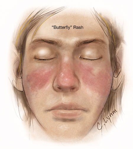
Image showing butterfly rash of SLE. Courtesy JAMA.
Image showing butterfly rash typical of SLE. Courtesy JAMA.

What Is Systemic Lupus Erythematosus?

[dropcap]S[/dropcap]ystemic lupus erythematosus (SLE) is a chronic autoimmune inflammatory disease that is characterized by involvement of multiple organs due to the production of antibodies to components of the cell nucleus.1 SLE has an unpredictable course of acute flare-ups and remissions.

Severity depends on the extent of organs affected with skin and nail involvement, called discoid lupus, being the least serious and inflammmation of the kidney, called lupus nephritis, being the worst.

Nevetheless, a classic presentation is development of a rash over the cheeks and nose that resembles a butterfy with wings spread hence the name “butterfly rash.”

Symptoms are many and varied depending on the tissues affected and are often not specific, for example hair loss has a variety of causes. Symptoms can be confused by co-existence with other autoimmune disease such as Sjogren’s syndrome.

Systemic lupus erythematosus should be managed by a specialist. Symptoms can be controlled with steroid therapy, but this disease can be a cause of premature death  mainly from active disease, organ failure (e.g., kidneys), infection, or cardiovascular disease from accelerated atherosclerosis.

Certain common medicines known to cause drug-induced lupus are:

  • Isoniazid
  • Hydralazine
  • Procainamide

Other less common drugs may also cause the condition. These may include:

  • Anti-seizure medications
  • Capoten
  • Chlorpromazine
  • Etanercept
  • Infliximab
  • Methyldopa
  • Minocycline
  • Penicillamine
  • Quinidine
  • Sulfasalazine

Symptoms tend to occur after taking the drug for at least 3 to 6 months.2

Although there is a strong familial aggregation, the disease is relatively uncommon and most cases are sporadic.1 According to the Center for Diseases (CDC), lupus most commonly affects women of childbearing age but also occurs in infants, children, adolescents, and men with peak occurrence between ages 15 and 40. Blacks (and possibly Hispanics, Asians, and Native Americans) are affected more than Whites.

What Is Systemic Lupus Erythematosus In Celiac Disease and/or Gluten Sensitivity?

Sources:
  1. http://www.cdc.gov/arthritis/basics/lupus.htm [] []
  2. www.nlm.nih.gov/medlineplus/ency/article/000446.htm []

Sjögren’s Syndrome 

Testing the Eyes for Sjogren's Syndrome.
Testing the Eye for Tear Production (L) and Damage to Conjunctiva from Dryness (R).

What Is Sjögren’s Syndrome?

[dropcap]S[/dropcap]jögren’s syndrome is a systemic inflammatory autoimmune disease with a chronic, progressive course that primarily attacks the lacrimal glands of the eye and the salivary glands of the mouth, which are exocrine glands. Exocrine glands secrete the substances they produce through a duct.

Sjögren’s syndrome is ordinarily characterized by dysfunction of the lacrimal glands to produce tears causing dry eye and the salivary glands to produce saliva causing dry mouth, but is not limited by or to these features.

Besides involvement of these exocrine glands, there may be involvement of other parts of the body, termed extraglandular, which may be more severe than eye or mouth features.

There is not yet agreement on classifying Sjögren’s syndrome. Primary and secondary are the two forms generally accepted.1 Both forms can cause mild to severe disease, called the spectrum:

  • Primary Sjögren syndrome. Disease occurs without involvement of other linked autoimmune disorders. In addition to the eyes and mouth, the nose, throat and skin may also be affected and joints, lungs, kidneys, blood vessels, digestive organs and nerves as well.2 Systemic manifestations (other than eyes and mouth) concern a third of patients, including lymphoma in 5% of the patients.3
  • Secondary Sjögren’s syndrome. Disease complicates other autoimmune disease such as systemic lupus erythematosus, rheumatoid arthritis, primary biliary cirrhosis, and celiac disease.

Diagnosis  of Sjögren’s syndrome is made by most doctors based on Schimer’s test for tears and unstimulated whole salivary flow to assess objective eye and oral involvement, since these are the tests most physicians use in clinical practice.4 Specific antibody tests would be  positive for anti-Ro (SSA)/anti-La (SSB) autoantibodies. Sjögren’s syndrome should also be considered when extraglandular manifestations such as vasculitis, polyneuropathy or arthritis occur, even when the patients do not complain of dry eyes and mouth.5

There is no cure for Sjögren’s syndrome. Treatment is aimed to diminish symptoms. For example, steroids and Ibupropen are used to decrease inflammation and pain in joints. Artificial tears and ointments are used for dry eye.

Most people who develop Sjogren’s syndrome are older than 40 years. Nine of ten people with Sjögren’s syndrome are women.2

What Is Sjögren’s Syndrome In Celiac Disease and/or Gluten Sensitivity?

Sources:
  1. Huang YF, Cheng Q, Jiang CM, An S, Xiao L, Gou YC, Yu WJ, Lei L, Chen QM, Wang Y, Wang J. The immune factors involved in the pathogenesis, diagnosis, and treatment of Sjogren’s syndrome. Clin Dev Immunol. 2013;2013:160491. doi: 10.1155/2013/160491. Epub 2013 Jul 9. []
  2. nlm.nih.gov [] []
  3. Fazaa A, Bourcier T, Chatelus E, Sordet C, Theulin A, Sibilia J, Gottenberg JE. Classification criteria and treatment modalities in primary Sjögren’s syndrome. Expert Rev Clin Immunol. 2014 Apr;10(4):543-51. doi: 10.1586/1744666X.2014.897230. []
  4. Cornec D, Saraux A, Cochener B, Pers JO, Jousse-Joulin S, Renaudineau Y, Marhadour T, Devauchelle-Pensec V. Level of agreement between 2002 American-European Consensus Group and 2012 American College of Rheumatology classification criteria for Sjogren’s syndrome and reasons for discrepancies. Arthritis Res Ther. 2014 Mar 19;16(2):R74. []
  5. Witte T. Pathogenesis and diagnosis of Sjögren’s syndrome. Z  Rheumatol. 2010 Feb;69(1):50-6. doi: 10.1007/s00393-009-0519-2. []

Intrauterine Growth Retardation (Failure to Grow Normally Before Birth)

intrauterine growth retardation gluten free
The twin on the right is much small than his brother on the left who has normal growth.

What Is Intrauterine Growth Retardation?

[dropcap]I[/dropcap]ntrauterine growth retardation (IUGR) is a fetal development abnormality characterized by failure to grow normally for gestational period. Specifically, it means the developing baby weighs less than 90% of other babies at the same age.

Intrauterine growth retardation puts the baby at increased risk for complications such as premature birth or that the baby will die inside the womb before birth.1

Intrauterine growth restriction  may be suspected if the size of the pregnant woman’s uterus is small. The condition is usually confirmed by ultrasound. Further tests may be needed to screen for infection or genetic problems if intrauterine growth retardation is suspected.

Q: Why would a baby not grow normally during pregnancy? A: An unborn baby cannot grow normally  if it does not obtain adequate oxygen and nutrition delivered through the placenta from the mother. Factors that impede adequate delivery of nutrition include:

  • Poor placenta placement. Conditions that limit or interfere with space for nutrient and oxygen absorption between the placenta and the uterine wall include 1) low attachment of the placenta near or over the cervix where maternal blood supply is poor, 2) pulling away or bleeding between the placenta and uterine wall, 3) multiple placentas (from multiple babies) sharing the uterine wall may limit blood supply to one or more of the fetuses, and 4) the presence of an hydatid mole,  (non-fertilized egg growing wildly), tumor or fibroids taking up space or growing under or into the placenta. 
  • Chromosomal abnormalities in the fetus. Conditions such as trisomy 22  have early onset  of  intrauterine growth retardation  in pregnancy. 
  • Poor health of the mother. These factors include 1) anemia which impairs the ability of the mother’s blood to deliver adequate oxygen, 2) preeclampsia which interferes with placenta function, 3) diabetes which impairs proper supply of energy, 4) kidney disease, 5) poor diet, 6) malabsorption, 7) high blood pressure or heart disease, 8) clotting disorders, and 9) toxins and infections during pregnancy that may harm the developing baby such as rubella, cytomegalovirus, toxoplasmosis, and syphilis. 
  • Risk factors in the mother. Any of the following may contribute to intrauterine growth retardation:1
  • Alcohol abuse.
  • Drug addiction.
  • Smoking.

What Is Intrauterine Growth Retardation In Celiac Disease and/or Gluten Sensitivity?

Sources:
  1. http://www.nlm.nih.gov/medlineplus/ency/article/001500.htm [] []

Obstetrical Complications

Inefficient Labor May Necessitate Ceasarian Section to Save the Baby.
Inefficient Labor May Necessitate Ceasarian Section to Save the Baby. Courtesy Wikipedia.org

What Are Obstetrical Complications?

[dropcap]O[/dropcap]bstetrical complications are reproductive disorders during pregnancy, labor and delivery that endanger the mother and unborn infant.

Complications may result from prolonged constipation, malnutriton, hormonal imbalance, infection, systemic disease such as diabetes, obesity, tumors of the uterus, medication adverse effects, drug abuse, smoking, and alcohol abuse.

What Are Obstetrical Complications In Celiac Disease and/or Gluten Sensitivity?

Candida Albicans Infection

Close-up shows inflammation and yellowish white patches of roof and back of the mouth caused by candidiasis. Uvula is greatly swollen, hanging near the level of the tongue. Courtesy: Wikipedia.
Close-up shows inflammation and infected patches on roof and back of the mouth caused by candidiasis. Uvula is swollen, hanging near the tongue. Courtesy: Wikimedia.

What Is Candida Albicans Infection?

[dropcap]C[/dropcap]andida albicans infection, called candidosis or candidiasis, is an opportunistic invasion of mucous membrane or skin by candida albicans, an endogenous yeast found in 40 to 80% of normal human beings. A former name for this small, budding fungus is monilia albicans.

Opportunistic means that yeast living on mucosal and skin surfaces does not invade (infect) unless these tissues become unhealthy and therefore cannot protect themselves.

Q: How does candida albicans cause infection?

A: Candida albicans lives on the mucosal surfaces and skin in most people without causing infection (colonizes) because of our normal defenses against invasion. In fact, candida albicans is a very effective colonizer of humans. For example, Russell and Lay found that 47% of 1-month-old infants were orally colonized with candida albicans, and 49% were colonized with other fungi.

During growth within the intestinal tract, the organism senses pH (acidity), oxygen, carbon sources, and the presence of surfaces allowing it to optimize gene expression for a particular environment. With these mechanisms for sensing, candida albicans is able to efficiently colonize humans in infancy.1

Candida Infection Of The Esophagus on X-ray. Courtesy Radiology Assistant.nl
Candida Infection Of The Esophagus (White Area) On X-ray. Courtesy Radiology Assistant.nl

Lowered host defenses allow yeast already present on mucosal and skin surfaces to take advantage and can grow rapidly, becoming pathogenic (disease producing) so that infection results.

Infection is characterized by superficial, irregular white patches on mucosal surfaces and possible invasion of the bloodstream by a filamentous form (thread-like structures) that can rapidly develop.

Candida albicans is unique among oral pathogens in its ability to invade cornified layers of stratified squamous epithelium of the tongue, mouth surfaces, hard and soft palate, esophagus, and gut. Stratified squamous epithelium is the tough surface cells that ordinarily protect underlying tissues from damage or invasion by microbes.

Candida albicans is also capable of invading the lungs and causing pneumonia and septicemia, which is the spread of infection into the bloodstream.

Here is a time honored simple do-it-youself test for infection of the mouth or throat: First thing in the morning before brushing your teeth or eating, fill a small see through glass with water then gently spit onto the surface. If after an hour the spit remains on the water surface, it is unlikely you have candida in the mouth. If it grows legs downward, it indicates that yeast is growing. If the spit sinks to the bottom, you have this problem. Yeast in the mouth can quickly travel down the esophagus and into the gut.

Medical diagnosis. Difinitive diagnosis for the oral cavity is made by your clinician by swabbing the areas of your mouth and/or throat and viewing under a microscope for evidence of candida.

Infections of the esophagus and gut require inspection by gastroscopy or endoscopy procedure and the taking of samples to be examined under microscope. This examination also give the opportunity to rule out other problems. Barium swallow can show the extent of infection and any disfiguration of the esophagus that results.

What Is Candida Albicans Infection In Celiac Disease and/or Gluten Sensitivity?

Sources:
  1. Rosenbach A, Dignard D, Pierce JV, Whiteway M, Kumamoto CA. Adaptations of Candida albicans for growth in the mammalian intestinal tract. Eukaryot Cell. 2010 Jul;9(7):1075-86. doi: 10.1128/EC.00034-10. Epub 2010 Apr 30. []

Chronic Fatigue Syndrome 

What Is Chronic Syndrome Fatigue? [dropcap]C[/dropcap]hronic fatigue syndrome is a debilitating illness characterized by persistent or relapsing overwhelming and incapacitating fatigue not relieved by rest, having a definite onset and often accompanied by numerous symptoms… 

Crohn’s Disease

Endoscopic image of Crohn'sDisease showing deep ulceration in sigmoid colon.
Endoscopic Image of Crohn’s Disease Showing Deep Ulceration in the Sigmoid Colon.

What Is Crohn’s Disease?

[dropcap]C[/dropcap]rohn’s disease is an inflammatory bowel disease characterized by patchy inflamed areas involving the full thickness of the intestinal wall that can occur anywhere in the intestinal tract, in addition to, mucosal disease.

In Crohn’s disease there is ongoing immune activation which produces inflammation and ulceration but the cause is not known and the severity varies among patients. At diagnosis of Crohn’s disease, factors predictive of subsequent 5-year aggressive disease are an age below 40 years, the presence of perianal disease, and the initial requirement for steroids.1

Dysbiosis is a factor that develops in and worsens Crohn’s disease and stress is a factor in both of these conditions. Psychological stress activates multiple physiological processes aimed at maintaining balance within the body. These physiological processes also have the capacity to influence the composition of microbial communities in the digestive tract, and research now indicates that exposure to stressful stimuli leads to gut microbiota dysbiosis.2

While the relative abundance of many different bacterial types can be altered during stressor exposure, findings in nonhuman primates and laboratory rodents, as well as humans, indicate that bacteria in the genus Lactobacillus are consistently reduced in the gut during stress.2

Q: Is there a cure for Crohn’s disease?

A: Presently, Crohn’s disease cannot be cured. This condition has a course of remissions, when symptoms subside, and flares, when symtpoms get worse. Treatment is aimed to reduce flares and promote remission.

What Is Crohn’s Disease In Celiac Disease and/or Gluten Sensitivity?

Sources:
  1. Beaugerie L, Seksik P, Nion-Larmurier I, Gendre JP, Cosnes J. Predictors of Crohn’s disease. Gastroenterology. 2006;130:650–656. []
  2. Galley JD, Bailey MT. Impact of stressor exposure on the interplay between commensal microbiota and host inflammation. Gut Microbes. 2014 May 1;5(3):390-396. Epub 2014 Apr 1. [] []

Addison’s Disease (Primary)

Underarm showing skin darkening, which is a feature of Addison's Disease
Underarm showing skin darkening, which is a feature of Addison’s Disease

What Is Primary Addison’s Disease?

[dropcap]A[/dropcap]ddison’s disease is an autoimmune destruction of the adrenal glands by autoantibodies that target the adrenal cortex, or outer part of these glands, and is characterized by a slow progressive failure of the adrenal glands to adequately produce its steroid hormones.

Symptoms of adrenal fatigue or failure may not develop until the majority of adrenal tissue is destroyed. When untreated, progression leads to coma, called Addisonian crisis, which is a medical emergency.

There are two adrenal glands each located on top of a kidney and enclosed in a connective tissue capsule. Each is a small, triangular shape that is made of two parts: the outer region and the inner region.

The inner region, called the adrenal medulla, produces epinephrine and norepinephrine chemicals that are needed to deal with stress.

The outer region, called the adrenal cortex, produces adrenocortical (steroid) hormones and releases them into the bloodstream in response to pituitary stimulating hormone from the brain.

Q: What is the function of steroid hormones produced by the adrenal glands?

A: Functions of the three steroid hormones produced by the adrenal glands are:

  1. Glucocorticoids restrain inflammation and metabolism of carbohydrates, fats and proteins to maintain a normal glucose blood level. The major glucocorticoid is hydrocortisone.
  2. Mineralocorticoids regulate the retention and excretion of fluids and electrolytes by the kidneys. The most important mineralocorticoid is aldosterone.
  3. Androgen (testosterone) is a male sex hormone.

Secondary adrenal insufficiency may develop from other causes that are not immune related such as chronic infections, tumor, and medications.

What Is Addison’s Disease In Celiac Disease and/or Gluten Sensitivity?

Colitis, Ulcerative

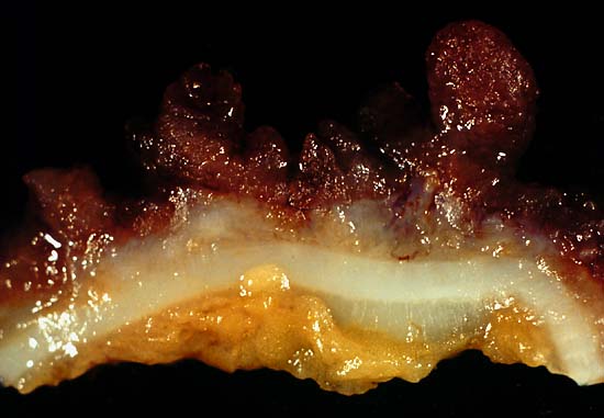
This photo was from a total colectomy done for clinically severe, intractable chronic ulcerative colitis. It shows a closer view of a longitudinal section through the colon wall. This demonstrates not only the angry red mucosa but also the tendency for the inflamed tissue to throw itself up into inflammatory pseudopolyps. Source: Ed Uthman, MD. Public domain.
This photo is from a total colectomy done for severe, intractable chronic ulcerative colitis. It shows a close view of a lengthwise section through the colon wall. This demonstrates not only the angry red mucosa, but also, the tendency for the inflamed tissue to throw itself up into inflammatory pseudopolyps.
Source: Ed Uthman, MD. Public domain.

What Is Ulcerative Colitis?

[dropcap]U[/dropcap]lcerative colitis is an inflammatory disorder of the colon characterized by continuous inflammation of the mucosa and submucosa usually with small ulcers, extending from the rectum and typically involving the distal colon, rectum, and anus and producing bloody diarrhea.

While the severity of ulcerative colitis varies among patients, iron deficiency anemia often develops due to blood loss especially when there are many bloody bowel movements in a day.

The onset of ulcerative colitis is most commonly in young adulhood.

Q: Is this disease painful?

A: Yes with the passage of stool.

Psychological stress and subsequent dysbiosis exacerbate ulcerative colitis.

Psychological stress activates multiple physiological processes aimed at maintaining balance within the body. These physiological processes also have the capacity to influence the composition of microbial communities in the digestive tract, and research now indicates that exposure to stressful stimuli leads to gut microbiota dysbiosis.1

While the relative abundance of many different bacterial types can be altered during stressor exposure, findings in nonhuman primates and laboratory rodents, as well as humans, indicate that bacteria in the genus Lactobacillus are consistently reduced in the gut during stress.2

Presently, ulcerative colitis cannot be cured. This condition has a course of remissions, when symptoms subside, and flares, when symtpoms get worse. Treatment is aimed to reduce flares and promote remission. In all cases, correction of dysbiosis improves the condition.

Ulcerative colitis is associated with increased incidence of cancer of the colon.3

What Is Ulcerative Colitis In Celiac Disease and/or Gluten Sensitivity?

Sources:
  1. Galley JD, Bailey MT. Impact of stressor exposure on the interplay between commensal microbiota and host inflammation. Gut Microbes. 2014 May 1;5(3):390-396. Epub 2014 Apr 1. []
  2. Galley JD, Bailey MT. Impact of stressor exposure on the interplay between commensal microbiota and host inflammation. Gut Microbes. 2014 May 1;5(3):390-396. Epub 2014 Apr 1. []
  3. Taber’s Cyclopedic Medical Dictionary. F. A. Davis. Philadelphia, PA []