Intestinal Edema of Duodenum in an 11 Month Old Baby. Courtesy: Nature.com
What Is Small Intestinal Edema?
S mall intestinal edema is characterized by fluid accumulation within the intestinal mucosa so that the intestinal wall appears thick and swollen.
Intestinal edema hampers peristalsis that can result in pain and gas build-up. Peristalsis is the normal rhythmic muscular wave-like action that moves residue along the gastrointestinal tract.
Q: What part of the small intestinal lining is swollen?
A: Any part of the small intestine may be affected. Some causes of edema include allergic reactions, enteropathies such as celiac disease, cow milk enteropthy, yeast infection, parasite infection, inflammatory bowel disease such as Crohn’s disease, and certain medications.
For example, the anti-hypertensive drugs known as Angiotension Converting Enzyme (ACE) inhibitors can cause intestinal angioedema and therefore the patient may present with gastrointestinal complaints.1
What Is Small Intestinal Edema In Celiac Disease and/or Gluten Sensitivity?
Sources:
LoCascio E J, Mahler S A, and Arnold TC. Intestinal Angioedema Misdiagnosed as Recurrent Episodes of Gastroenteritis. West J Emerg Med. Sep 2010; 11(4): 391–394. [↩]
P ostbulbar duodenal ulceration is a chronic inflammatory disorder characterized by thickening of the intestinal lining with excoriation penetrating the muscularis mucosae (muscle layer of the intestine) associated with villous atrophy.
The ulcer is well-defined. Stenosis due to thickening of the area narrows the hollow inside of the duodenum which impairs passage of foodstuffs and may lead to obstruction.
Q: Is postbulbar duodenal ulceration painful?
A: Postbulbar duodenal ulceration may not cause symptoms but usually causes gnawing pain that is relieved by eating food. After eating there develops nausea and bloating.
Complications include obstruction, bleeding, and perforation of the duodenal lining. Ulcers do not usually become malignant.
What Is Postbulbar Duodenal Ulceration/Stenosis In Celiac Disease and/or Gluten Sensitivity?
A therosclerosis is a disease of arteries involving the buildup of fatty material called plaque along the walls of medium and large arteries characterized by patchy subintimal thickening, hardening, and loss of elasticity of blood vessels.
The intima is the innermost layer of an artery, having contact with blood. The subintima is beneath it.
Q: What happens when arteries become narrowed and less flexible?
A: Narrowing of the inside diameter of blood vessels and hardening of their walls reduce or obstruct blood flow through them which impairs their ability to supply tissues of the body with oxygen and nourishment.
When tissues are deprived of oxygen, pain and dysfunction results such as angina pectoris involving heart muscle because the heart continually needs oxygen never being able to rest.
It is thought that atherosclerosis develops from 1) epithelial cell dysfunction of the intima, and 2) lipid (fat) accumulation in smooth muscle cells and in foam cells, causing buildup of fatty deposits on the inside walls progressing to fibrous plaque formation. That is, intimal smooth muscle cells are surrounded by connective tissue and intracellular and extracellular lipids (fat build-up inside and outside of these cells).
What Is Atherosclerosis In Celiac Disease and/or Gluten Sensitivity?
What Is Intestinal Dysbiosis? Intestinal dysbiosis is an imbalance of the composition and quantity of microbe populations (called the microbiota), that naturally inhabit our human gut. Dysbiosis causes altered gut immunity, abnormal fermentation of undigested foodstuffs,…
Microscopic Image Showing a Pink Collagen Band in Collagenous Colitis.
What Is Collagenous Colitis?
Collagenous colitis is a disease of the large intestine (colon) that is characterized by microscopic inflammation of the surface mucosal lining and an abnormally thickened collagen band of tissue that develops wthin the lining of the colon.
The thicker than normal layer of collagen of at least 10 µm (reference value: 2–7 µm) can vary in different locations. Inflammation occurs with increased numbers of lymphocytes (white blood cells) and plasma cells and epithelial (surface cell) damage. These changes can only be seen under microscopic examination of multiple biopsied tissue samples taken during a colonoscopy procedure.
Q: What is collagen?
A:Collagen is a strong, fibrous protein found in connective tissue of the colon and many other tissues such as tendons. The normal basement membrane in the bowel consists mainly of collagen type IV, laminin, and fibronectin. The increased collagen band observed in collagenous colitis consists basically of collagen type I and III, which are the subtypes produced by repair functions, indicating a reactive origin to some irritant or drug.1
The biopsies should preferably be taken from the ascending colon, since the pathological hallmarks may be absent in the descending colon, and in the normally occurring thicker collagen layer in the rectosigmoid region.1 Inflammation of the ileum (last segment of the small intestine next to colon) is common.2
Endoscopy and radiological (x-ray) examinations are usually normal.3
Autoimmune disorders are frequently seen in adult patients with collagenous colitis.4 In the study below by Koskela et al. concomittent autoimmune diseases were present in 53% of patients with collagenous colitis.5
Importantly, the finding of collagenous colitis in patients with autoimmune diseases may reflect the treatment with NSAIDs (non-steroidal anti-inflammatory drugs), such as Ibuprofin and aspirin, PPIs (proton pump inhibitors), and other drugs. However, if secondary forms of collagenous colitis are not taken into consideration, underlying, treatable diseases may be overlooked, while only the gastrointestinal symptoms are treated symptomatically or with budesonide (a steroid).6
Treatment with budesonide steroid is efficacious irrespective of bile acid malabsorption.7
Budesonide at a mean dose of 4.5 mg/day maintained clinical remission for at least 1 year in the majority of patients with collagenous colitis and preserved health-related quality of life without safety concerns. Treatment extension with low-dose budesonide beyond 1 year may be beneficial given the high relapse rate after budesonide discontinuation.8
See below for nutritional deficiency problems caused by steroid usage and steps to be taken for correction.
What Is Collagenous Colitis In Celiac Disease and/or Gluten Sensitivity?
Sources:
Ohlsson B. New insights and challenges in microscopic colitis. Therap Adv Gastroenterol. 2015 Jan;8(1):37-47. doi: 10.1177/1756283X14550134. [↩] [↩]
Bjørnbak C, Engel PJ, Nielsen PL, Munck LK. Microscopic colitis: clinical findings, topography and persistence of histopathological subgroups. Aliment Pharmacol Ther. 2011 Nov;34(10):1225-34. doi: 10.1111/j.1365-2036.2011.04865.x. [↩]
Abdo AA, Urbanski SJ, Beck PL. Lymphotcytic and collagenous colitis: the emerging entity of microscopic colitis. An update on pathophysiology, diagnosis and management. Canadian Journal of Gastroenterology. Jul 2003;17(7):425-32. [↩]
Leung ST, Chandan VS, Murray JA, Wu TT. Collagenous gastritis: histopathologic features and association with other gastrointestinal diseases. Am J Surg Pathol. 2009 May;33(5):788-98. doi: 10.1097/PAS.0b013e318196a67f. [↩]
Koskela RM, Niemela SE, Karttunen TJ, Lehtola JK. Clinical characteristics of collagenous and lymphocytic colitis. Scandanavian Journal of Gastroenterology. Sep 2004;39(9):837-45. [↩]
Ohlsson B. New insights and challenges in microscopic colitis. Therap Adv Gastroenterol. 2015 Jan;8(1):37-47. doi: 10.1177/1756283X14550134. [↩]
Bjørnbak C, Engel PJ, Nielsen PL, Munck LK. Microscopic colitis: clinical findings, topography and persistence of histopathological subgroups. Aliment Pharmacol Ther. 2011 Nov;34(10):1225-34. doi: 10.1111/j.1365-2036.2011.04865.x. [↩]
Münch A, Bohr J, Miehlke S, et al. Low-dose budesonide for maintenance of clinical remission in collagenous colitis: a randomised, placebo-controlled, 12-month trial. Gut. 2014 Nov 25. pii: gutjnl-2014-308363. doi: 10.1136/gutjnl-2014-308363. [↩]
Microscopic Slide of Biopsy Sample Showing Lymphocytic Colitis. Courtesy Quizlet.com
What Is Lymphocytic Colitis?
Lymphocytic colitis is a microscopic inflammation of the large intestinal mucosa with infiltration of lymphocytes (IELs) that is characterized by non-bloody secretory diarrhea.
Secretory diarrhea describes bowel movements that consist of a large volume of liquid stool.
Q: What are IELs?
A: IELs is an abbreviation for intraepithelial lymphocytes, which are white blood cells that infiltrate within epithelial cells or between them. Epithelial cells form the surface mucosa of the large intestine also called the colon.
The histopathological criteria (biopsy) for lymphocytic colitis are a density of at least 20 IELs per 100 surface epithelial cells; chronic inflammatory infiltrate of mononuclear cells in the lamina propria; epithelial damage; and a subepithelial collagen layer of less than 10 µm. The increased collagen band consists basically of collagen type I and III, which are the subtypes produced by repair functions, indicating a reactive origin.1That is, the mucosa is reacting to some irritative substance.
Up to 10% of adults undergoing colonoscopy for investigation of chronic diarrhea and having visibily normal appearing mucosa may have lymphocytic colitis.2
Bile acid malabsorption has been shown to coexist in 60% of patients with lymphocytic colitis.1
Lymphocytic colitis (LC) is categorized as primary or secondary. Primary LC is a clinical and histopathological disease of unknown cause. Secondary LC may develop as the result of iritating factors acting on the colon such as smoking or many medications. In one study, the most common drug treatments as a percentage of the study group were corticosteroids (32.1%), proton pump inhibitors (26.0%), antidepressant drugs, specifically selective serotonin reuptake inhibitors (21.4%), angiotensin-converting enzyme inhibitors or angiotensin II receptor antagonists (18.3%), statins (17.6%), thyroid hormones (17.6%), and beta-blockers (16.0%).3
Secondary lymphocytic colitis is associated with several concomitant diseases including celiac disease. This is why lymphocytic changes must be interpreted with caution before considering them as a separate entity of autoimmune origin, instead of secondary reactions to ischemia and toxic stimulants. Efforts must be made to better classify and diagnose patients with real, primary lymphocytic colitis to avoid over-prescription of corticosteroids for treatment.3
What Is Lymphocytic Colitis In Celiac Disease and/or Gluten Sensitivity?
Sources:
Ohlsson B. New insights and challenges in microscopic colitis. Therap Adv Gastroenterol. 2015 Jan;8(1):37-47. doi: 10.1177/1756283X14550134. [↩] [↩]
Abdo AA, Urbanski SJ, Beck PL. Lymphotcytic and collagenous colitis: the emerging entity of microscopic colitis. An update on pathophysiology, diagnosis and management. Canadian Journal of Gastroenterology. Jul 2003;17(7):425-32. [↩]
Roth B, Manjer J, Ohlsson B. Drug Target Insights. 2013 Aug 11;7:19-25. doi: 10.4137/DTI.S12109. [↩] [↩]
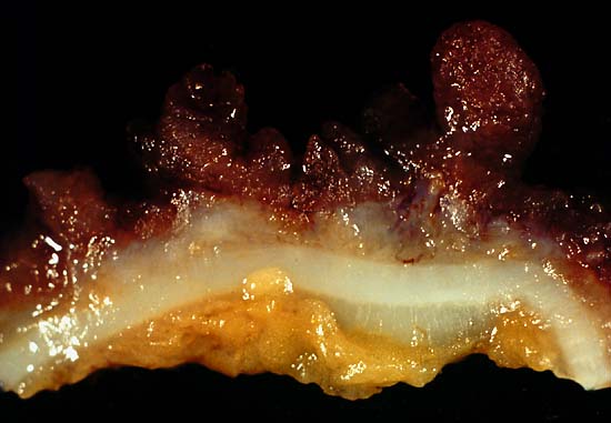
This photo is from a total colectomy done for severe, intractable chronic ulcerative colitis. It shows a close view of a lengthwise section through the colon wall. This demonstrates not only the angry red mucosa, but also, the tendency for the inflamed tissue to throw itself up into inflammatory pseudopolyps. Source: Ed Uthman, MD. Public domain.
What Is Ulcerative Colitis?
Ulcerative colitis is an inflammatory disorder of the colon characterized by continuous inflammation of the mucosa and submucosa usually with small ulcers, extending from the rectum and typically involving the distal colon, rectum, and anus and producing bloody diarrhea.
While the severity of ulcerative colitis varies among patients, iron deficiency anemia often develops due to blood loss especially when there are many bloody bowel movements in a day.
The onset of ulcerative colitis is most commonly in young adulhood.
Q: Is this disease painful?
A: Yes with the passage of stool.
Psychological stress and subsequent dysbiosis exacerbate ulcerative colitis.
Psychological stress activates multiple physiological processes aimed at maintaining balance within the body. These physiological processes also have the capacity to influence the composition of microbial communities in the digestive tract, and research now indicates that exposure to stressful stimuli leads to gut microbiota dysbiosis.1
While the relative abundance of many different bacterial types can be altered during stressor exposure, findings in nonhuman primates and laboratory rodents, as well as humans, indicate that bacteria in the genus Lactobacillus are consistently reduced in the gut during stress.2
Presently, ulcerative colitis cannot be cured. This condition has a course of remissions, when symptoms subside, and flares, when symtpoms get worse. Treatment is aimed to reduce flares and promote remission. In all cases, correction of dysbiosis improves the condition.
Ulcerative colitis is associated with increased incidence of cancer of the colon.3
What Is Ulcerative Colitis In Celiac Disease and/or Gluten Sensitivity?
Sources:
Galley JD, Bailey MT. Impact of stressor exposure on the interplay between commensal microbiota and host inflammation. Gut Microbes. 2014 May 1;5(3):390-396. Epub 2014 Apr 1. [↩]
Galley JD, Bailey MT. Impact of stressor exposure on the interplay between commensal microbiota and host inflammation. Gut Microbes. 2014 May 1;5(3):390-396. Epub 2014 Apr 1. [↩]
Taber’s Cyclopedic Medical Dictionary. F. A. Davis. Philadelphia, PA [↩]
M ultiple gastric ulcerations denote a stomach disorder that involves damage to the superficial mucosa characterized by many sores of the stomach antrum that are marked by inflammation, necrosis (death of the affected cells) and sloughing of destroyed tissue.
Q: Where is the stomach antrum?
A: The antrum is the lower region of the stomach before the pylorus which is nearest the duodenum (first part of the small intestine). The stomach and duodenum are separated by the powerful pyloric sphincter.
The stomach antrum propels food in the stomach against the pylorus which resists passage of food until it is turned into chyme. Chyme is highly acidic liquified food that has been thoroughly mixed with stomach juices.
Gastric ulcerations are typically associated with widespread gastritis (inflammation), inflammatory involvement of acid producing cells, and atrophy of acid and pepsin producing cells.1
The primary causes of gastric ulcerations are H. pylori infection, use of Aspirin and non-steroidal drugs (NSAIDS), and stress.1
What Are Multiple Gastric Ulcerations In Celiac Disease and/or Gluten Sensitivity?
What Is Helicobacter Pylori (H. Pylori) Infection? Helicobacter pylori infection is a potentially deadly stomach disease characterized by chronic superficial inflammation and ulcerations in 100% of infected patients. This infection disrupts normal defense and repair…
Obesity is an inflammatory metabolic disorder that is characterized by body mass index greater than 30% resulting from excessive body fat stored in adipose tissue.
Q: What is body fat?
A: Body fat is part of the body that functions as a reserve of stored energy. It is composed of fat cells, called adipocytes, having thin membranes between these cells. Adipocytes expand to store fat and shrink as fat is released as needed into the bloodstream for other body cells to use for metabolizing energy.
Each adipocyte contains a drop of triglyceride which is a type of lipid (fat). Triglycerides are a normal component in the bloodstream and, as such, are transported wherever needed as a form of energy. Excess triglycerides are the form of fat that is stored.
Initially, fat that is eaten in the diet is changed by digestive enzymes into the triglyceride form which is a molecule composed of three fatty acids and glycerol. Triglycerides are then absorbed through the small intestinal wall to be delivered to the liver. Of note, the liver can make triglycerides from excess protein and carbohydrates eaten in a meal, especially sugar and alcohol. The liver on the other hand makes cholesterol from triglycerides.
Triglyceride levels in the blood generally increase as weight increases. It is thought that an elevated blood triglyceride level hampers the body’s ability to feel full or satisfied with food that is eaten. Elevated triglyceride levels also increase the risk of clot formation because they cause the blood to become thicker. A normal triglyceride blood level is 150 mg/dL.
The causes of obesity are complex and varied. Those related to gluten sensitivity are discussed below.
What Is Obesity In Celiac Disease and/or Gluten Sensitivity?