Skip to content

Antidiarrheals: definition

bronchoalveolitisWhat Is Lymphocytic Bronchoalveolitis?

L ymphocytic bronchoalveolitis is a pulmonary disorder characterized by lymphocytic airway inflammation of the bronchi (main passageways branching from the windpipe), bronchioles (small passageways), and alveoli (air sacs) at the end.

The chronic, productive cough that develops and worsens with progression is due to increased mucus secretion in the large and small airways.

If infection develops in the bronchioles, it is call bronchopneumonia. If infection develops in the alveoli, it is called interstitial pneumonia. The most common organism causing infection is staphylcoccus aureus.

Q: What effect does inflammation have on the lungs?

A: Alveoli are air sacs, each only one cell thick, where oxygen is obtained by the bloodsteam from air breathed into them and carbon dioxide is released from the bloodstream to air that is breathed out of air sacs. Two things happen:

  1. Inflammation  impairs exchange of gases in alveoli, resulting in lack of sufficient oxygen (O2) for body cell functions, called hypoxia, and build-up of carbon dioxide (CO2) in blood, called CO2 retention.
  2. Inflammation narrows passageways, which reduces the movement of air to and from the alveoli, and this puts stress on the right side of the heart.

Lymphocytes, which are a type of white blood cell, can be collected and assessed by a procedure called bronchoalveolar lavage to determine the degree of involvement.

What Is Lymphocytic Bronchoalveolitis In Celiac Disease and/or Gluten Sensitivity?

Antiemetics: definition

Autopsy View of Lung Abcess.
Autopsy View Of Lung Cavity.

What Is Abscess Formation Causing Lung Cavities?

A bscess formation and subsequent lung cavities that develop from necrosis (death) of affected lung tissue constitute severe life-threatening respiratory disease  of the lung.

Q: What is the difference between lung abscess and lung cavity?

A: Lung abscess is characterized by localized pus formation in the lung that develops from uncontrolled infection from encapsulated bacteria such as streptococcus pneumoniae and klebsiella. The capsules that surround bacteria protect them from phagocytosis by macrophages and neutrophils, white blood cells that are specialized for this normal defensive action. Phagocytosis is a seek and destroy mission.

Lung cavities are holes that form from dead tissue during the destructive abscess development, which involves significant inflammation and edema of the area. Lung cavities often cause death of the patient. The outlook is very poor.

What Is Abscess Formation Causing Lung Cavities In Celiac Disease and/or Gluten Sensitivity?

Antispasmodics: definition

Pneumococcus bacteria
Pneumococcal Bacteria Occuring in Pairs.

What Is Pneumoccocal Septicemia?

P neumococcal septicemia (sepsis) is a life-threatening syndrome characterized by a cascade of systemic (body-wide) inflammatory responses to lung infection caused by bacterial infection spread to the bloodstream.

It is fatal in about 40% of cases, showing inadequate blood flow to internal organs. About 400,000 people die of sepsis each year in the United States.

Q: What is a pneumococcus?

A: A pneumococcus is an oval shaped , encapsulated, non-spore forming, gram positive bacterium of the genus Streptococcus. These bacteria usually occur in pairs.1

Infection can be prevented with pneumococcal vaccination.

What Is Pneumococcal Septicemia In Celiac Disease and/or Gluten Sensitivity?

Sources:
  1. Taber’s Encyclopedic Medical Dictionary. 10th ed., Phila. []

Antigen, Immunogen Type: definition

Pulmonary Hemosiderosis. Courtesy Quizlet.com
Lung from a Patient who Had Pulmonary Hemosiderosis. Courtesy Quizlet.com

What Is Idiopathic Pulmonary Hemosiderosis?

P ulmonary hemosiderosis (IPH) is a rare and severe pulmonary syndrome characterized by a triad of recurrent episodes of alveolar hemorrhage (bleeding into air sacs), hemoptysis (coughing blood), and iron deficiency anemia.

Q: What is the prognosis for pulmonary hemosiderosis?

A: Untreated pulmonary hemosiderosis ends in death. Idiopathic is a term that means the cause of a condition is not known.

What Is Pulmonary Hemosiderosis In Celiac Disease and/or Gluten Sensitivity?

Antibody: definition

Image shows
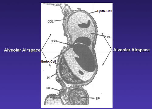
Image Showing  Blood-Air Barrier in the Lung. Courtesy quizlet.com

What Is Increased Pulmonary Permeability?

I ncreased pulmonary permeability is a mucosal defect in the lung characterized by alteration of the normally tight epithelial  blood-air barrier in the lung apparently caused by inflammation.

Q: What is the blood-air barrier in the lung?

A: This blood-air barrier in the lung consists of the alveolar epithelium (surface cells of the alveoli), the underlying capillary endothelium (surface cells of the capillaries), their basement membranes and the interstitial space between the cell layers.

Alveoli are also called “air sacs.” The exchange of oxygen breathed into air sacs from air on inspiration and carbon dioxide breathed out from capillary blood on expiration occurs between  air sacs and capillaries.

The capillary endothelium prevents proteins in blood from leaking (permeating) into the air sacs while allowing water and small molecules to pass. This is why breath has moisture in it.

Little is known about the interactions between the alveolar and the blood compartment.1

What Is Increased Pulmonary Permeability In Celiac Disease and/or Gluten Sensitivity?

Sources:
  1. Neuhaus W, Samwer F, Kunzmann S, Muellenbach RM, Wirth M, Speer CP, Roewer N, Förster CY. Lung endothelial cells strengthen, but brain endothelial cells weaken barrier properties of a human alveolar epithelium cell culture model. Differentiation. 2012 Nov;84(4):294-304. doi: 10.1016/j.diff.2012.08.006. Epub 2012 Sep 27. []

Antibody, Immunoglobulin A: definition

Bacteria that causes tuberculosis. Courtesy Wikimedia.

What Is Increased Susceptibility To Tuberculosis?

T uberculosis (TB), is an infectious disease caused by a bacteria called mycobacterium tuberculosis. It is characterized by chronic bacterial infection most commonly affecting lungs that develops in stages.

Increased susceptibility to tuberculosis menas the person’s defense mechanisms against developing infection are inadequate. Tuberculosis may be dormant or active.

Q: What happens in active tuberculosis?

A: Active tuberculosis  produces inflammation and formation of tubercles, necrosis (death of tissues), abcess, fibrosis, and calcification. Calcification is the body’s action to encapsulate the bacterial invasion. Active tuberculosis is life-threatening and may result in death.

About one third of the world’s population is infected with tuberculosis bacteria. In 2012 the number reached a staggering 8.6 million people. Of these, 1.3 million people died from tuberulosis.  About 95% of tuberulosis deaths occur in low- and middle-income countries and it is among the top three causes of death among women aged 15 to 44.1

People with weakened immune systems have a much greater risk of falling ill from tuberculosis. For example, a person living with HIV is about 20 to 30 times more likely to develop active tuberculosis.2 The combination of tuberculosis, HIV coinfection, and malnutrition has been commonly termed as “triple trouble.”3

What Is Increased Susceptibility To Tuberculosis In Celiac Disease and/or Gluten Sensitivity?

Sources:
  1. http://www.who.int/features/factfiles/tb_facts/en/index.html []
  2. http://www.who.int/features/factfiles/tuberculosis/en/ []
  3. Steinbrenner H, Al-Quraishy S, Dkhil MA, Wunderlich F, Sies H. Dietary selenium in adjuvant therapy of viral and bacterial infections. Adv Nutr. 2015 Jan 15;6(1):73-82. doi: 10.3945/an.114.007575. Print 2015 Jan. []

Antibody, Immunoglobulin G: definition

Chest x-ray showing tuberculosis. Courtesy wikipedia.
Chest x-ray showing tuberculosis with consolidation of left lung (white area). Courtesy wikipedia.

What Is Non-Response to Tuberculosis Treatment?

N on-response to treatment for tuberculosis means that proper medical treatment failed to control active disease. Tuberculosis is an infection that may be dormant or active.

Q: What happens in active tuberculosis?

A: Tuberculosis is an infectious disease caused by a bacteria called mycobacterium tuberculosis. It is characterized by chronic bacterial infection most commonly affecting lungs that develops in stages.

Active tuberculosis  produces inflammation and formation of tubercles, necrosis, abcess, fibrosis, and calcification. Calcification is the body’s action to encapsulate the bacterial invasion. Active tuberculosis is life-threatening and may result in death.

About one third of the world’s population is infected with tuberculosis bacteria. In 2012 the number reached a staggering 8.6 million people. Of these, 1.3 million people died from tuberulosis.  About 95% of tuberulosis deaths occur in low- and middle-income countries and it is among the top three causes of death among women aged 15 to 44.1

People with weakened immune systems have a much greater risk of falling ill from tuberculosis. For example, a person living with HIV is about 20 to 30 times more likely to develop active tuberculosis.2

What Is Non-Response To Tuberculosis Treatment In Celiac Disease and/or Gluten Sensitivity?

Sources:
  1. http://www.who.int/features/factfiles/tb_facts/en/index.html []
  2. http://www.who.int/features/factfiles/tuberculosis/en/ []

Antibody, Immunoglobulin E: definition

Classic Bitot's spot in a 29-year-old man that shrunk dramatically with vitamin A therapy. (Sommer A: Nutritional Blindness: Xerophthalmia and Keratomalacia. New York, Oxford University Press, 1982)
Classic Bitot’s spot in a 29-year-old man that shrunk dramatically with vitamin A therapy.*

What Are Bitot’s Spots?

B itot’s spots are superficial foamy patches that develop on the exposed bulbar conjunctiva (white of the eye) as a manifestation of advanced vitamin A deficiency.

This painless eye disorder is reversible only with vitamin A therapy. It is named after Charles Bitot, who first described it.

Q: What makes up these foamy alterations of the conjunctiva?

A: Bitot’s spots are composed of epithelial debris (dead surface cells) and secretions.1

Bitot’s spots may develop in malnutrition, reduced intake including alcoholism, medication adverse effect, old age, low stomach acid, and disease causing vitamin A deficiency such as celiac disease, Crohn’s disease, pancreatic insufficiency, and short bowel syndrome.  Other conditions associated with vitamin A deficiency may include disordered transport (Abetalipoprotenemia, a genetic disorder) and reduced liver storage caused by liver disease.

What Are Bitot’s Spots In Celiac Disease and/or Gluten Sensitivity?

Sources:
  1. Sadowski B, Rohrback JM, Steuhl KP, Weidle EG, Castrillon-Obendorfer WL. Corneal manifestations in Vitamin A deficiency. Klinische Monatsblatter fur Augenheilkunde. Aug 1994;205(2)76-85. []

Antrum: definition

Photo showing swollen eyelids in blepharitis. Courtesy Wikimedia
Swollen Eyelids in Blepharitis. Courtesy Wikimedia

What Is Non-Infectious Blepharitis?

N on-infectious blepharitis is an inflammatory condition of the eyelids that is NOT caused by an invading organism but rather nutritional deficiencies. It is characterized by non-ulcerative inflammation of the hair follicles and lacrimal glands along the eyelid edges.

Q: What  is the difference between inflammation and an invading organism which causes inflammation?

A: An invading organism such as Staphylococcus aureus, a common bacteria on the body’s surface, is called a stye. Styes are treated with antibiotics and resolve. Whereas, non-infectious blepharitis does not respond to antibiotics because there is no infection.

The eyelids are an important structural defense against infection and threats to the eye through blinking and tear production. Their continual pattern of blinking every few seconds sweeps away debris and keeps the eye moistened with tears.

Tears are made by the meibomian glands in the eyelids and consist of fatty acids, water and mucous and also contain anti-microbial substances.

Failure of the meibomian glands to function properly, called meibomian gland dysfunction or MGD, is a chronic, diffuse abnormality characterized by terminal duct obstruction (plugs) and/or changes in the amount or quality of the glandular secretion called meibom (tears). This may result in alteration and/or insufficiency of the tear film, symptoms of eye irritation, clinically apparent inflammation, and disease of the eye surface.

What Is Non-Infectious Blepharitis In Celiac Disease and/or Gluten Sensitivity?

Anus: definition

Dry Eye Due to Riboflavin Deficiency. GFW

What Is Chronic Dry Eye Syndrome?

C hronic dry eye syndrome is an inflammatory condition of the conjunctiva with inadequate tear production or distribution over the eye surface. It is characterized by surface damage to the conjunctiva, giving the appearance of redness.

This complex condition involves the lacrimal glands, eyelids, and tear film, as well as a variety of eye surface tissues, including epithelial, inflammatory, immune, and goblet cells.1

Q: What is the conjunctiva?

A: The conjunctiva is the mucous membrane that lines the inside of the eyelids and covers the eyeball surface, also called the white of eye. The conjunctiva has a rich blood supply that can quickly bring microbe fighting cells to prevent invasion and overcome infection.

While infection and inflammation go hand-in-hand, they are not the cause of, but may result from, chronic dry eyes.

Dry eye syndrome develops from poor health of the conjunctiva that is associated with inadequate nutrition and poor quality of the tears that steadily lubricate the eye surface. These tears are called the tear film and are not the same as tears produced when crying or that result from irritating substances such as onions. Blinking spreads the tears that protect the surface from drying and flushes away unwanted matter.

The tear film is made up of three layers: mucous layer produced by goblet cells in the conjunctiva, aqueous (water) layer produced by the lacrimal glands, and lipid (fat) layer produced by the meibomian glands in the eyelids. Dysfunction of any of these layers impairs the the tear film. Without an adequate tear film the conjunctiva becomes dry and inflamed.

When the meibomian glands fail to produce sufficient lipid that is clear and fluid but rather cloudy and thick, the condition is called meibomian gland dysfunction (MGD). The main cause of MGD is hyperkeratinization (thickening of the glands) and its related pathogenesis (for example, ductal dilatation and acinar atrophy). Other disorders such as atopy, pemphigoid acne, rosacea, and seborrhea are related to MGD and may result in a chronic inflammation of the eye surface.2

MGD is “the most underrecognized, underappreciated and undertreated disease in ophthalmic care. It is so common as to be taken as ‘normal’ in many clinical practices,” according to Joseph Tauber, MD, an anterior segment subspecialist and refractive surgeon in Kansas City, Mo.3

Untreated dry eye progresses to xerophthalmia, a condition of extreme drying and thickening of the conjunctiva characterized by hazy, dry cornea. Other causes of dry eye, beside celiac disease, include certain systemic conditions such as Sjogren’s syndrome, an autoimmune disease.

What Is Chronic Dry Eye Syndrome In Celiac Disease and/or Gluten Sensitivity?

Sources:
  1. Liu A, Ji J. Omega-3 essential fatty acids therapy for dry eye syndrome: a meta-analysis of randomized controlled studies. Med Sci Monit. 2014 Sep 6;20:1583-9. doi: 10.12659/MSM.891364. []
  2. Oleñik A, Jiménez-Alfaro I, Alejandre-Alba N, Mahillo-Fernández I. A randomized, double-masked study to evaluate the effect of omega-3 fatty acids supplementation in meibomian gland dysfunction. Clin Interv Aging. 2013;8:1133-8. doi: 10.2147/CIA.S48955. Epub 2013 Aug 30. []
  3. Rethinking Meibomian Gland Dysfunction: How to Spot It, Stage It and Treat It. Linda Roach, PhD. OPHTHALMIC PEARLS. []