Skip to content

Turner’s Syndrome 

Hyperkeratosis Back of Arm. GFW Photo.
Hyperkeratosis Back of Arm. GFW Photo

What Is Follicular Hyperkeratosis?

F ollicular hyperkeratosis is an abnormal skin condition characterized by disordered overgrowth of the horny layer of the epidermis with horny plugs filling the openings of hair follicles. The plugs look like bumps in hair follicles.

Follicular hyperkeratosis with corkscrew hairs (coiled hairs in the follicles) appears in scurvy, which is advanced vitamin C deficiency.1

Typical  hyperkeratotic papules first appear on the extensor surfaces of the extremities (e.g. the back of upper arms), shoulders, and buttocks.

Phrynoderma is a distinctive form of follicular hyperkeratosis with papule formation in dilated hair follicles believed to be a manifestation of severe malnutrition.

Q: Why does hyperkeratosis develop?

A: Follicular hyperkeratosis develops from lack of necessary nutrients to support healthy skin. Although originally thought to represent only vitamin A deficiency or vitamin C in the case of scurvy, several studies have demonstrated deficiencies of B vitamins and vitamin E. General malnutrition seems to be the strongest association. The clinical picture typically improves with enhanced nutritional status.2

What Is Follicular Hyperkeratosis In Celiac Disease and/or Gluten Sensitivity?

Sources:
  1. Krause’s Food, Nutrition, & Diet Therapy. 10th Edition. Kathleen Mahan, Sylvia Escott-Stump. 2000. W.B. Saunders Company. []
  2. Maronn M, Allen DM, Esterly NB. Phrynoderma: a manifestation of vitamin A deficiency?… The rest of the story. Pediatr Dermatol. 2005 Jan-Feb;22(1):60-3. []

Short Stature

What Is Vitiligo? V itiligo is a pigmentation disorder of the skin characterized by permanent loss of melanocytes in defined areas and, in some patients, antibodies to melanin. Vitiligo has significant psychological impact if occurring before… 

Smell, Loss of  

CT Scan of Parathyroid Carcinoma Displacing the Trachea. Courtesy Orphanet Journal of Rare Diseases
CT Scan of Parathyroid Carcinoma Displacing the Trachea by Doina Piciu. Courtesy Orphanet Journal of Rare Diseases

What Is Parathyroid Carcinoma?

P arathyroid carcinoma is a slow growing rare malignancy involving overactive parathyroid glands and is characterized by profound hypercalcemia (elevated blood calcium level), parathyroid hormone levels of more than 3 times upper normal limits, and palpable neck mass.1

Parathyroid carcinoma is a cause of severe hyperparathyroidism. Hyperparathyroidism results from overproduction of parathyroid hormone by the parathyroid glands.

Research comparing patients with carcinoma to patients with primary hyperparathyroidism showed that patients with parathyroid carcinoma had significantly higher serum parathyroid hormone and calcium levels compared with patients with primary hyperparathyroidism.2

Q: Why is the blood calcium level elevated?

A: Blood calcium level is elevated due to overproduction of parathyroid hormone by the diseased gland(s).

The function of parathyroid hormone (PTH) is to keep calcium blood levels normal, which it does by drawing calcium out of bones as needed. The problem of too much PTH is that bone is constantly robbed of calcium and this depletion causes bone demineralization, resulting in osteopenia progressing to osteoporosis. Of course, weakened bones are subject to breakage with light trauma.

On the other hand, too much calcium in the blood can cause kidney stone formation because urine production is the way the body excretes excess calcium. That is, highly concentrated calcium precipitates out of the urine solution to form stones.

With an estimated incidence of 0.015 per 100,000 population and an estimated prevalence of .005% in the United States, parathyroid cancer is one of the rarest of all human cancers according to the National Cancer Institute.

What Is Parathyroid Carcinoma In Celiac Disease and/or Gluten Sensitivity?

Sources:
  1. Schaapveld M, Jorna FH, Aben KK, Haak HR, Plukker JT, Links TP. Incidence and prognosis of parathyroid gland carcinoma: a population-based study in The Netherlandsestimating the preoperative diagnosis. Am J Surg. 2011 Nov;202(5):590-7. doi: 10.1016/j.amjsurg.2010.09.025. Epub 2011 Aug 20. []
  2. Schaapveld M, Jorna FH, Aben KK, Haak HR, Plukker JT, Links TP. Incidence and prognosis of parathyroid gland carcinoma: a population-based study in The Netherlandsestimating the preoperative diagnosis. Am J Surg. 2011 Nov;202(5):590-7. doi: 10.1016/j.amjsurg.2010.09.025. Epub 2011 Aug 20. []

Sperm Abnormalities 

This 21 year-old woman (left) appears as old as her 70 year-old granny (right). Courtesy Prof Dr Chua Chung Nen
This 21 year-old woman (right) appears as old as her 70 year-old grandmother (left). Courtesy Prof Dr Chua Chung Nen

What Is Acquired Cutis Laxa?

A cquired cutis laxa is an uncommon skin disorder characterized by abnormal reduction and degeneration of elastic fibers of the skin that can appear simply as thick, saggy skin with loose folds to severe involvement showing a premature aged appearance.

Q: What are elastic fibers of the skin?

A: Elastic fibers of the skin are connective tissue found in the dermis, which is the layer of skin under the epidermis, or surface layer. They hold the shape of skin and are important for wound healing in the development of scars.

What Is Cutis Laxa In Celiac Disease and/or Gluten Sensitivity?

Delayed Puberty in Boys

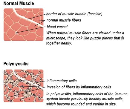
Drawing of Biopsy Showing Muscle Fibers Invaded by Immune Cells. Courtesy MDA.org

What Is Polymyositis?

P olymyositis is a body-wide connective tissue disease resulting from autoimmune attack of skeletal muscles that is characterized by inflammatory and degeneratory changes. The course is unpredictable being marked by spontaneous flare-ups and remissions.

Polymyositis can begin slowly or abruptly according to the factor that is triggering the onset such as infection, medications like phenytoin, and autoimmune disease.

Progressive muscle weakness starts in the proximal skeletal muscles (muscles closest to the trunk of the body).

Skeletal muscles, also called voluntary, are muscles that move the body as we want, such as walking and  lifting objects, as opposed to those we cannot voluntarily control, such as the muscles of digestion. 

Q: What are the degeneratory changes in skeletal muscles?

A: In polymyositis, degeneratory changes in skeletal muscles means that muscles are being destroyed (called necrosis), resulting in fibrosis, or scarring. When scar tissue takes the place of lost muscle tissue, it cannot act like muscle to contract and relax.   Muscle destruction is what causes muscle pain and weakness.

After the clinical work-up of exams and blood studies to determine muscle damage, the diagnosis of polymyositis is confirmed by muscle biopsy. See image at above left. The black dots are inflammatory cells. Edema (fluid) between cells caused by inflammation pushes muscle fibers apart.

There is no cure for polymyositis, but the symptoms can be treated.  Options include medication, physical therapy, exercise, heat therapy (including microwave and ultrasound), orthotics and assistive devices, and rest.  The standard treatment for polymyositis is a corticosteroid drug, given either in pill form or intravenously.  Immunosuppressant drugs, such as azathioprine and methotrexate, may reduce inflammation in people who do not respond well to prednisone. 

Periodic treatment using intravenous immunoglobulin can also improve recovery.  Other immunosuppressive agents used to treat the inflammation associated with polymyositis include cyclosporine A, cyclophosphamide, and tacrolimus.  Physical therapy is usually recommended to prevent muscle atrophy and to regain muscle strength and range of motion.1

Diagnosis is based on elevated muscles enzymes, increased urinary creatine level, and electromyograph abnormalities.

Polymyositis can affect people at any age. It is most common in adults between ages 50 and 70, and in children ages 5 to 15. It affects women twice as often as men and is more common in African Americans than Caucasians.2 The major causes of death from polymyositis are cancer and lung disease, including pneumonia.  The 5-year mortality rate can be as high as 1 in 5 patients.2

What Is Polymyositis In Celiac Disease and/or Gluten Sensitivity?

Sources:
  1. National Institute of Neurological Disorders and Stroke []
  2. http://www.nlm.nih.gov/medlineplus/ency/article/000428.htm [] []

Down Syndrome

Crohnie_sores_4[1]What Is Erythema Nodosum?

E rythema nodosum is an inflammatory disorder involving the deep dermis layer of skin and subcutaneous fat septa that underlies the skin. It is characterized by eruptions of recurrent or persistent multiple painful, red nodules under the skin that leave a bruised appearance when healing and do not scar.

The lower legs are most affected, but sores can appear anywhere there is subcutaneous fat.

Q: How do the nodules develop in erythema nodosum?

A: The edges of nodules are poorly defined, and the nodules vary from 2-6 cm.

During the first week of eruption, nodules become tense, hard, and painful. During the second week, they change color from bright red to bluish or livid and may become soft, but do not ulcerate. As absorption progresses, the color gradually fades to a yellowish hue, resembling a bruise. This disappears in 1 or 2 weeks as the overlying skin sloughs off and is replaced.1

The eruptive phase of erythema nodosum begins with flulike symptoms of fever and generalized aching followed by a painful rash within 1-2 days.  Aching legs and swelling ankles may occur and precede the eruption or appear during the eruptive phase and may persist for weeks.2

Currently, the most common cause of erythema nodosum is streptococcal infection in children and streptococcal infection and sarcoidosis in adults.3 Most sores in infection-induced erythema nodosum heal within 7 weeks, but active disease may last up to 18 weeks.

In contrast, 30% of idiopathic erythema nodosum cases may last more than 6 months. Idiopathic means that the cause is not known.

What Is Erythema Nodosum In Celiac Disease and/or Gluten Sensitivity?

Sources:
  1. http://emedicine.medscape.com/article/1081633-clinical#a0217 []
  2. http://emedicine.medscape.com/article/1081633-clinical []
  3. http://emedicine.medscape.com/article/1081633-overview#a0199 []

Congenital Anomalies 

Hangnail on lowest finger. Courtesy Wikipedia.org

What Is Hangnail?

H angnail is a broken strip of epidermis (piece of skin) at root or lateral (side) edge of fingernail or toenail that causes sharp pain.

A hangnail develops because the skin around the nail is unhealthy due to inadequate nutrition. Injury from trauma including biting the skin and pushing back the cuticles or exposure to excessive detergents and water that remove protective oils promote the development of hangnail.

All ages and both sexes can be affected.

Q: Can a hangnail become infected?

A: Infection, called paronychia, may develop from invasion of sore skin by any of these pathogens: bacteria, fungus, or yeast (Candida). Infected skin is red, swollen, and painful. Topical ointment is required to treat the infection.1

What Is Hangnail In Celiac Disease and/or Gluten Sensitivity?

Sources:
  1. www.nlm.nih.gov/medlineplus/ency/article/001444.htm []

Failure To Thrive And Growth Retardation

Hyde's Prurigo. Courtesy quizlet.com
Hyde’s Prurigo. Courtesy quizlet.com

What Is Prurigo Nodularis (Hyde’s Prurigo)?

P rurigo nodularis is a chronic dermatitis characterized by hard, dry, deep seated, intensely itchy papules (small bumps like pimples) and/or nodules (large bumps) that erupt most commonly on the arms, legs, and back.

Papules and nodules vary in number and may become infected after picking or scratching.

Q: Does the itching go away?

A: New nodules develop from time to time, and existing nodules may remain itchy indefinitely, although some may regress spontaneously and leave scars. In most cases, the disease runs a very protracted course with exacerbations and remissions.1

Prurigo nodularis is an unusual disorder of unknown etiology, which is notoriously resistant to therapy. A variety of systemic conditions have been reported to be associated with prurigo nodularis. However, the mechanism by which these disorders may trigger prurigo nodularis is unknown.2

It has been shown to be associated with malnutriton and infection such as tonsillitis, which resolved after removal of tonsils.3

What Is Prurigo Nodularis In Celiac Disease and/or Gluten Sensitivity?

Sources:
  1. Katotomichelakis M, Balatsouras DG, Bassioukas K, Kontogiannis N, Simopoulos K, Danielides V. Recurrent prurigo nodularis related to infected tonsils: a case report. J Med Case Rep. 2008 Jul 24;2:243. doi: 10.1186/1752-1947-2-243. []
  2. Lee MR, Shumack S. Prurigo nodularis: a review. Australas J Dermatol. 2005 Nov;46(4):211-18; quiz 219-20. []
  3. Katotomichelakis M, Balatsouras DG, Bassioukas K, Kontogiannis N, Simopoulos K, Danielides V. Recurrent prurigo nodularis related to infected tonsils: a case report. J Med Case Rep. 2008 Jul 24;2:243. doi: 10.1186/1752-1947-2-243. []