Skip to content

Smell, Loss of  

CT Scan of Parathyroid Carcinoma Displacing the Trachea. Courtesy Orphanet Journal of Rare Diseases
CT Scan of Parathyroid Carcinoma Displacing the Trachea by Doina Piciu. Courtesy Orphanet Journal of Rare Diseases

What Is Parathyroid Carcinoma?

P arathyroid carcinoma is a slow growing rare malignancy involving overactive parathyroid glands and is characterized by profound hypercalcemia (elevated blood calcium level), parathyroid hormone levels of more than 3 times upper normal limits, and palpable neck mass.1

Parathyroid carcinoma is a cause of severe hyperparathyroidism. Hyperparathyroidism results from overproduction of parathyroid hormone by the parathyroid glands.

Research comparing patients with carcinoma to patients with primary hyperparathyroidism showed that patients with parathyroid carcinoma had significantly higher serum parathyroid hormone and calcium levels compared with patients with primary hyperparathyroidism.2

Q: Why is the blood calcium level elevated?

A: Blood calcium level is elevated due to overproduction of parathyroid hormone by the diseased gland(s).

The function of parathyroid hormone (PTH) is to keep calcium blood levels normal, which it does by drawing calcium out of bones as needed. The problem of too much PTH is that bone is constantly robbed of calcium and this depletion causes bone demineralization, resulting in osteopenia progressing to osteoporosis. Of course, weakened bones are subject to breakage with light trauma.

On the other hand, too much calcium in the blood can cause kidney stone formation because urine production is the way the body excretes excess calcium. That is, highly concentrated calcium precipitates out of the urine solution to form stones.

With an estimated incidence of 0.015 per 100,000 population and an estimated prevalence of .005% in the United States, parathyroid cancer is one of the rarest of all human cancers according to the National Cancer Institute.

What Is Parathyroid Carcinoma In Celiac Disease and/or Gluten Sensitivity?

Sources:
  1. Schaapveld M, Jorna FH, Aben KK, Haak HR, Plukker JT, Links TP. Incidence and prognosis of parathyroid gland carcinoma: a population-based study in The Netherlandsestimating the preoperative diagnosis. Am J Surg. 2011 Nov;202(5):590-7. doi: 10.1016/j.amjsurg.2010.09.025. Epub 2011 Aug 20. []
  2. Schaapveld M, Jorna FH, Aben KK, Haak HR, Plukker JT, Links TP. Incidence and prognosis of parathyroid gland carcinoma: a population-based study in The Netherlandsestimating the preoperative diagnosis. Am J Surg. 2011 Nov;202(5):590-7. doi: 10.1016/j.amjsurg.2010.09.025. Epub 2011 Aug 20. []

Anti-Endomysium Antibodies (EMA) Present In Blood

Smooth muscle. Courtesy pathguy.com
Smooth Muscle. Courtesy pathguy.com

What Are Anti-Endomysium Antibodies?

Anti-endomysium antibodies (EmA) are connective tissue autoantibodies produced in persons who have inherited the genes for celiac disease, an autoimmune disease, and are reacting to gluten in their diet.

Autoantibodies are abnormal in that they attack the body’s own tissue.

Q: What is endomysium?

A: Endomysium is the delicate connective tissue that surrounds individual muscle fibers. The autoantigen, or target, that stimulates the autoimmune response is the naturally occuring enzyme in endomysium called tissue transglutaminase (tTG), or more specifically tranglutaminase-2 (TG2). Anti-tissue transglutaminase antibodies recognize the same antigen as EmA, from which they differ in terms of detection method.

Anti-endomysium antibodies (EmA) are tested by the indirect immunofluorescence method and directed against “reticulin-like” fibres in connective tissue around smooth muscle fibres in the esophagus, liver, stomach, and bladder of monkeys, in the sections of the jejunum and kidneys of rats and in sections of the human umbilical cord. In comparison, for the determination of anti-tissue transglutaminase IgA and IgG antibodies, ELISA with human extractive or recombinant transglutaminase is recommended.1

EmA‐binding patterns in serum samples from patients with celiac disease have proved to be exclusively TG2‐targeted and the correlation between EmA and TG2 antibodies is therefore good. Evidence shows that celiac autoantibodies are produced in the small‐bowel mucosa.2

What Are Anti-Endomysium Antibodies In Celiac Disease and/or Gluten Sensitivity?

Sources:
  1. Trigoni E, Tsirogianni A, Pipi E, Mantzaris G, Papasteriades C. Celiac disease in adult patients: specific autoantibodies in the diagnosis, monitoring, and screening. Autoimmune Dis. 2014;2014:623514. doi: 10.1155/2014/623514. []
  2. Salmi TT, Collin P, Korponay-Szabó IR, Laurila K, Partanen J, Huhtala H, Király R, Lorand L, Reunala T, Mäki M, Kaukinen K. Endomysial antibody‐negative coeliac disease: clinical characteristics and intestinal autoantibody deposits.Gut. 2006 Dec;55(12):1746-53. []

Anti-Gliadin Antibodies Present

This is a model of Immunoglobulin-G Antibody (IgG)

What Are Anti-Gliadin Antibodies?

Anti-gliadin antibodies (AGA) are produced by the body as an immune response against partially digested gliadin peptides (fragments) that have abnormally entered the body from the intestinal tract. Gliadin is a protein in wheat grain.

Anti-gliadin antibodies circulating in blood are measured by laboratory testing on blood that is drawn. The antibodies are both immunoglobulin A (IgA) and immunoglobulin G (IgG) types, which are termed AGA-IgA and AGA-IgG.

The presence of anti-gliadin antibodies in blood is an abnormal laboratory finding. That is, when the intestinal lining is healthy and able to keep out undigested matter from the body, there are no molecules of gliadin present to trigger the immune system for defense.

Q: Who produces anti-gliadin antibodies?

A: Persons, with or without celiac disease, can produce anti-gliadin antibodies if and when leaky gut (increased intestinal permeability) develops that then allow gliadin peptides to get into the bloodstream.

Gliadin itself causes leaky gut, but there are other causes such as various foods, intestinal infections, medications, and other diseases of the gastrointestinal tract. Whatever the cause of leaky gut, if poorly digested gliadin is present, it can slip through the leaky lining and enter the bloodstream where it can trigger an antibody response by anti-gliadin antibodies.

What Are Anti-Gliadin Antibodies In Celiac Disease and/or Gluten Sensitivity Or Non-Celiac Gluten Sensitivity?

Anti-tissue Transglutaminase Antibodies (tTG) Present In Blood

Image depicts tissue transglutaminase (tTg).
Image Depicts Tissue Transglutaminase (tTG).

What Are Anti-tissue Transglutaminase Antibodies?

Anti-tissue transglutaminase antibodies (anti-tTG) are connective tissue autoantibodies and can be detected in blood samples from affected persons who are reacting to gluten in the diet.

Autoantibodies are abnormal because they attack the body’s own tissue, which in the case of these antibodies is tissue transglutaminase 2 (TG2).

Q: What is tissue transglutaminase 2 (TG2)?

A: Tissue transglutaminase 2 (TG2) is an enzyme that appears in many cell locations and is particularly abundant in endothelial cells that line the small intestine. It has been implicated in a variety of cellular processes, such as differentiation, cell death, inflammation, cell migration and wound healing.

The cell appears to adapt the dynamics of this enzyme to meet specific sub-cellular needs or to respond to stress or other stimuli. Substantial evidence indicates that the location of TG2 within cells is critical for the regulation of its various biochemical activities, which subsequently trigger diverse downstream events,1

Although initially studied as an enzyme within cells, TG2 is now known to be secreted also into the extracellular space (between cells) or onto the cell surface.1

Abnormal activation of TG2 or deregulation of its function(s) is involved in a variety of human diseases, such as celiac disease, diabetes, neurodegenerative diseases, multiple sclerosis and rheumatoid arthritis. A role in inflammatory disorders and septic shock has also been shown. Moreover, multiple studies have revealed elevated TG2 expression in many types of cancer cells.1

What Are Anti-tissue Transglutaminase Antibodies In Celiac Disease and/or Gluten Sensitivity?

Sources:
  1. Piacentini M, D’Eletto M, Farrace MG, Rodolfo C, Del Nonno F, Ippolito G, Falasca L. Characterization of distinct sub-cellular location of transglutaminase type II: changes in intracellular distribution in physiological and pathological states. Cell Tissue Res. 2014 Dec;358(3):793-805. doi: 10.1007/s00441-014-1990-x. [] [] []

Bone Alkaline Phosphatase (BALP), Elevated

Vertebrae. Courtesy FreePik.com

What Is Elevated Bone Alkaline Phosphatase?

Elevated bone alkaline phosphatase (BALP) is a laboratory result that indicates an abnormal blood level of this bone enzyme.

A bone alkaline phosphatase blood level is one of the most frequently used biochemical markers of bone formation.

Q: Why is the purpose of  bone alkaline phosphatase?

A: Bone alkaline phosphatase is produced by bone cells called osteoclasts in normal bone maintenance for the purpose of breaking down old or damaged bone so that other bone cells called osteoblasts can fill in the excavated areas with new bone. This process keeps bone stong and healthy.

Elevated bone alkaline phosphatase shows that more bone is being broken down than is being replaced. It can be caused by hyperparathyroidism, bone tumors from cancer, and malnutrition.

What Is Elevated Bone Alkaline Phosphatase (BALP) In Celiac Disease and/or Gluten Sensitivity?

Delayed Puberty in Boys

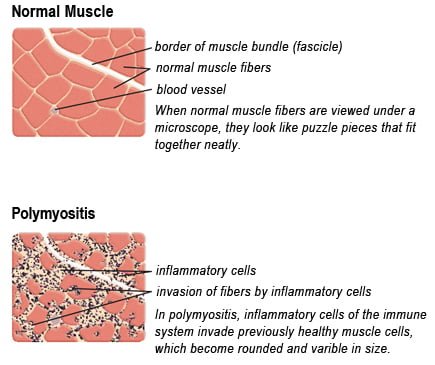
Drawing of Biopsy Showing Muscle Fibers Invaded by Immune Cells. Courtesy MDA.org

What Is Polymyositis?

P olymyositis is a body-wide connective tissue disease resulting from autoimmune attack of skeletal muscles that is characterized by inflammatory and degeneratory changes. The course is unpredictable being marked by spontaneous flare-ups and remissions.

Polymyositis can begin slowly or abruptly according to the factor that is triggering the onset such as infection, medications like phenytoin, and autoimmune disease.

Progressive muscle weakness starts in the proximal skeletal muscles (muscles closest to the trunk of the body).

Skeletal muscles, also called voluntary, are muscles that move the body as we want, such as walking and  lifting objects, as opposed to those we cannot voluntarily control, such as the muscles of digestion. 

Q: What are the degeneratory changes in skeletal muscles?

A: In polymyositis, degeneratory changes in skeletal muscles means that muscles are being destroyed (called necrosis), resulting in fibrosis, or scarring. When scar tissue takes the place of lost muscle tissue, it cannot act like muscle to contract and relax.   Muscle destruction is what causes muscle pain and weakness.

After the clinical work-up of exams and blood studies to determine muscle damage, the diagnosis of polymyositis is confirmed by muscle biopsy. See image at above left. The black dots are inflammatory cells. Edema (fluid) between cells caused by inflammation pushes muscle fibers apart.

There is no cure for polymyositis, but the symptoms can be treated.  Options include medication, physical therapy, exercise, heat therapy (including microwave and ultrasound), orthotics and assistive devices, and rest.  The standard treatment for polymyositis is a corticosteroid drug, given either in pill form or intravenously.  Immunosuppressant drugs, such as azathioprine and methotrexate, may reduce inflammation in people who do not respond well to prednisone. 

Periodic treatment using intravenous immunoglobulin can also improve recovery.  Other immunosuppressive agents used to treat the inflammation associated with polymyositis include cyclosporine A, cyclophosphamide, and tacrolimus.  Physical therapy is usually recommended to prevent muscle atrophy and to regain muscle strength and range of motion.1

Diagnosis is based on elevated muscles enzymes, increased urinary creatine level, and electromyograph abnormalities.

Polymyositis can affect people at any age. It is most common in adults between ages 50 and 70, and in children ages 5 to 15. It affects women twice as often as men and is more common in African Americans than Caucasians.2 The major causes of death from polymyositis are cancer and lung disease, including pneumonia.  The 5-year mortality rate can be as high as 1 in 5 patients.2

What Is Polymyositis In Celiac Disease and/or Gluten Sensitivity?

Sources:
  1. National Institute of Neurological Disorders and Stroke []
  2. http://www.nlm.nih.gov/medlineplus/ency/article/000428.htm [] []

Vitamin B12 Deficiency Anemia

What Is Vitamin B12 Deficiency Anemia? Vitamin B12 deficiency anemia is a megaloblastic anemia that is characterized by defective DNA synthesis of red blood cells due to a lack of vitamin B12. Vitamin B12 is essential… 

Anemia, Severe Iron Deficiency In Pregnancy 

Dermatitis Herpetiformis. Blisters Opened Giving Relief From Pain and Itching.
Dermatitis Herpetiformis On Forearm. Skin Is Darkened Where Old Blisters Healed.

What Is Dermatitis Herpetiformis?

D ermatitis herpetiformis (DH) is an autoimmune extremely itchy, painful bullous skin rash (blistering eruptions) arising from the underlying dermis layer of skin as a consequence of gluten sensitivity.

Dermatitis herpetiformis is characterized by multiple intensely itchy, red blisters appearing on the elbows which can extend down the forearm to the wrist and the knees. Less usual areas involve the back, buttocks, scalp, and abdomen.

Q: Do the blisters leave a mark when healed?

A: Crops of skin eruptions begin with itching or a burning sensation in reddened papules. There are grouped vesicles and tense blisters. The blister contents may be serous or bloody, with symmetrical distribution (eg, both knees or both elbows). Fluid filled elements rupture leaving denuded areas of sore skin and crust. Subsequently, there is residual hypopigmentation (a white area) or hyperpigmentation (dark area).1

Rupture of blisters begins relief from intense burning and itching.

Dermatitis Herpetiformis Eruptions On Knees.
Dermatitis Herpetiformis Eruptions On Knees. Notice White Areas Showing Loss of Pigmentation From Healed Blisters.

What Is Dermatitis Herpetiformis In Celiac Disease and/or Gluten Sensitivity?

Primary care providers should be aware of this skin condition, as they are more likely than a gastroenterologist to be confronted with this type of presentation of celiac disease.2

Sources:
  1. Mendes FB, Hissa-Elian A, de Abreu MA, Gonçalves VS. Review: dermatitis herpetiformis. An Bras Dermatol. 2013 Jul-Aug;88(4):594-9. []
  2. Robinson BL, Davis SC, Vess J, Lebel, J. Primary care management of celiac disease. Autoimmune Disorders. Nurse Practitioner. February 2015: Vol 40 – Issue 2; 28–34. []