Skip to content

Cheilosis (Cracks in corners of mouth)

cheilosisWhat Is Cheilosis?

Cheilosis is a painful feature of nutritional deficiencies, especially riboflavin and/or pyridoxine deficiency, and less frequently, from vitamin B12, folic acid, and iron. It is characterized by redness of the lips with cracking and weeping in the corner of the mouth.

Q: What is the consequence of weeping at the corners of the mouth?

A: The open sores that develop at the corners of the mouth invite infection by candida albicans, a yeast organism, and less commonly by bacteria normally present on the skin such as staphylococcus aureus.

These sores make opening the mouth very painful and so restrict eating, talking, socializing, and dental care.

What Is Cheilosis In Celiac Disease and/or Gluten Sensitivity?

Bleeding / Swollen Gums

Vitamin C Deficiency: Clean Teeth with Swollen Gums.
Vitamin C Deficiency: Swollen Gums.

What Are Bleeding / Swollen Gums?

Bleeding and/or swollen gums is  an inflammatory disorder characterized by changes in gum integrity that may result in tooth loss.

Sponginess of the gum (swelling) indicates inflammation, called gingivitis. 

Q: What causes gingivitis?

A: Gingivitis that is caused by inadequate dental hygiene shows as pockets of swollen gum between or along the base of teeth where sticky plaque was allowed to build up. Plaque holds bacteria that inflames the gums where it occurs.

Gingivitis that is caused by vitamin C deficiency, pregnancy, certain drugs such as the anti-epileptic drug phenytoin, and leukemia affects the whole gum irrespective of mouth care.

What Are Bleeding / Swollen Gums In Celiac Disease and/or Gluten Sensitivity?

Oral Mucosal Lesions, Chronic (Mouth Soreness)

Canker Sore Inside Mouth. Notice The White Spot on This Person's Nail Showing Zinc Deficiency.
Inflammation/Sore Inside Mouth. Notice White Spot on Fingernail Nail Showing Zinc Deficiency.

What Are Chronic Oral Mucosal Lesions?

Chronic lesions of the oral mucosa are disorders of the mouth that are characterized by soreness and sores of the soft mucosal lining of the mouth.

What Are Chronic Oral Mucosal Lesions In Celiac Disease and/or Gluten Sensitivity?

Hypophosphatemia (Low Phosphate Blood Level)

Testing Thigh Strength. Courtesy Charlie Goldberg, M.D., UCSD School of Medicine

What Is Hypophosphatemia?

Hypophosphatemia means the level of phosphates in the bloodstream is too low to meet metabolic needs of the body for this mineral.

Q: How important is phosphorus in metabolism?

A: Phosphorus is crucial to  life, being present in every cell of the body and constitutes 45% of skeletal bone weight along with 40% calcium needed to support the body as a framework.

A low blood phosphate level is characterized by alterations in blood acid-alkaline balance and serious neuromuscular, hematologic, renal, skeletal, and dental abnormalities.

Symptoms result primarily from decreased production of adenosine triphosphate (ATP), the main energy source in cells, and phosphocreatine, a secondary energy source for muscle contraction.

  • Acute phosphorus deficiency may precipitate rhabdomyolysis which is destruction of muscle.
  • Nervous system dysfunction is observed in severe hypophosphatemia.
  • Chronic phosphorus deficiency causes proximal myopathy (upper arms and thighs).1
  • Severe phosphorus deficiency has widespread and ultimately fatal consequences.

What Is Hypophosphatemia In Celiac Disease and/or Gluten Sensitivity?

Sources:
  1. Takeda E, Ikeda S, Nakahashi O. Lack of phosphorus intake and nutrition. Clin Calcium. 2012 Oct;22(10):1487-91. [↩]

Macroamylasemia

Depiction of pancreatic amylase molecule.

What Is Macroamylasemia?

Macroamylasemia is an acquired enzyme disorder that causes elevated levels of the enzyme amylase (hyperamylasaemia) in the bloodstream. It is characterized by altered amylase molecules that have become abnormally bound with plasma proteins in the bloodstream, commonly IgG (immunoglobulin G) and/or IgA (immunoglobulin A).

The resulting molecule is too large to be properly filtered by the kidneys and excreted in the urine as would be normal unbound amylase, causing sustained elevation of amylase levels in the plasma.

With normal kidney function, a hyperamylasemia without an increase in urine amylase suggests the diagnosis of macroamylasemia, and is confirmed by identifying the macromolecular components.1

Amylase is a an enzyme produced by the pancreas and the parotid glands to digest starch in the diet. Comparatively small amounts are also produced by other organs.

Q: How do the pancreas and parotid glands deliver their amylase for digestion of starch?

A: The parotid glands deliver their amylase into the mouth. The action of amylase in the mouth starts the breakdown of starch as it is chewed and moistened with saliva.

After the starchy food is liquified in the stomach, the mass empties into the duodenum. Here pancreatic amylase is delivered through the common bile duct into the duodenum where it turns starch into maltose sugar. The next step to final digestion of starch is carried out by enzymes in the small intestinal lining that split maltose into its component sugar which is glucose.

Glucose is a simple sugar that can be absorbed into the bloodstream for use in the body. Humans cannot live without adequate glucose.

Distinquishing macroamylasemia from hyperamylasemia due to pancreatic disease is necessary to prevent needless treatment and investigation for pancreatitis.2

What Is Macroamylasemia In Celiac Disease and/or Gluten Sensitivity?

Sources:
  1. Torrent Vernetta A, Segarra Cantón O, Soler Palacín P, Segura Cardona RM, Infante Pina D. Macroamylasaemia in paediatrics. An Pediatr (Barc). 2008 Nov;69(5):439-41. [↩]
  2. Isham CA, Ridgeway NA, Hedrick R, Cate JC 4th. Screening for macroamylase in a community hospital. Clin Chem. 1984 May;30(5):741-2. [↩]

Macrolipasemia

Lipase Enzyme. Courtesy johnsonmatthey.
Lipase Enzyme. Courtesy Johnson Matthey Catalysts.

What Is Macrolipasemia?

Macrolipasemia is a rare enzyme disorder characterized by altered molecules of lipase, a pancreatic enzyme needed to digest fats, that are abnormally bound with serum antibody proteins. These antibodies are commonly immunoglobulin G (IgG) and/or less likely immunoglobulin A (IgA).

Q: What happens when lipase is bound to immunoglobulins (IgA and/or IgG)?

A: The resulting molecule is too large to be filtered by the kidneys and excreted in the urine, consequently these abnormal molecules build up in the plasma causing sustained elevation of lipase levels called macrolipasemia.

Macrolipasemia occurs with or without macroamylasemia, which is the binding of immunoglobulin A and/or G to amylase, a pancreatic enzyme needed to digest starches.1

What Is Macrolipasemia In Celiac Disease and/or Gluten Sensitivity?

Sources:
  1. La Villa G, Pantaleo P, Tarquini R, Cirami L, Perfetto F, Mancuso F, Laffi G. Multiple immune disorders in unrecognized celiac disease: a case report. World J Gastroenterol. 2003;9(6):1377-1380, Available at: http://www.wjgnet.com/1007-9327/9/1377.asp. Accessed Jan 3, 2005. [↩]

Abdominal Distention, Chronic  (Bloating)

chronic abdominal distention celiac disease gluten symptomWhat Is Chronic Abdominal Distention?

Abdominal distention, or bloating, is characterized by enlargement in normal size of abdomen not due to fatty tissue.

Q: Why does the abdomen enlarge?

A: The abdomen enlarges because the bowel is dysfunctional. Loops of bowel usually lack normal peristalsis (rhythmic wave-like muscle movement) trapping gas in stagnant loops of bowel, inflammation swells loops of bowel also trapping gas, and dysbiosis overproduces gas. All these factors distend the abdomen.

What Is Chronic Abdominal Distention In Celiac Disease and/or Gluten Sensitivity?

Hyperprolactinemia (Excess Prolactin Hormone)

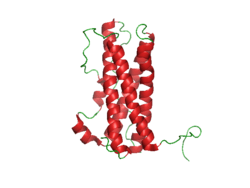
Prolactin Structure. Courtesy Boris TM at Wikipedia.

What Is Hyperprolactinemia?

Hyperprolactinemia is an excess secretion of prolactin hormone causing an elevated blood level that is characterized by its effects on reproduction: in females causing  amenorrhea and in males causing reduced sexual potency.

Q: What is prolactin?

A: Prolactin is a hormone produced by the the anterior pituitary gland that is transported by the bloodstream to stimulate androgen hormone production in male and estrogen hormone production in female sex glands. However, too much prolactin inhibits hormone production of estrogen in women and testosterone in men.

In pregnant females upon delivery of a baby, prolactin stimulates breast development and milk production for the purpose of feeding the infant. Sucking by the infant stimulates the release of prolactin which keeps the milk supply adequate.

Prolactin also promotes autoimmune mechanisms. Prolactin interferes specifically with B cell tolerance induction, enhances proliferative response to antigens and mitogens and increases the production of immune globulins, cytokines and autoantibodies.1

Other conditions that can stimulate prolactin release include nutritional deficiencies, thyroid stimulating hormone (TSH) produced by the pituitary gland, stress, exercise, and anterior pituitary tumors.

What Is An Elevated Prolactin Level In Celiac Disease and/or Gluten Sensitivity?

Sources:
  1. Shelly S, Boaz M, Orbach H. Prolactin and autoimmunity. Autoimmun Rev. 2012 May;11(6-7):A465-70. doi: 10.1016/j.autrev.2011.11.009. [↩]

Hangnail: definition

vitamin c gluten celiac disease symptomWhat Is Vitamin C?

V itamin C, also called ascorbic acid or ascorbate, is an essential water soluble vitamin.

Fresh supplies of vitamin C are required every day to perform vital roles throughout the body among which are the production of connective tissue such as skin, blood vessels and bone, anti-inflammatory responses and anti-oxidant performance. See below for all roles.

Vitamin C fights for us! It has an anti-cancer effect stemming from its role as a potent anti-oxidant in blood plasma and within cells. It also protects nucleic acids (DNA) from oxidative damage and inhibits the formation of nitrosamines (carcinogenic compounds formed in the digestive tract).

Importantly, vitamin C can regenerate vitamin E from an oxidized state after vitamin E performs its own anti-oxidant role against free radicals and vitamin C works together with other anti-oxidants, such as beta-carotene and glutathione, to increase their overall anti-oxidant effect.

A study investigating the specific anti-inflammatory and anti-oxidant micronutrients that reduce oxidative stress found that in adolescents with metabolic syndrome (MetS) the lower the vitamin C level in blood the worse the MetS status and blood uric acid level.  MetS prevalence was estimated at 7% among boys and 3% among girls.1

Vitamin C reverses osteoporosis by reducing or preventing oxidative stress that destroys living bone tissue. Osteoporosis is a disorder of bone inflammation that results in thin, weak bones that may easily fracture. In normal bone structure, vitamin C is required to form collagen, which is living bone tissue.

In humans, vitamin C reduces the duration of common cold symptoms, even if its effect is not clear.2

Vitamin C must be obtained from food daily because our bodies, unlike monkeys and other animals, cannot produce it.

Urinary excretion of vitamin C cannot be detected when vitamin intake is below the required levels. On the other hand, when intake exceeds saturation in the body, the vitamin and/or its metabolites are actively excreted into urine to prevent excessive toxicity of the vitamins.3

What Is Vitamin C Deficiency In Celiac Disease and/or Gluten Sensitivity?

Sources:
  1. TGBeydoun MA, Canas JA, Beydoun HA, Chen X, Shroff MR, Zonderman AB. Serum antioxidant concentrations and metabolic syndrome are associated among U.S. adolescents in recent national surveys. J Nutr. 2012 Sep;142(9):1693-704. doi: 10.3945/jn.112.160416. [↩]
  2. Shaik-Dasthagirisaheb YB, Varvara G, Murmura G, Saggini A, Caraffa A, Antinolfi P, Tete’ S, Tripodi D, Conti F, Cianchetti E, Toniato E, Rosati M, Speranza L,Pantalone A, Saggini R, Tei M, Speziali A, Conti P, Theoharides TC, Pandolfi F. Role of vitamins D, E and C in immunity and inflammation. J Biol Regul Homeost Agents. 2013 Apr-Jun;27(2):291-5. [↩]
  3. Shibata K, Hirose J, Fukuwatari T. Relationship Between Urinary Concentrations of Nine Water-soluble Vitamins and their Vitamin Intakes in Japanese Adult Males. Nutr Metab Insights. 2014 Aug 5;7:61-75. doi: 10.4137/NMI.S17245. eCollection 2014. [↩]