Skip to content

Candida Albicans Infection

Close-up shows inflammation and yellowish white patches of roof and back of the mouth caused by candidiasis. Uvula is greatly swollen, hanging near the level of the tongue. Courtesy: Wikipedia.
Close-up shows inflammation and infected patches on roof and back of the mouth caused by candidiasis. Uvula is swollen, hanging near the tongue. Courtesy: Wikimedia.

What Is Candida Albicans Infection?

[dropcap]C[/dropcap]andida albicans infection, called candidosis or candidiasis, is an opportunistic invasion of mucous membrane or skin by candida albicans, an endogenous yeast found in 40 to 80% of normal human beings. A former name for this small, budding fungus is monilia albicans.

Opportunistic means that yeast living on mucosal and skin surfaces does not invade (infect) unless these tissues become unhealthy and therefore cannot protect themselves.

Q: How does candida albicans cause infection?

A: Candida albicans lives on the mucosal surfaces and skin in most people without causing infection (colonizes) because of our normal defenses against invasion. In fact, candida albicans is a very effective colonizer of humans. For example, Russell and Lay found that 47% of 1-month-old infants were orally colonized with candida albicans, and 49% were colonized with other fungi.

During growth within the intestinal tract, the organism senses pH (acidity), oxygen, carbon sources, and the presence of surfaces allowing it to optimize gene expression for a particular environment. With these mechanisms for sensing, candida albicans is able to efficiently colonize humans in infancy.1

Candida Infection Of The Esophagus on X-ray. Courtesy Radiology Assistant.nl
Candida Infection Of The Esophagus (White Area) On X-ray. Courtesy Radiology Assistant.nl

Lowered host defenses allow yeast already present on mucosal and skin surfaces to take advantage and can grow rapidly, becoming pathogenic (disease producing) so that infection results.

Infection is characterized by superficial, irregular white patches on mucosal surfaces and possible invasion of the bloodstream by a filamentous form (thread-like structures) that can rapidly develop.

Candida albicans is unique among oral pathogens in its ability to invade cornified layers of stratified squamous epithelium of the tongue, mouth surfaces, hard and soft palate, esophagus, and gut. Stratified squamous epithelium is the tough surface cells that ordinarily protect underlying tissues from damage or invasion by microbes.

Candida albicans is also capable of invading the lungs and causing pneumonia and septicemia, which is the spread of infection into the bloodstream.

Here is a time honored simple do-it-youself test for infection of the mouth or throat: First thing in the morning before brushing your teeth or eating, fill a small see through glass with water then gently spit onto the surface. If after an hour the spit remains on the water surface, it is unlikely you have candida in the mouth. If it grows legs downward, it indicates that yeast is growing. If the spit sinks to the bottom, you have this problem. Yeast in the mouth can quickly travel down the esophagus and into the gut.

Medical diagnosis. Difinitive diagnosis for the oral cavity is made by your clinician by swabbing the areas of your mouth and/or throat and viewing under a microscope for evidence of candida.

Infections of the esophagus and gut require inspection by gastroscopy or endoscopy procedure and the taking of samples to be examined under microscope. This examination also give the opportunity to rule out other problems. Barium swallow can show the extent of infection and any disfiguration of the esophagus that results.

What Is Candida Albicans Infection In Celiac Disease and/or Gluten Sensitivity?

Sources:
  1. Rosenbach A, Dignard D, Pierce JV, Whiteway M, Kumamoto CA. Adaptations of Candida albicans for growth in the mammalian intestinal tract. Eukaryot Cell. 2010 Jul;9(7):1075-86. doi: 10.1128/EC.00034-10. Epub 2010 Apr 30. []

Dementia

DementiaWhat Is Dementia?

[dropcap]D[/dropcap]ementia is the term used to describe a group of symptoms that show significant deterioration of an individual’s intellectual and social abilities.

The deterioration in intellectual function is progressive and is characterized by memory and cognitive impairment involving deficits in reasoning, judgment, abstract thought, comprehension, learning, use of language, and task execution.

Some types of dementia can be reversed,  while most types of dementia are degenerative or nonreversible.

Q: What causes dementia?

A: There are  many differing causes of dementia.  Here are some causes according to nonreversible and reversible:

  • Nonreversible dementia may not be turned back due to these conditions:
  • Alzheimer’s disease is the most common type of degenerative dementia caused by abnormal protein structures in certain areas of the brain. 
  • Lewy body disease is a leading cause of dementia in elderly adults.
  • Vascular dementia due to many small strokes.
  • Medical conditions: Huntington’s disease, multiple sclerosis, infections that can affect the brain, such as HIV/AIDS and Lyme disease, Parkinson’s disease, Pick’s disease, and progressive supranuclear palsy.
  • Reversible dementia may be stopped or reversed if these conditions are found soon enough:
  • Brain injury.
  • Brain tumors.
  • Chronic alcohol abuse.
  • Changes in blood sugar, sodium, and calcium levels.
  • Changes that can occur with celiac disease, diabetes, thyroid disease, and other metabolic disorders.
  • Nutritional deficiencies.
  • Use of certain medications, including cimetadine and some cholesterol-lowering medications.1

What Is Dementia In Celiac Disease and/or Gluten Sensitivity?

Sources:
  1. http://www.ncbi.nlm.nih.gov/pubmedhealth/PMH0001748/ []

Crohn’s Disease

Endoscopic image of Crohn'sDisease showing deep ulceration in sigmoid colon.
Endoscopic Image of Crohn’s Disease Showing Deep Ulceration in the Sigmoid Colon.

What Is Crohn’s Disease?

[dropcap]C[/dropcap]rohn’s disease is an inflammatory bowel disease characterized by patchy inflamed areas involving the full thickness of the intestinal wall that can occur anywhere in the intestinal tract, in addition to, mucosal disease.

In Crohn’s disease there is ongoing immune activation which produces inflammation and ulceration but the cause is not known and the severity varies among patients. At diagnosis of Crohn’s disease, factors predictive of subsequent 5-year aggressive disease are an age below 40 years, the presence of perianal disease, and the initial requirement for steroids.1

Dysbiosis is a factor that develops in and worsens Crohn’s disease and stress is a factor in both of these conditions. Psychological stress activates multiple physiological processes aimed at maintaining balance within the body. These physiological processes also have the capacity to influence the composition of microbial communities in the digestive tract, and research now indicates that exposure to stressful stimuli leads to gut microbiota dysbiosis.2

While the relative abundance of many different bacterial types can be altered during stressor exposure, findings in nonhuman primates and laboratory rodents, as well as humans, indicate that bacteria in the genus Lactobacillus are consistently reduced in the gut during stress.2

Q: Is there a cure for Crohn’s disease?

A: Presently, Crohn’s disease cannot be cured. This condition has a course of remissions, when symptoms subside, and flares, when symtpoms get worse. Treatment is aimed to reduce flares and promote remission.

What Is Crohn’s Disease In Celiac Disease and/or Gluten Sensitivity?

Sources:
  1. Beaugerie L, Seksik P, Nion-Larmurier I, Gendre JP, Cosnes J. Predictors of Crohn’s disease. Gastroenterology. 2006;130:650–656. []
  2. Galley JD, Bailey MT. Impact of stressor exposure on the interplay between commensal microbiota and host inflammation. Gut Microbes. 2014 May 1;5(3):390-396. Epub 2014 Apr 1. [] []

Nails, Dry And Brittle (Chip, Peel, Crack, Break Easily)

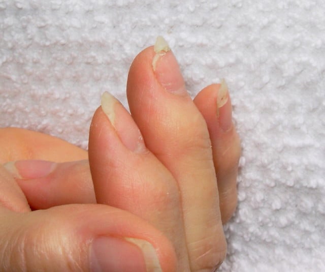
DSCN4758aWhat Are Brittle Nails?

[dropcap]B[/dropcap]rittle nails are abnormalities of the nail plate that are characterized by poor nail structure affecting all fingernails and toenails seen as thin, dry nails that easily chip, split, and are difficult to maintain a clean edge. Usually longitudinal ridging occurs from the nail base to the tips.

Q: What is the nail plate?

A: The nail plate is the hard keratin cover protecting the finger tip and toe tip. The nail plate (non-living tissue) is produced by the nail matrix (living tissue) at the base of the nail plate under the lunula (moon), which is the site of brittle nail development.

Nail Anatomy. A. Nail plate; B. lunula; C. root; D. sinus; E. matrix; F. nail bed; G. hyponychium; H. free margin. Courtesy Wikipedia.org

Poor nail structure affecting all nails may be a feature of nutritional deficiency in poor diet or malabsorption such as occurs in celiac disease.

Some other causes are: idiopathic (unknown cause), the result of aging, the effects of certain drugs, or an association with systemic autoimmune disorders such as vitiligo, alopecia areata (with pitting), psoriasis (with pitting), and lichen planus. 

External (non-nutritional or disease) causes of dry, brittle nails, such as detergents and cleaners, would only affect fingernails but not toenails.

Note: It has been shown that working with your hands in water or soaking them through activities like swimming does not cause dry, brittle nails but will worsen them.

What Are Brittle Nails In Celiac Disease and/or Gluten Sensitivity?

Colitis, Collagenous

Collagenous Colitis.
Microscopic Image Showing a Pink Collagen Band in Collagenous Colitis.

What Is Collagenous Colitis?

[dropcap]C[/dropcap]ollagenous colitis is a disease of the large intestine (colon) that is characterized by microscopic inflammation of the surface mucosal lining and an abnormally thickened collagen band of tissue that develops wthin the lining of the colon.

The thicker than normal layer of collagen of at least 10 µm (reference value: 2–7 µm) can vary in different locations. Inflammation occurs with increased numbers of lymphocytes (white blood cells) and plasma cells and epithelial (surface cell) damage. These changes can only be seen under microscopic examination of multiple biopsied tissue samples taken during a colonoscopy procedure.

Q: What is collagen?

A: Collagen is a strong, fibrous protein found in connective tissue of the colon and many other tissues such as tendons. The normal basement membrane in the bowel consists mainly of collagen type IV, laminin, and fibronectin. The increased collagen band observed in collagenous colitis consists basically of collagen type I and III, which are the subtypes produced by repair functions, indicating a reactive origin to some irritant or drug.1

The biopsies should preferably be taken from the ascending colon, since the pathological hallmarks may be absent in the descending colon, and in the normally occurring thicker collagen layer in the rectosigmoid region.1 Inflammation of the ileum (last segment of the small intestine next to colon) is common.2

Endoscopy and radiological (x-ray) examinations are usually normal.3

Autoimmune disorders are frequently seen in adult patients with collagenous colitis.4 In the study below by Koskela et al. concomittent autoimmune diseases were present in 53% of patients with collagenous colitis.5

Importantly, the finding of collagenous colitis in patients with autoimmune diseases may reflect the treatment with NSAIDs (non-steroidal anti-inflammatory drugs), such as Ibuprofin and aspirin, PPIs (proton pump inhibitors), and other drugs. However, if secondary forms of collagenous colitis are not taken into consideration, underlying, treatable diseases may be overlooked, while only the gastrointestinal symptoms are treated symptomatically or with budesonide (a steroid).6

Treatment with budesonide steroid is efficacious irrespective of bile acid malabsorption.7

Budesonide at a mean dose of 4.5 mg/day maintained clinical remission for at least 1 year in the majority of patients with collagenous colitis and preserved health-related quality of life without safety concerns. Treatment extension with low-dose budesonide beyond 1 year may be beneficial given the high relapse rate after budesonide discontinuation.8

See below for nutritional deficiency problems caused by steroid usage and steps to be taken for correction.

What Is Collagenous Colitis In Celiac Disease and/or Gluten Sensitivity?

Sources:
  1. Ohlsson B. New insights and challenges in microscopic colitis. Therap Adv Gastroenterol. 2015 Jan;8(1):37-47. doi: 10.1177/1756283X14550134. [] []
  2. Bjørnbak C, Engel PJ, Nielsen PL, Munck LK. Microscopic colitis: clinical findings, topography and persistence of histopathological subgroups. Aliment Pharmacol Ther. 2011 Nov;34(10):1225-34. doi: 10.1111/j.1365-2036.2011.04865.x. []
  3. Abdo AA, Urbanski SJ, Beck PL. Lymphotcytic and collagenous colitis: the emerging entity of microscopic colitis. An update on pathophysiology, diagnosis and management. Canadian Journal of Gastroenterology. Jul 2003;17(7):425-32. []
  4. Leung ST, Chandan VS, Murray JA, Wu TT. Collagenous gastritis: histopathologic features and association with other gastrointestinal diseases. Am J Surg Pathol. 2009 May;33(5):788-98. doi: 10.1097/PAS.0b013e318196a67f. []
  5. Koskela RM, Niemela SE, Karttunen TJ, Lehtola JK. Clinical characteristics of collagenous and lymphocytic colitis. Scandanavian Journal of Gastroenterology. Sep 2004;39(9):837-45. []
  6. Ohlsson B. New insights and challenges in microscopic colitis. Therap Adv Gastroenterol. 2015 Jan;8(1):37-47. doi: 10.1177/1756283X14550134. []
  7. Bjørnbak C, Engel PJ, Nielsen PL, Munck LK. Microscopic colitis: clinical findings, topography and persistence of histopathological subgroups. Aliment Pharmacol Ther. 2011 Nov;34(10):1225-34. doi: 10.1111/j.1365-2036.2011.04865.x. []
  8. Münch A, Bohr J, Miehlke S, et al. Low-dose budesonide for maintenance of clinical remission in collagenous colitis: a randomised, placebo-controlled, 12-month trial. Gut. 2014 Nov 25. pii: gutjnl-2014-308363. doi: 10.1136/gutjnl-2014-308363. []

Colitis, Lymphocytic

Microscopic Slide of Lymphocytic Colitis. Courtesy Quizlet.com
Microscopic  Slide of Biopsy Sample Showing Lymphocytic Colitis. Courtesy Quizlet.com

What Is Lymphocytic Colitis?

[dropcap]L[/dropcap]ymphocytic colitis is a microscopic inflammation of the large intestinal mucosa with infiltration of lymphocytes (IELs)  that is characterized by non-bloody secretory diarrhea.

Secretory diarrhea describes bowel movements that consist of a large volume of liquid stool.

Q: What are IELs?

A: IELs is an abbreviation for intraepithelial lymphocytes, which are white blood cells that infiltrate within epithelial cells or between them. Epithelial cells form the surface mucosa of the large intestine also called the colon.

The histopathological criteria (biopsy) for lymphocytic colitis are a density of at least 20 IELs per 100 surface epithelial cells; chronic inflammatory infiltrate of mononuclear cells in the lamina propria; epithelial damage; and a subepithelial collagen layer of less than 10 µm. The increased collagen band consists basically of collagen type I and III, which are the subtypes produced by repair functions, indicating a reactive origin.1That is, the mucosa is reacting to some irritative substance.

Up to 10% of adults undergoing colonoscopy for investigation of chronic diarrhea and having visibily normal appearing mucosa may have lymphocytic colitis.2

Bile acid malabsorption has been shown to coexist in 60% of patients with lymphocytic colitis.1

Lymphocytic colitis (LC) is categorized as primary or secondary.  Primary LC is a clinical and histopathological disease of unknown cause. Secondary LC may develop as the result of iritating factors acting on the colon such as smoking or many medications.  In one study, the most common drug treatments as a percentage of the study group were corticosteroids (32.1%), proton pump inhibitors (26.0%), antidepressant drugs, specifically selective serotonin reuptake inhibitors (21.4%), angiotensin-converting enzyme inhibitors or angiotensin II receptor antagonists (18.3%), statins (17.6%), thyroid hormones (17.6%), and beta-blockers (16.0%).3

Secondary lymphocytic colitis is associated with several concomitant diseases including celiac disease. This is why lymphocytic changes must be interpreted with caution before considering them as a separate entity of autoimmune origin, instead of secondary reactions to ischemia and toxic stimulants. Efforts must be made to better classify and diagnose patients with real, primary lymphocytic colitis to avoid over-prescription of corticosteroids for treatment.3

What Is Lymphocytic Colitis In Celiac Disease and/or Gluten Sensitivity?

Sources:
  1. Ohlsson B. New insights and challenges in microscopic colitis. Therap Adv Gastroenterol. 2015 Jan;8(1):37-47. doi: 10.1177/1756283X14550134. [] []
  2. Abdo AA, Urbanski SJ, Beck PL. Lymphotcytic and collagenous colitis: the emerging entity of microscopic colitis. An update on pathophysiology, diagnosis and management. Canadian Journal of Gastroenterology. Jul 2003;17(7):425-32. []
  3. Roth B, Manjer J, Ohlsson B. Drug Target Insights. 2013 Aug 11;7:19-25. doi: 10.4137/DTI.S12109. [] []

Colitis, Ulcerative

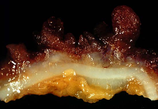
This photo was from a total colectomy done for clinically severe, intractable chronic ulcerative colitis. It shows a closer view of a longitudinal section through the colon wall. This demonstrates not only the angry red mucosa but also the tendency for the inflamed tissue to throw itself up into inflammatory pseudopolyps. Source: Ed Uthman, MD. Public domain.
This photo is from a total colectomy done for severe, intractable chronic ulcerative colitis. It shows a close view of a lengthwise section through the colon wall. This demonstrates not only the angry red mucosa, but also, the tendency for the inflamed tissue to throw itself up into inflammatory pseudopolyps.
Source: Ed Uthman, MD. Public domain.

What Is Ulcerative Colitis?

[dropcap]U[/dropcap]lcerative colitis is an inflammatory disorder of the colon characterized by continuous inflammation of the mucosa and submucosa usually with small ulcers, extending from the rectum and typically involving the distal colon, rectum, and anus and producing bloody diarrhea.

While the severity of ulcerative colitis varies among patients, iron deficiency anemia often develops due to blood loss especially when there are many bloody bowel movements in a day.

The onset of ulcerative colitis is most commonly in young adulhood.

Q: Is this disease painful?

A: Yes with the passage of stool.

Psychological stress and subsequent dysbiosis exacerbate ulcerative colitis.

Psychological stress activates multiple physiological processes aimed at maintaining balance within the body. These physiological processes also have the capacity to influence the composition of microbial communities in the digestive tract, and research now indicates that exposure to stressful stimuli leads to gut microbiota dysbiosis.1

While the relative abundance of many different bacterial types can be altered during stressor exposure, findings in nonhuman primates and laboratory rodents, as well as humans, indicate that bacteria in the genus Lactobacillus are consistently reduced in the gut during stress.2

Presently, ulcerative colitis cannot be cured. This condition has a course of remissions, when symptoms subside, and flares, when symtpoms get worse. Treatment is aimed to reduce flares and promote remission. In all cases, correction of dysbiosis improves the condition.

Ulcerative colitis is associated with increased incidence of cancer of the colon.3

What Is Ulcerative Colitis In Celiac Disease and/or Gluten Sensitivity?

Sources:
  1. Galley JD, Bailey MT. Impact of stressor exposure on the interplay between commensal microbiota and host inflammation. Gut Microbes. 2014 May 1;5(3):390-396. Epub 2014 Apr 1. []
  2. Galley JD, Bailey MT. Impact of stressor exposure on the interplay between commensal microbiota and host inflammation. Gut Microbes. 2014 May 1;5(3):390-396. Epub 2014 Apr 1. []
  3. Taber’s Cyclopedic Medical Dictionary. F. A. Davis. Philadelphia, PA []

DHA (Docosahexaenoic Acid) Deficiency

Smoked Salmon...Brain Food that's Good for the Eyes!
Salmon…Brain Food That’s Good for Your Eyes, Heart, Blood Vessels, Blood Sugar, Liver, Muscles, and Fights Inflammation!

What Is DHA?

[dropcap]D[/dropcap]ocosahexaenoic acid (DHA) is an essential omega-3 fatty acid that is abundant in the brain, being crucial in brain structure. As such DHA is a key component of neuronal membranes together with arachidonic acid (a major opposing omega-6 fatty acid), making up 15-20% of the brain’s dry mass.

This polyunsaturated fatty acid is obtained from fish sources of food.

In healthy human volunteers, positron emission tomography (PET) has shown that the normal human brain consumes 4.6 mg/day of DHA.1

DHA is particularly concentrated in highly active membranes such as nerve synapses (junctions) and photoreceptors in the eye (retina).

Q: How much DHA is in the retina?

A: DHA makes up more than 30% of the retina.2

In other roles, DHA is an  important building material for the eicosanoids, a large group of highly bioactive hormone-like substances including prostaglandins, leukotrienes, and thromboxanes that are involved in blood clotting, inflammation, and vasoconstriction.

DHA has been shown to increase insulin sensitivity as opposed to the opposite problem of insulin resistance, to improve muscle mass, and protect against non-alcoholic fatty liver disease.3

Egert et al. in a study of people aged 19 to 43 years with normal cholesterol showed that DHA intake significantly increased serum HDL (good) cholesterol. Also, DHA significantly decreased fasting serum triglycerides.4

What Is DHA Deficiency In Celiac Disease and/or Gluten Sensitivity?

Sources:
  1. Rapoport SI. Brain arachidonic and docosahexaenoic acid cascades are selectively altered by drugs, diet and disease. Prostaglandins Leukot Essent Fatty Acids. 2008 Sep-Nov;79(3-5):153-6. Epub 2008 Oct 29. []
  2. Richardson AJ. The importance of omega-3 fatty acids for behavior, cognition, and mood. Scandinavian Journal of Nutrition. 2003;47(2):92-8 []
  3. Espinosa A, Valenzuela R, González-Mañán D, D’Espessailles A, Guillermo Gormaz J, Barrera C, Tapia G. Prevention of liver steatosis through fish oil supplementation: correlation of oxidative stress with insulin resistance and liver fatty acid content. Arch Latinoam Nutr. 2013 Mar;63(1):29-36. []
  4. Egert S, Kannenberg F, Somoza V, et al. Dietary alpha-linolenic acid, EPA, and DHA have differential effects on LDL fatty acid composition but similar effects on serum lipid profiles in normolipidemic humans. J Nutr. 2009;139:861–868. doi: 10.3945/jn.108.103861 []

EPA (Eicosapentaenoic Acid) Deficiency

Macherel...
Macherel…EPA Rich!

What Is EPA?

[dropcap]E[/dropcap]PA (eicosapentaenoic acid) is an essential omega-3 fatty acid that is crucial for fetal brain and retina development and the child’s subsequent neurodevelopment among very many other activities in people of all ages.

Omega-3 fatty acids  are polyunsaturated long chain fatty acids which must be obtained from animal foods since they do not occur in plants.

In all ages, EPA is essential for normal brain function.

Q: Why is EPA essential to the way the brain works?

A: EPA helps nerve cells in the brain to communicate with each other.

In pregnancy, EPA may also play a role in determining the length of gestation and in preventing perinatal depression in the mother.1 and is essential for normal growth in children.

EPA is important building material for the eicosanoids, a large group of highly bioactive hormone-like substances including prostaglandins, leukotrienes, and thromboxanes that are involved in blood clotting, inflammation, and vasoconstriction. Its many important functions are described below.

What Is EPA Deficiency?

Sources:
  1. Coletta JM, Bell SJ, and Roman AS. Omega-3 Fatty Acids and Pregnancy. Rev Obstet Gynecol. 2010 Fall; 3(4): 163–171 []

Alpha-Linolenic Acid Deficiency

Flaxseed Crispbread is Loaded with Alpha-Linolenic Acid.
Flaxseed Crispbread…Loads of Alpha-Linolenic Acid.

What Is Alpha-Linolenic Acid?

[dropcap]A[/dropcap]lpha-linolenic acid is an essential (need/can’t make) polyunsaturated omega-3 fatty acid that must be obtained in the diet. Alpha-linolenic acid is found in plant sources only.

Alpha-linolenic acid is required for normal brain function and nervous system health. It is an important building material for the eicosanoids.

Q: What are eicosanoids?

A: Eicosanoids are a large group of highly bioactive hormone-like substances including prostaglandins, leukotrienes, and thromboxanes that are involved in blood clotting, inflammation, and vasoconstriction (constriction of blood vessels).

Alpha-linolenic acid is a “parent” fatty acid because if sufficient amounts are present in the body, it can be used to produce both eicosapentanoic acid (EPA) and docosahexaenoic acid (DHA), two other critical omega-3 fatty acids.1 However, this conversion is limited. In healthy individuals, the conversion rate of ALA to EPA is less than 5 to 10% and ALA to DHA is only 2 to 5%.

Additionally, a deficient level of the mineral selenium can interfere with the conversion of alpha-linolenic acid into EPA and DHA. Also, folic acid supplement has been shown to increase omega-3 status and, conversely, to decrease omega-3 when folic acid is in deficient in animal models.

What Is Alpha-Linolenic Acid Deficiency?

Sources:
  1. Richardson AJ. The importance of omega-3 fatty acids for behavior, cognition, and mood. Scandinavian Journal of Nutrition. 2003; 47(2):92-8 []