What Is Helicobacter Pylori (H. Pylori) Infection? [dropcap]H[/dropcap]elicobacter pylori infection is a potentially deadly stomach disease characterized by chronic superficial inflammation and ulcerations in 100% of infected patients. This infection disrupts normal defense and repair…
Enlarged heart showing dilated cardiomyopathy at autopsy.
What Is Idiopathic Dilated Cardiomyopathy?
[dropcap]I[/dropcap]diopathic dilated cardiomyopathy (IDCM) is a disorder of myocardial (heart muscle) function characterized by dilation or enlargement of the cardiac chambers and reduction in the ability of the ventricles (lower chambers) to contract with the required force needed to pump blood out of the heart to the body.
Idiopathic means the cause is not known.
Q: What is the prognosis for idiopathic dilated cardiomyopathy?
A: This disorder often results in symptomatic heart failure due to the inability of the heart to pump blood as required to supply the body with oxygen and meet metabolic needs.
What Is Idiopathic Dilated Cardiomyopathy In Celiac Disease and/or Gluten Sensitivity?
[dropcap]S[/dropcap]troke in childhood is a medical emergency during which blood flow to an area of the brain is stopped, causing infarction or death of the area of cells in the brain fed by the blocked artery. Stroke is characterized by loss of muscle and brain function according to the location of the lost cells.
The brain is a highly active metabolic and complex organ of our body that performs important functions, therefore, any disruption in its normal functioning can have devastating effects on whole body.
After stroke, an immune response is initiated that leads to production of proinflammatory cytokines (chemicals) and gathering of various inflammatory cells like neutrophils, T-cells, macrophage, and monocytes to the affected area that exacerbate or worsen the ischemic (oxygen starved) injury.1
Arterial ischemic stroke is an important cause of acquired brain injury in children.2 This stroke results from loss of adequate blood flow through an artery that supplies the affected area of the brain with oxygen and nutrients.
A recent study found the majority of children (85%) had focal features (most commonly one sided weakness or paralysis) at presentation to their medical practitioner. Seizures were more common in younger children a year old or younger and headache was more common in children 5 years or older.3
Who is at Risk in the General Population?
A British study in 2014 found the crude incidence of childhood arterial ischemic stroke was 1.60 per 100,000 per year.
The incidence of arterial ischemic stroke was highest in children aged under 1 year (4.14 per 100,000 per year).
There was no difference in the risk of arterial ischemic stroke between sexes.
Asian and black children were at higher risk than were white children.3
What Is Stroke in Childhood In Celiac Disease and/or Gluten Sensitivity?
Sources:
Andrabi SS, Parvez S, Tabassum H. Melatonin and Ischemic Stroke: Mechanistic Roles and Action. Adv Pharmacol Sci. 2015;2015:384750. doi: 10.1155/2015/384750. Epub 2015 Sep 7. [↩]
Mallick AA, Ganesan V, Kirkham FJ, et al. Childhood arterial ischaemic stroke incidence, presenting features, and risk factors: a prospective population-based study. Lancet Neurol. 2014 Jan;13(1):35-43. doi: 10.1016/S1474-4422(13)70290-4. Epub 2013 Dec 2. [↩]
Mallick AA, Ganesan V, Kirkham FJ, et al. Childhood arterial ischaemic stroke incidence, presenting features, and risk factors: a prospective population-based study. Lancet Neurol. 2014 Jan;13(1):35-43. doi: 10.1016/S1474-4422(13)70290-4. Epub 2013 Dec 2. [↩] [↩]
This photo taken during laparoscopy shows the gall bladder (small white organ in middle) surrounded by yellow fat. Liver (dark red organ) is overlying.
What Is Impaired Gall Bladder Motility?
[dropcap]I[/dropcap]mpaired gall bladder motility means the gall bladder is slow to empty or is dysfunctional.
The functional disorder of the gallbladder is caused initially either by metabolic abnormalities or by an alteration in its muscular ability to contract (motility dysfunction).
The diagnostic criteria based on symptoms of motility dysfunction of the gallbladder are episodes of moderate to severe steady pain located in the epigastrium and right upper abdominal quadrant that last at least 30 minutes.
Gallbladder motility disorder is suspected after gallstones and other structural abnormalities have been excluded.1
Q: What does the gallbladder do?
A: The gallbladder is a small pouch-like organ about the size of a pear that receives bile produced by the liver and stores it until needed during digestion. It lies just under the liver.
Bile is a complex fluid containing water, electrolytes and many organic molecules including bile acids, cholesterol, phospholipids and bilirubin. Bile acids are critical for digestion and absorption of fats and fat-soluble vitamins in the small intestine. Many waste products, including bilirubin, are eliminated from the body by secretion into bile and elimination in feces.2
Before a meal, the gallbladder is usually full of bile. In response to fat in the diet, the gallbladder squeezes stored bile into the small intestine through a series of ducts. When emptied after meals, the gallbladder is flat.
What Is Impaired Gall Bladder Motility In Celiac Disease and/or Gluten Sensitivity?
Sources:
Behar J, Corazziari E, Guelrud M, Hogan W, Sherman S, Toouli J. Functional gallbladder and sphincter of oddi disorders. Gastroenterology. 2006 Apr;130(5):1498-509. [↩]
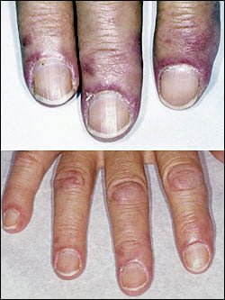
[dropcap]D[/dropcap]ermatomyositis is a rare autoimmune systemic disease of the connective tissue that is characterized by inflammatory and debilitatingdegenerative changes in the muscles and in the skin.
Dermatomyositis results in symmetric, proximal muscle weakness of limbs (upper arms and legs), and skin manifestations. 50-70% of patients have circulating myositis-specific auto-antibodies.
The course of dermatomyositis is unpredictable being marked by spontaneous flare-ups and remissions. It can begin slowly or abruptly according to the factor that is triggering the onset such as infection, medications like phenytoin, and autoimmune disease.
Q: What are the skin manifestations of dermatomyositis?
A: Classic skin manifestations of dermatomyositis include these features:
The heliotrope rash (lilac color) on upper eyelids.
Rash on face, neck, shoulders, upper chest, elbows, knees, knuckles, and back.
Gottron’s papules (scaly, red eruptions or patches over the knuckles, elbows, and knees).
The V-sign (rash front of neck and chest).
The shawl sign (rash distribution on shoulders and back).1
Additional cutaneous manifestations are described below under symptoms.
Dermatomyositis is associated with an increased risk of cancer, other autoimmune diseases, such as lupus and psoriasis, and it can be a complication of interferon-α therapy.About 1 person in 100,000 are affected according to various studies. While it affects all ages, women have twice the occurence of men.
There is no cure for dermatomyositis, but the symptoms can be treated. Options include medication, physical therapy, exercise, heat therapy (including microwave and ultrasound), orthotics and assistive devices, and rest. The standard treatment for dermatomyositis is a corticosteroid drug, given either in pill form or intravenously. Immunosuppressant drugs, such as azathioprine and methotrexate, may reduce inflammation in people who do not respond well to prednisone.2
What Is Dermatomyositis In Celiac Disease and/or Gluten Sensitivity?
Sources:
Marvi U, Chung L, Fiorentino DF. Clinical presentation and evaluation of dermatomyositis. Indian J Dermatol. 2012 Sep;57(5):375-81. doi: 10.4103/0019-5154.100486. [↩]
National Institute of Neurological Disorders and Stroke. [↩]
[dropcap]R[/dropcap]eversible hypertension is a pressure disorder of arteries associated with increased systemic (body wide) blood vessel resistance to blood flow due to endothelial (cell) dysfunction of arterial blood vessels that can improve with nutritional treatment.
Hypertension itself is defined as a systolic blood pressure (SBP) of 140 mm Hg (mercury) or greater and/or diastolic blood pressure (DBP) of 90 mm Hg or greater.
Q: What is blood vessel (vascular) resistance to blood flow?
A: Vascular resistance to blood flow means the arteries carrying blood away from the heart cannot relax or dilate when needed to lower blood pressure but stay constricted, which in turn, keeps the pressure high.
Here’s an analogy: if you replace your garden hose having a one inch inside diameter with one that has a smaller half inch diameter and open the water valve as usual, the result would be water shooting out with more force.
What Is Reversible Hypertension In Celiac Disease and/or Gluten Sensitivity?
[dropcap]C[/dropcap]hronic dry eye syndrome is an inflammatory condition of the conjunctiva with inadequate tear production or distribution over the eye surface. It is characterized by surface damage to the conjunctiva, giving the appearance of redness.
This complex condition involves the lacrimal glands, eyelids, and tear film, as well as a variety of eye surface tissues, including epithelial, inflammatory, immune, and goblet cells.1
Q: What is the conjunctiva?
A: The conjunctiva is the mucous membrane that lines the inside of the eyelids and covers the eyeball surface, also called the white of eye. The conjunctiva has a rich blood supply that can quickly bring microbe fighting cells to prevent invasion and overcome infection.
While infection and inflammation go hand-in-hand, they are not the cause of, but may result from, chronic dry eyes.
Dry eye syndrome develops from poor health of the conjunctiva that is associated with inadequate nutrition and poor quality of the tears that steadily lubricate the eye surface. These tears are called the tear film and are not the same as tears produced when crying or that result from irritating substances such as onions. Blinking spreads the tears that protect the surface from drying and flushes away unwanted matter.
The tear film is made up of three layers: mucous layer produced by goblet cells in the conjunctiva, aqueous (water) layer produced by the lacrimal glands, and lipid (fat) layer produced by the meibomian glands in the eyelids. Dysfunction of any of these layers impairs the the tear film. Without an adequate tear film the conjunctiva becomes dry and inflamed.
When the meibomian glands fail to produce sufficient lipid that is clear and fluid but rather cloudy and thick, the condition is called meibomian gland dysfunction (MGD). The main cause of MGD is hyperkeratinization (thickening of the glands) and its related pathogenesis (for example, ductal dilatation and acinar atrophy). Other disorders such as atopy, pemphigoid acne, rosacea, and seborrhea are related to MGD and may result in a chronic inflammation of the eye surface.2
MGD is “the most underrecognized, underappreciated and undertreated disease in ophthalmic care. It is so common as to be taken as ‘normal’ in many clinical practices,” according to Joseph Tauber, MD, an anterior segment subspecialist and refractive surgeon in Kansas City, Mo.3
Untreated dry eye progresses to xerophthalmia, a condition of extreme drying and thickening of the conjunctiva characterized by hazy, dry cornea. Other causes of dry eye, beside celiac disease, include certain systemic conditions such as Sjogren’s syndrome, an autoimmune disease.
What Is Chronic Dry Eye Syndrome In Celiac Disease and/or Gluten Sensitivity?
Sources:
Liu A, Ji J. Omega-3 essential fatty acids therapy for dry eye syndrome: a meta-analysis of randomized controlled studies. Med Sci Monit. 2014 Sep 6;20:1583-9. doi: 10.12659/MSM.891364. [↩]
Oleñik A, Jiménez-Alfaro I, Alejandre-Alba N, Mahillo-Fernández I. A randomized, double-masked study to evaluate the effect of omega-3 fatty acids supplementation in meibomian gland dysfunction. Clin Interv Aging. 2013;8:1133-8. doi: 10.2147/CIA.S48955. Epub 2013 Aug 30. [↩]
Rethinking Meibomian Gland Dysfunction: How to Spot It, Stage It and Treat It. Linda Roach, PhD. OPHTHALMIC PEARLS. [↩]
[dropcap]E[/dropcap]czema, or atopic dermatitis, is a chronic recurring inflammatory skin disorder characterized by itching, eczematous plaques and a defective epidermal barrier (surface layer of skin cells) that fails to hold moisture, with affected areas becoming dry then inflamed.
Q: Why does eczema develop?
A: Patients with atopic dermatitis have genetically determined risk factors that affect the barrier function of the skin and immune responses that interact with environmental factors.1
Atopic eczema is associated both with an abnormal skin matrix and impaired systemic immune response. Therefore, isolated topical treatment may have suboptimal effect.2
Atopic eczema is a common presentation of immune response towards food proteins in infancy.3
Global evidences reflect a marked increase in prevalence, which has tripled since 1960. In the United States, the current prevalence rates range from 10% to 20% in children and 1 to 3% in adults.4
What Is Eczema In Celiac Disease and/or Gluten Sensitivity?
Sources:
Mesquita Kde C, Igreja AC, Costa IM. Atopic dermatitis and vitamin D: facts and controversies. An Bras Dermatol. 2013 Nov-Dec;88(6):945-53. doi: 10.1590/abd1806-4841.20132660. [↩]
Caffarelli C, Santamaria F, Vottero A, Bernasconi S. Progress in Pediatrics in 2012: choices in allergy, endocrinology, gastroenterology, hematology, infectious diseases, neurology, nutrition and respiratory tract illnesses. Ital J Pediatr. 2013 May 8;39(1):26. [↩]
Ho MH, Wong WH, Chang C. Clinical Spectrum of Food Allergies: a Comprehensive Review. Clin Rev Allergy Immunol. 2012 Nov 16. [↩]
Mesquita Kde C, Igreja AC, Costa IM. Atopic dermatitis and vitamin D: facts and controversies. An Bras Dermatol. 2013 Nov-Dec;88(6):945-53. doi: 10.1590/abd1806-4841.20132660. [↩]
X-ray of Lungs in Sacroidosis Showing Honeycoming. Courtesy Wikimedia.
What Is Sarcoidosis?
[dropcap]S[/dropcap]arcoidosis is a multisystem granulomatous disease characterized by hard granulomas and inflammation of the alveoli in lungs that occurrs in 80% of patients.
Alveoli are the tiny one cell thick round structures that are clustered at the ends of air passages in the lungs where oxygen from air inhaled is exchanged for carbon dioxide from blood which is breathed out.
Q: What are granulomas?
A: Granulomas are collections of macrophages (white blood cells) that targeted and enclosed but failed to destroy a foreign substance that entered the body. In turn, the macrophages become encased by other immune cells and fibroblasts and collagen. They can occur in any organ.
Presently, the etiology, or cause, of sarcoidosis is unknown, although it is thought the predisposition is genetic with a local immune mechanism. This disease has flares when active and remissions when symptoms subside.
Referral to a specialist is important for proper treatment and monitoring. The main treatment for severe active sarcoidosis is prednisone, a type of steroid that reduces inflammation in the body. In most people, prednisone relieves symptoms within a couple of months, although most people need to take prednisone for 12 months or longer. Long-term use of prednisone, especially at high doses, can cause serious side effects. Side effects include bone loss, elevated blood sugar, and high blood pressure.
When wheezing and coughing are a problem, inhaler devices are used to deliver medicine into the lungs in order to help open the airways.1
Ibuprofen is prescribed if sarcoidosis is causing joint pain. Steroid creams are prescribed for skin problems.
Sarcoidosis affects 10 to 20 person per 100,000.
What Is Sarcoidosis In Celiac Disease and/or Gluten Sensitivity?
What Is Peripheral Neuropathy? [dropcap]P[/dropcap]eripheral neuropathy is a syndrome involving damage to one or more peripheral nerves characterized by impaired nerve transmission. Peripheral nerves are nerves of the body outside the brain and spinal cord. Q:…


![Grade_1_hypertension[1]](https://glutenfreeworks.com/health/wp-content/uploads/sites/10/Grade_1_hypertension1.jpg)


