Skip to content

Blurred Vision, Unexplained

Microscopic view of pancreatic islet cells. Courtesy Dr. José Sánchez Gonzales

What Is Type I Diabetes Mellitus?

T ype 1 diabetes mellitus (T1DM), also termed type 1A, is an inherited autoimmune disorder in which anti-islet autoantibodies destroy the islet cells of the pancreas that secrete insulin hormone. Type 1 diabetes mellitus was formerly called juvenile diabetes because it usually afflicts persons under the age of 25 years.

Loss of insulin production results in failure to metabolize glucose. Glucose is a simple sugar that is a required source of energy for the body, especially the brain and muscles.

Type 1 diabetes mellitus is characterized by sustained fasting blood glucose levels above 126 mg/dL (hyperglycemia) with subsequent loss of glucose from the body by removal through the urine (glucosuria) as the body attempts to lower blood glucose, and cell starvation that follows.

That is, while glucose accumulates in blood, the body cannot access it. Without insulin treatment, this disorder quickly produces coma and ultimately results in death. In fact, it is 5th leading cause of death in the United States.

Q: How does insulin work?

A: Insulin moves glucose from the bloodstream into body cells where it is used or reformulated for high energy storage. For example, muscles can use glucose for immediate work or store it in the form of glygogen for later work, depending on need. Healthy insulin production keeps an 8 hour fasting blood glucose level to less than 100 mg/dL. Upon eating carbohydrate food, glucose is digested and absorbed from the small intestine into the bloodstream which then raises blood glucose levels. The elevated level is controlled by prompt action of insulin to lower it to below 140 mg/dL  within 2 hours of eating.

Insulin does not work alone. The islets of Langerhans manage glucose in the body. The islets are specialized formations located on the outer surface of the pancreas. The islets are composed of two different types of cells known as alpha and beta cells. These cells make the competing hormones that keep blood glucose within a healthy range.

Alpha cells secrete glucagon to raise blood glucose levels by triggering the body to release stored energy in the form of glycogen. In the opposite, beta cells secrete insulin to lower blood glucose by opening body cells so that glucose in blood can enter. Without insulin, glucose cannot enter cells but remains in the bloodstream where it accumulates.

Insulin is also needed to move magnesium into cells from the bloodstream. On the other side, magnesium is needed to produce insulin. Insulin has other functions such as building muscle and helping regulate cholesterol which directly impacts the sex hormones, estrogen, progesterone, and testosterone.

Onset of symptoms usually occurs over a period of days or weeks, although beta cell destruction can begin years earlier. The SEARCH for Diabetes in Youth multicenter study, funded by the Centers for Disease Control and Prevention (CDC) and the National Institutes of Health (NIH), has determined that based on data from 2002 to 2003, a total of 15,000 youth in the United States were newly diagnosed with type 1 diabetes each year. Non-Hispanic white youth had the highest rate of new cases of type 1 diabetes according to NIH.

Type 1A diabetes mellitus has become one of the most intensively studied autoimmune disorders. It is now possible to predict its development, beginning with HLA-encoded genetic susceptibility, followed by the development of a series of anti-islet autoantibodies.1

What Is Type I Diabetes Mellitus In Celiac Disease and/or Gluten Sensitivity?

Sources:
  1. Liu E, Eisenbarth GS. Type 1A diabetes mellitus-associated autoimmunity. Endocrinology and Metabolism Clinics of North America. Jun 2002;31(2):391-410, vii-viii. []

Dysgeusia (Impaired Taste)

DyspareuniaWhat Is Autoimmune Thyroiditis (Hypothyroidism)?

A utoimmune thyroiditis, also called Hashimoto’s thyroiditis or Hashimoto’s Disease, is an autoimmune destruction of thyroid tissue characterized by insufficient thyroid hormone circulating in the body that causes formation of a goiter (enlarged thyroid gland) and hypothyroidism.

Hypothroidism refers to the condition of markedly reduced secretion of thyroid hormone. There are other causes of hypothyroidism besides Hashimoto’s thyroiditis.

Hashimoto’s thyroiditis is often associated with other autoimmune diseases such as celiac disease.

In Hashimoto’s thyroiditis, a profusion of antibodies are produced, which build up in the blood. Left untreated so that much of the thyroid gland is destroyed, this condition may progress to the very serious and life-threatening condition called myxedema.

Note: In myxedema, protein, electrolytes, and water abnormally accumulate in between cells which produce firm, inelastic puffy skin that is cool, dry, rough, scaly, and may appear yellow; in some people, areas such as the ankles become crusty with a look of tree bark. Many systemic changes develop shown by significant slowing of mental and physical functions. Please see below.

Q: What thyroid tissue is targeted for destruction?

A: In Hashimoto’s thyroiditis, high levels of autoantibodies target thyroglobulin and thyroid peroxidase, leading to inflammation and destruction of the thyroid gland. The resulting fibrosis or scarring of the gland results in lack of thyroid hormone production.

The thyroid gland consists of a large number of closed vesicles that contain a homogenous substance called colloid, which contains the thyroglobulin. Thyroglobulin is an iodine-containing protein secreted by the thyroid gland and stored within its colloid, from which the thyroid hormones thyroxine (T4) and triiodothyroinine (T3) are derived.1

T3 is the active hormone and is made from T4. Thyroid hormones affect metabolism, brain development, breathing, heart and nervous system functions, body temperature, muscle strength, skin dryness, menstrual cycles, weight, and cholesterol levels.

Thyroid hormone production is regulated by thyroid-stimulating hormone (TSH), which is made by the pituitary gland in the brain. Normally, when thyroid hormone levels in the blood are low, the pituitary releases more TSH. When thyroid hormone levels are high, the pituitary decreases TSH production.

Hashimoto’s disease, with or without the development of hypothyroidism, is treated with synthetic thyroxine, which is man-made T4. Health care providers prefer to use synthetic T4, such as Synthroid® (Levothyroxine), rather than synthetic T3, because T4 stays in the body longer, ensuring a steady supply of thyroid hormone throughout the day. The thyroid preparations made with animal thyroid are not considered as consistent as synthetic thyroid.2

What Is Autoimmune Thyroiditis In Celiac Disease and/or Gluten Sensitivity?

Sources:
  1. Taber’s Cyclopedic Medical Dictionary. 19th ed. F.A. Davis Company. Philadelphia, PA. []
  2. National Endocrine and Metabolic Diseases Information Service. []

Bladder Infection (Cystitis)

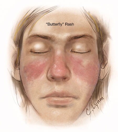
Image showing butterfly rash of SLE. Courtesy JAMA.
Image showing butterfly rash typical of SLE. Courtesy JAMA.

What Is Systemic Lupus Erythematosus?

S ystemic lupus erythematosus (SLE) is a chronic autoimmune inflammatory disease that is characterized by involvement of multiple organs due to the production of antibodies to components of the cell nucleus.1 SLE has an unpredictable course of acute flare-ups and remissions.

Severity depends on the extent of organs affected with skin and nail involvement, called discoid lupus, being the least serious and inflammmation of the kidney, called lupus nephritis, being the worst.

Nevetheless, a classic presentation is development of a rash over the cheeks and nose that resembles a butterfy with wings spread hence the name “butterfly rash.”

Symptoms are many and varied depending on the tissues affected and are often not specific, for example hair loss has a variety of causes. Symptoms can be confused by co-existence with other autoimmune disease such as Sjogren’s syndrome.

Systemic lupus erythematosus should be managed by a specialist. Symptoms can be controlled with steroid therapy, but this disease can be a cause of premature death  mainly from active disease, organ failure (e.g., kidneys), infection, or cardiovascular disease from accelerated atherosclerosis.

Certain common medicines known to cause drug-induced lupus are:

  • Isoniazid
  • Hydralazine
  • Procainamide

Other less common drugs may also cause the condition. These may include:

  • Anti-seizure medications
  • Capoten
  • Chlorpromazine
  • Etanercept
  • Infliximab
  • Methyldopa
  • Minocycline
  • Penicillamine
  • Quinidine
  • Sulfasalazine

Symptoms tend to occur after taking the drug for at least 3 to 6 months.2

Although there is a strong familial aggregation, the disease is relatively uncommon and most cases are sporadic.1 According to the Center for Diseases (CDC), lupus most commonly affects women of childbearing age but also occurs in infants, children, adolescents, and men with peak occurrence between ages 15 and 40. Blacks (and possibly Hispanics, Asians, and Native Americans) are affected more than Whites.

What Is Systemic Lupus Erythematosus In Celiac Disease and/or Gluten Sensitivity?

Sources:
  1. http://www.cdc.gov/arthritis/basics/lupus.htm [] []
  2. www.nlm.nih.gov/medlineplus/ency/article/000446.htm []

Inflammatory Vaginitis

The tiny brown streak is a splinter hemorrhage.

What Are Splinter Hemorrhages In Nails?

S ubungual (under the nail) splinter hemorrhage is characterized by fragility of the underlying capillaries resulting in bleeding under the nail . Bleeding that results looks like a brown streak or splinter.1

Because the streak of blood is not in the nail but under the nail, it does not grow out with the nail. It can be a feature of vitamin C deficiency which resolves with adequate nutrition.

Nail Anatomy. Nail Anatomy. A. Nail plate; B. lunula; C. root; D. sinus; E. matrix; F. nail bed; G. hyponychium; H. free margin. Courtesy Wikipedia.org.

A. Nail plate; B. lunula; C. root; D. sinus; E. matrix; F. nail bed; G. hyponychium; H. free margin. Courtesy Wikipedia.

Splinter hemorrhage can be seen in a diet poor in vitamin C, disorders of food absorption, systemic disease such as psoriasis, nail lichen planus, an aggressive disease that may destroy the nail plate, and endocarditis. Some drugs, such as oral contraceptives, cause vitamin C deficiency.

Who is Affected in the General Population? Both sexes are commonly affected, though it occurs significantly more frequently in males. Where present in females, they were confined to a single digit.2

What Are Splinter Hemorrhages In Nails In Celiac Disease and/or Gluten Sensitivity?

Sources:
  1. Krause’s Food, Nutrition, & Diet Therapy. 10th Edition. Kathleen Mahan, Sylvia Escott-Stump. 2000. W.B. Saunders Company. []
  2. Monk BE. The prevalence of splinter haemorrhages. Br J Dermatol. 1980 Aug;103(2):183-5. []

Impotence

This White Spot Is A Feature of Zinc Deficiency.

What Are White Spots And White Bands In Nails?

W hite spots in the nails is a feature of zinc deficiency and white bands signify protein deficiency characterized by abnormal appearance anywhere in one or more nails.

White spots and bands can appear separately or together in nails. There may be just one mark or many marks depending on the level of deficiency.

Q: What part of the nail is affected by zinc and/or protein deficiency?

A: Zinc and protein deficiencies affect the nail plate.

The nail plate is the hard keratin cover of the finger tip and toe tip, simply called “the nail.” The nail plate is produced by the living nail tissue at its base. Here is an illustration of the parts that make up a fingernail:

Nail Anatomy. Nail Anatomy. A. Nail plate; B. lunula; C. root; D. sinus; E. matrix; F. nail bed; G. hyponychium; H. free margin. Courtesy Wikipedia.org.

A. Nail plate; B. lunula; C. root; D. sinus; E. matrix; F. nail bed; G. hyponychium; H. free margin. Courtesy Wikipedia.

What Are White Spots And White Bands In Nails In Celiac Disease and/or Gluten Sensitivity?

Infertility In Males

Cutaneous Vasculitis. Courtesy quizlet.com

What Is Cutaneous Vasculitis?

Cutaneous vasculitis is a vascular disease of small blood vessels serving the skin that is characterized by segmental (spotty) inflammation of affected vessel walls resulting in their necrosis (destruction). Scarring is seen in the affected skin that lies over the destroyed blood vessels.

Skin appearance varies from small flat, purplish spots to raised purplish areas over damaged vessels.

In most cases, cutaneous vasculitis results from deposits of toxic immune complexes in the affected vessel walls that is caused by autoimmune activity targeting small blood vessels.1

Cutaneous vasculitis can also result from infection, drug reactions, or malignancies.

Who is Affected in the general Population? Cutaneous vasculitis affects all ages.

The overall annual incidence of cutaneous vasculitis was 38.6 persons per million in 1998.2

What Is Cutaneous Vasculitis In Celiac Disease and/or Gluten Sensitivity?

Sources:
  1. Abenavoli L, Proietti I, Leggio L, Ferrulli A, Vonghia L, Capizzi R, Rotoli M, Amerio PL, Gasbarrini G, Addolorato G. Cutaneous manifestations in celiac disease. World J Gastroenterol. 2006 Feb 14;12(6):843-52. []
  2. Watts RA, Jolliffe VA, Grattan CE, Elliott J, Lockwood M, Scott DG. Cutaneous vasculitis in a defined population–clinical and epidemiological associations. J Rheumatol. 1998 May;25(5):920-4. []

Irritability

Small Bowel Volvulus, Courtesy  African Journal of Pediatric Surgery.
Small Bowel Volvulus Caused the  Enlarged Dark Loops. Courtesy African Journal of Pediatric Surgery.

What Is Volvulus?

V olvulus is twisting of a loop(s) of bowel onto itself which effectively closes it thus preventing digested matter from passing and causing engorgement of the closed loop with gas and fluid.

Q: How does the bowel get untwisted?

A: This condition usually necessitates surgical correction although some do return to their proper position.

The danger is that there may develop necrosis of the twisted loop(s) in which case, if left untreated, death will ensue.

What Is Volvulus In Celiac Disease and/or Gluten Sensitivity?

Constipation Alternating with Diarrhea

Bristol Stool Chart Showing Normal and Abnormal Stool.

What Is Chronic Constipation Alternating With Diarrhea?

Chronic constipation alternating with diarrhea is an intestinal motility disorder, or irregularity, characterized by alteration in stool formation, consistency, and evacuation which results in a bowel movement that consists of some hard or balled stool along with some loose stool that can cause leakage.

Q: How do irregular movement patterns develop in the colon?

A: The colon produces irregular movements as a result of problems that originate in  the colon (large intestine) itself and/or the small intestine which then affects function of the colon.

Here are listed the many types of problems or diseases that cause these abnormal bowel movements:

  • Disorders that adversely affect the colon, an organ which must propel stool, remove excess water, absorb electrolytes, ferment undigested food material that passes into it, and produce nutrients from the fermentation process:
  • Poor diet that does not contain adequate nutrition, fiber, probiotics, prebiotics, and water to form normal stool.
  • Diet that contains irritating, toxic or allergenic food that cause spasms.
  • Diseases that inflame the mucosa lining such as collagenous colitis, altering the proper absorption of water and electrolytes.
  • Diseases that damage and swell the colon walls, such as Crohn’s disease, ulcerative colitis, and diverticulitis.
  • Diseases that obstruct the lumen or passageway so that stool passes with difficulty.
  • Diseases that hamper normal peristalsis (muscle action), such as irritable bowel syndrome (IBS), diabetes and thyroid disease.
  • Disorders that adversely affect the small intestine, an organ which must digest and absorb nutrients needed by the body while passing unabsorbed food material to the colon:  
  • Diet that conatins too much fat, sugar or artifical sweeteners, causing diarrhea.
  • Disorders that result in malabsorption, such as gluten enteropathy, milk enteropathy, steatorrhea (fat malabsorption), lactose intolerance, sucrose intolerance, maltose intolerance, and bacterial overgrowth, passing abnormal amounts of undigested food material to the colon where it is fermented producing excessive gas, diarrhea and spasm.
  • Disorders that impair peristalsis, such as active celiac disease, diabetes, scleroderma, and thyroid disease.
  • Tumors like cancer and lymphoma impair regular passage of material to colon.
  • Drugs that impair peristalsis, such as iron supplements, aluminum containing antacids, narcotics, some anti-depressants, some anti-seizure, and some diuretics.

What Is Chronic Constipation Alternating With Diarrhea In Celiac Disease and/or Gluten Sensitivity?

Bleeding, Occult Gastrointestinal

Erosions Of The Esophagus Can Be A Cause Of Gastrointestinal Bleeding.
Erosions Resulting From Esophagitis Can Be A Cause Of Gastrointestinal Bleeding.

What Is Occult Gastrointestinal Bleeding?

Occult gastrointestinal bleeding is characterized by unseen or minute quantities of blood in stool. The origin of bleeding is from mucosa that lines the inside of the digestive tract at a site that must be found by medical testing and procedures to look at the lining.

Q: What tests and procedures are performed to locate the bleeding?

A: The simplest test to discover blood that cannot be seen is the stool test. This consists of a card with 3 spaces for 3 separate  stool collection over 3 separate days. A tiny smear of stool is placed on a test card space on each of three days. Then the card is examined for a reaction that indicates the existence of blood in the stool.

If the stool test is positive, the origin of bleeding must be located. This search requires certain procedures that look at the mucosal lining directly by a gastroenterologist, usually under light sedation.

  • Gastroscopy procedure can visualize the upper gastrointestinal tract, which includes the esophagus and stomach.
  • Endoscopy procedure can also visualize the upper small intestine. However, endoscopy procedure is limited in that it cannot visualize the middle small intestine (jejunum).
  • Colonoscopy can visualize the end of the small intestine and colon all the way to the rectum.
  • Capsule endoscopy procedure.  If  gastroendoscopy and colonoscopy fail to discover the source of bleeding, the physician may administer a capsule endoscopy which visualizes the entire gastrointestinal tract. In this procedure, the patient swallows a capsule size camera (encased) which takes constant pictures over the course of a day until expelled through the rectum.  Of course, the patient must be able to swallow the large size capsule.

What Is Occult Gastrointestinal Bleeding In Celiac Disease and/or Gluten Sensitivity?

Ataxia, Gait

Normal Liver Lobule. Courtesy almostadoctor.com
Figure 1: Normal Liver Lobule. Courtesy almostadoctor.com

What Is Autoimmune Cholangitis?

A utoimmune cholangitis is a rare inflammatory self-targeted disease of the biliary tract produced by high levels of immunoglobulin G (IgG) antibodies and antinuclear antibodies (ANA).

Autoimmune cholangitis is characterized by progressive inflammation and scarring that leads to narrowing and destruction of the bile ducts and development of biliary cirrhosis of the liver.

Autoimmune cholangitis is also termed anti-mitochondrial antibody-negative primary biliary cirrhosis because there is no production of anti-mitochondrial antibodies.

Q: What happens when bile ducts can no longer transport bile out of the liver?

A: Bile that cannot be removed from the liver by the biliary tract backs up and damages the liver, causing cirrhosis. In Figure 1, bile back up would directly damage the hepatic (liver) cells seen here as grey “blocks.” 

Bile is a greenish brown liquid continually made by the liver cells (hepatocytes) from phospholipids salt, cholesterol, and aging blood cells that it removes from circulation. 

Bile also carries away waste products produced by normal metabolism and toxic substances that are removed by the liver for eventual elimination in stool. As such, bile must continually flow out of the liver to prevent build-up in the liver. 

Bile from the hepatic cells drains into small tubes called canniculi and then into larger bile ducts to be carried out of the liver to the gall bladder for storage until needed to aid in the digestion and absorption of fat from the small intestine.   Bile emulsifies fat eaten in the diet so that the pancreatic enzyme called lypase can break it down into its fatty acid and glycerol components. 

Unfortunately, autoimmune cholangitis is difficult to diagnose because it mimics many other diseases such as primary sclerosing cholangitis, sarcoidosis, cancer and lymphoma.

What Is Autoimmune Cholangitis In Celiac Disease and/or Gluten Sensitivity?