Skip to content

Delayed Puberty in Boys
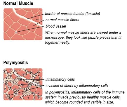
Drawing of Biopsy Showing Muscle Fibers Invaded by Immune Cells. Courtesy MDA.org

What Is Polymyositis?

P olymyositis is a body-wide connective tissue disease resulting from autoimmune attack of skeletal muscles that is characterized by inflammatory and degeneratory changes. The course is unpredictable being marked by spontaneous flare-ups and remissions.

Polymyositis can begin slowly or abruptly according to the factor that is triggering the onset such as infection, medications like phenytoin, and autoimmune disease.

Progressive muscle weakness starts in the proximal skeletal muscles (muscles closest to the trunk of the body).

Skeletal muscles, also called voluntary, are muscles that move the body as we want, such as walking and  lifting objects, as opposed to those we cannot voluntarily control, such as the muscles of digestion. 

Q: What are the degeneratory changes in skeletal muscles?

A: In polymyositis, degeneratory changes in skeletal muscles means that muscles are being destroyed (called necrosis), resulting in fibrosis, or scarring. When scar tissue takes the place of lost muscle tissue, it cannot act like muscle to contract and relax.   Muscle destruction is what causes muscle pain and weakness.

After the clinical work-up of exams and blood studies to determine muscle damage, the diagnosis of polymyositis is confirmed by muscle biopsy. See image at above left. The black dots are inflammatory cells. Edema (fluid) between cells caused by inflammation pushes muscle fibers apart.

There is no cure for polymyositis, but the symptoms can be treated.  Options include medication, physical therapy, exercise, heat therapy (including microwave and ultrasound), orthotics and assistive devices, and rest.  The standard treatment for polymyositis is a corticosteroid drug, given either in pill form or intravenously.  Immunosuppressant drugs, such as azathioprine and methotrexate, may reduce inflammation in people who do not respond well to prednisone. 

Periodic treatment using intravenous immunoglobulin can also improve recovery.  Other immunosuppressive agents used to treat the inflammation associated with polymyositis include cyclosporine A, cyclophosphamide, and tacrolimus.  Physical therapy is usually recommended to prevent muscle atrophy and to regain muscle strength and range of motion.1

Diagnosis is based on elevated muscles enzymes, increased urinary creatine level, and electromyograph abnormalities.

Polymyositis can affect people at any age. It is most common in adults between ages 50 and 70, and in children ages 5 to 15. It affects women twice as often as men and is more common in African Americans than Caucasians.2 The major causes of death from polymyositis are cancer and lung disease, including pneumonia.  The 5-year mortality rate can be as high as 1 in 5 patients.2

What Is Polymyositis In Celiac Disease and/or Gluten Sensitivity?

Sources:
  1. National Institute of Neurological Disorders and Stroke []
  2. http://www.nlm.nih.gov/medlineplus/ency/article/000428.htm [] []

Angina Pectoris

What Is Angina Pectoris?Coronary Artery Lesion

Angina pectoris, or simply angina, is a coronary syndrome characterized by an oppressive substernal pain (pain under breastbone) or pressure brought on by exertion and relieved by rest that results from failure of coronary arteries to deliver adequate oxygen to heart tissue due to ischemic heart disease.

Q: Why do coronary arteries fail to deliver adequate oxygen to heart tissue?

A: Coronary arteries are the blood vessels that serve the heart. In angina, these vessels fail to deliver adequate oxygen to heart tissue because they are narrowed or blocked by fatty buildups, called atherosclerotic plaques or by a blood clot which impair their ability to carry adequate blood that carries the oxygen. Diseased coronary arteries cannot deliver adequate oxygenated blood pumped by the heart to its own muscle cells.

The heart is a muscular organ that is working all the time without rest, so it needs a constant supply of oxygen. When heart muscle has to work harder, it needs more oxygen. Lack of oxygen causes pain which makes the affected person stop activity and rest.

Angina can be stable or unstable. Unstable angina is much more serious and can be life-threatening.

  • Stable angina produces predictable pain and responds to rest and/or medication. It is less serious than unstable angina but can be very painful or uncomfortable. Anything that makes the heart muscle need more oxygen can cause an angina attack in someone with heart disease, including: smoking, cold weather, exercise, emotional stress, obesity, and large meals. Other causes of angina include: abnormal heart rhythms (usually ones that cause the heart to beat quickly), anemia, coronary artery spasm, heart failure, heart valve disease, and hyperthyroidism (overactive thyroid).1
  • Unstable angina produces unpredictable pain that may occur at rest, lasting more than 20 minutes. It is more severe than stable angina and less responsive to medication. Atherosclerosis is by far the most common cause of unstable angina. Oxidized low-density lipoprotein, so-called bad cholesterol, and oxysterols play an important role in atherogenesis, the development of atherosclerosis. Coronary arteries that are narrowed by atherosclerotic plaques can rupture causing injury to the coronary blood vessel resulting in blood clotting which blocks the flow of blood to the heart muscle. Blood clots may form, partially dissolve, and later form again and angina can occur each time a clot blocks blood flow in an artery. People with unstable angina are at increased risk of having a heart attack.2

What Is Angina In Celiac Disease and/or Gluten Sensitivity?

Sources:
  1. http://www.ncbi.nlm.nih.gov/pubmedhealth/PMH0001247/ []
  2. http://www.heart.org/HEARTORG/Conditions/HeartAttack/SymptomsDiagnosisofHeartAttack/Unstable-Angina_UCM_437513_Article.jsp# []

Antiphospholipid Syndrome

Testing Thigh Strength. Courtesy Charlie Goldberg, M.D., UCSD School of Medicine

What Is Hypophosphatemia?

H ypophosphatemia means the level of phosphates in the bloodstream is too low to meet metabolic needs of the body for this mineral.

Q: How important is phosphorus in metabolism?

A: Phosphorus is crucial to  life, being present in every cell of the body and constitutes 45% of skeletal bone weight along with 40% calcium needed to support the body as a framework.

A low blood phosphate level is characterized by alterations in blood acid-alkaline balance and serious neuromuscular, hematologic, renal, skeletal, and dental abnormalities.

Symptoms result primarily from decreased production of adenosine triphosphate (ATP), the main energy source in cells, and phosphocreatine, a secondary energy source for muscle contraction.

  • Acute phosphorus deficiency may precipitate rhabdomyolysis which is destruction of muscle.
  • Nervous system dysfunction is observed in severe hypophosphatemia.
  • Chronic phosphorus deficiency causes proximal myopathy (upper arms and thighs).1
  • Severe phosphorus deficiency has widespread and ultimately fatal consequences.

What Is Hypophosphatemia In Celiac Disease and/or Gluten Sensitivity?

Sources:
  1. Takeda E, Ikeda S, Nakahashi O. Lack of phosphorus intake and nutrition. Clin Calcium. 2012 Oct;22(10):1487-91. []

Abscess Formation Causing Lung Cavities

Bristol Stool Chart Showing Normal and Abnormal Stool.

What Is Chronic Constipation Alternating With Diarrhea?

C hronic constipation alternating with diarrhea is an intestinal motility disorder, or irregularity, characterized by alteration in stool formation, consistency, and evacuation which results in a bowel movement that consists of some hard or balled stool along with some loose stool that can cause leakage.

Q: How do irregular movement patterns develop in the colon?

A: The colon produces irregular movements as a result of problems that originate in  the colon (large intestine) itself and/or the small intestine which then affects function of the colon.

Here are listed the many types of problems or diseases that cause these abnormal bowel movements:

  • Disorders that adversely affect the colon, an organ which must propel stool, remove excess water, absorb electrolytes, ferment undigested food material that passes into it, and produce nutrients from the fermentation process:
  • Poor diet that does not contain adequate nutrition, fiber, probiotics, prebiotics, and water to form normal stool.
  • Diet that contains irritating, toxic or allergenic food that cause spasms.
  • Diseases that inflame the mucosa lining such as collagenous colitis, altering the proper absorption of water and electrolytes.
  • Diseases that damage and swell the colon walls, such as Crohn’s disease, ulcerative colitis, and diverticulitis.
  • Diseases that obstruct the lumen or passageway so that stool passes with difficulty.
  • Diseases that hamper normal peristalsis (muscle action), such as irritable bowel syndrome (IBS), diabetes and thyroid disease.
  • Disorders that adversely affect the small intestine, an organ which must digest and absorb nutrients needed by the body while passing unabsorbed food material to the colon:  
  • Diet that conatins too much fat, sugar or artifical sweeteners, causing diarrhea.
  • Disorders that result in malabsorption, such as gluten enteropathy, milk enteropathy, steatorrhea (fat malabsorption), lactose intolerance, sucrose intolerance, maltose intolerance, and bacterial overgrowth, passing abnormal amounts of undigested food material to the colon where it is fermented producing excessive gas, diarrhea and spasm.
  • Disorders that impair peristalsis, such as active celiac disease, diabetes, scleroderma, and thyroid disease.
  • Tumors like cancer and lymphoma impair regular passage of material to colon.
  • Drugs that impair peristalsis, such as iron supplements, aluminum containing antacids, narcotics, some anti-depressants, some anti-seizure, and some diuretics.

What Is Chronic Constipation Alternating With Diarrhea In Celiac Disease and/or Gluten Sensitivity?

Coronary Artery Disease

Image on left shows how atherosclerosis impedes blood flow through coronary arteries while blood clots block blood flow. Courtesy Google.
Figure on right shows how atherosclerosis impedes blood flow through coronary arteries while blood clots block blood flow. Courtesy Google.

What Is Coronary Artery Disease (CAD)?

Coronary artery disease (CAD), also called ischemic heart disease, is a gradual narrowing of medium and large arteries of the heart by fatty buildups, called atherosclerotic plaques.

It is characterized by slowly developing interference with blood flow to heart tissue itself, resulting in oppressive chest pain called angina and, ultimately, thrombosis (clot) causing heart attack.  

The heart is a muscular organ that is working all the time, so it needs a constant supply of oxygen. Oxygen is brought to the working heart tissue by the coronary arteries with each beat of the heart. When heart muscle has to work harder, it needs more oxygen delivered to itself. Lack of oxygen causes pain.

In fact, failure of diseased coronary arteries to deliver adequate oxygen to heart tissue is the most common cause of angina pectoris – substernal pain (under breastbone) or pressure brought on by exertion and relieved by rest. 

Thrombosis, or clot formation, occurs when blood cells within a narrowed artery can no longer get through. Trapped, blood cells pile up and block the artery thus triggering a cascade of events called heart attack. Coronary arteries that are narrowed by atherosclerotic plaques can rupture causing injury to the coronary blood vessel resulting in blood clotting which blocks the flow of blood to the heart muscle. Blood clots may form, partially dissolve, and later form again and angina can occur each time a clot blocks blood flow in an artery.1

Q: How does coronary artery disease develop?

A: Coronary artery disease slowly develops from this combination of events:

  • Dysfunction of epithelial cells that line the inside of arteries cause the vessels to stiffen, and subsequently

  • Accumulation of lipid (fat) in smooth muscle cells beneath the inside lining of arteries and in foam cells cause buildup of fatty deposits on the inside walls progressing to fibrous plaque formation.

Oxidized low-density lipoprotein (oxLDL), so-called bad cholesterol, and oxysterols play important roles in the development of  atherosclerosis. OxLDL triggers the immune system to produce autoantibodies against oxLDL that are detectable in serum. These antibodies are called anti-oxLDL. Anti-oxLDL antibody and oxysterol concentrations are associated with coronary artery stenosis. Oxidative stress may be greatly increased in unstable angina.2 and Chronic inflammation in the general population is a major risk factor for ischemic heart disease.

The pathophysiology of atherosclerosis is, clearly, different in women when compared to the men. The women have a higher risk of blood coagulability making them at high risk for the blood clot formation. In a large number of women endothelial dysfunction, small vessel size and diffuse atherosclerosis have been identified as causes of ischemia without evidence of blockade in the coronary arteries.3

Also, atherosclerotic plaque in women is less fibrotic and contains more lipid filled foam cells, implying greater potential for reversibility but also potentially greater vulnerability for plaque rupture and thrombosis.4

Who is Affected in the General Population?

  • Coronary artery disease remains the leading cause of death in developed countries despite significant progress in primary prevention and treatment strategies.

  • It is the leading cause of death in women, as well as an important cause of disability.

  • Older patients are at particularly high risk of poor outcomes following acute coronary syndrome.5

What Is Coronary Artery Disease In Celiac Disease and/or Gluten Sensitivity?

Ischemic heart disease is the leading cause of death in the United States, making cardiovascular risk assessments and potential interventions or treatments imperative for patients with celiac disease.6

Sources:
  1. http://www.heart.org/HEARTORG/Conditions/HeartAttack/SymptomsDiagnosisofHeartAttack/Unstable-Angina_UCM_437513_Article.jsp# []
  2. Yasunobu Y, Hayashi K, Shingu T, Yamagata T, Kajiyama G, Kambe M. Coronary atherosclerosis and oxidative stress as reflected by autoantibodies against oxidized low-density lipoprotein and oxysterosis. Atherosclerosis. Apr 2001;155(2):445-53. []
  3. Kunadian V, Ford GA, Bawamia B, Qiu W, Manson JE. Vitamin D deficiency and coronary artery disease: A review of the evidence. Am Heart J. 2014 Mar;167(3):283-291. doi: 10.1016/j.ahj.2013.11.012. Epub 2013 Dec 19. []
  4. Kunadian V, Ford GA, Bawamia B, Qiu W, Manson JE. Vitamin D deficiency and coronary artery disease: A review of the evidence. Am Heart J. 2014 Mar;167(3):283-291. doi: 10.1016/j.ahj.2013.11.012. Epub 2013 Dec 19. []
  5. Kunadian V, Ford GA, Bawamia B, Qiu W, Manson JE. Vitamin D deficiency and coronary artery disease: A review of the evidence. Am Heart J. 2014 Mar;167(3):283-291. doi: 10.1016/j.ahj.2013.11.012. []
  6. Robinson BL, Davis SC, Vess J, Lebel, J. Primary care management of celiac disease. Nurse Practitioner. February 2015: Vol 40 – Issue 2; 28–34. []

Congenital Anomalies 

Hangnail on lowest finger. Courtesy Wikipedia.org

What Is Hangnail?

H angnail is a broken strip of epidermis (piece of skin) at root or lateral (side) edge of fingernail or toenail that causes sharp pain.

A hangnail develops because the skin around the nail is unhealthy due to inadequate nutrition. Injury from trauma including biting the skin and pushing back the cuticles or exposure to excessive detergents and water that remove protective oils promote the development of hangnail.

All ages and both sexes can be affected.

Q: Can a hangnail become infected?

A: Infection, called paronychia, may develop from invasion of sore skin by any of these pathogens: bacteria, fungus, or yeast (Candida). Infected skin is red, swollen, and painful. Topical ointment is required to treat the infection.1

What Is Hangnail In Celiac Disease and/or Gluten Sensitivity?

Sources:
  1. www.nlm.nih.gov/medlineplus/ency/article/001444.htm []

Spina Bifida 

pityriasis rubraWhat Is Pityriasis Rubra Pilaris?

P ityriasis rubra pilaris (PRP) is a chronic generalized exfoliative dermatitis (sloughing skin) characterized by erythema (redness), scaling, dilated plugged hair follicles, and keratoderma (thickened skin) of the hands and feet that is often associated with anemia and low serum albumin.

It may manifest either as Type I classical adult onset PRP, Type II atypical adult (onset) PRP, or Type VI PRP (HIV-associated PRP pityriasis rubra pilaris) in contrast to classical juvenile (Type III) and circumscribed juvenile (Type IV) encountered among children.1

Q: Who is affected in the general population?

A: All ages are affected. Pityriasis rubra pilaris occurs all over the world but with racial variations – it is 1 in 5,000 in Great Britain and 1 in 50,000 in India.2

What Is Pityriasis Rubra Pilaris In Celiac Disease and/or Gluten Sensitivity?

Sources:
  1. Sehgal VN, Srivastava G, Dogra S. Adult onset pityriasis rubra pilaris. Indian J Dermatol Venereol Leprol. 2008 Jul-Aug;74(4):311-21. []
  2. Sehgal VN, Srivastava G, Dogra S. Adult onset pityriasis rubra pilaris. Indian J Dermatol Venereol Leprol. 2008 Jul-Aug;74(4):311-21. []

Failure To Thrive And Growth Retardation

Hyde's Prurigo. Courtesy quizlet.com
Hyde’s Prurigo. Courtesy quizlet.com

What Is Prurigo Nodularis (Hyde’s Prurigo)?

P rurigo nodularis is a chronic dermatitis characterized by hard, dry, deep seated, intensely itchy papules (small bumps like pimples) and/or nodules (large bumps) that erupt most commonly on the arms, legs, and back.

Papules and nodules vary in number and may become infected after picking or scratching.

Q: Does the itching go away?

A: New nodules develop from time to time, and existing nodules may remain itchy indefinitely, although some may regress spontaneously and leave scars. In most cases, the disease runs a very protracted course with exacerbations and remissions.1

Prurigo nodularis is an unusual disorder of unknown etiology, which is notoriously resistant to therapy. A variety of systemic conditions have been reported to be associated with prurigo nodularis. However, the mechanism by which these disorders may trigger prurigo nodularis is unknown.2

It has been shown to be associated with malnutriton and infection such as tonsillitis, which resolved after removal of tonsils.3

What Is Prurigo Nodularis In Celiac Disease and/or Gluten Sensitivity?

Sources:
  1. Katotomichelakis M, Balatsouras DG, Bassioukas K, Kontogiannis N, Simopoulos K, Danielides V. Recurrent prurigo nodularis related to infected tonsils: a case report. J Med Case Rep. 2008 Jul 24;2:243. doi: 10.1186/1752-1947-2-243. []
  2. Lee MR, Shumack S. Prurigo nodularis: a review. Australas J Dermatol. 2005 Nov;46(4):211-18; quiz 219-20. []
  3. Katotomichelakis M, Balatsouras DG, Bassioukas K, Kontogiannis N, Simopoulos K, Danielides V. Recurrent prurigo nodularis related to infected tonsils: a case report. J Med Case Rep. 2008 Jul 24;2:243. doi: 10.1186/1752-1947-2-243. []

Delayed Puberty In Girls 

sick woman on bed, symptom of cold, flu, insomnia, stress, headache, hangover, dizzinessWhat Is Hypokalemic Rhabdomyolysis?

H ypokalemic rhabdomyolysis is an acute and sometimes fatal disease due to its rapid progression of muscle destruction when untreated.

It is characterized by the accumulation of by-products of skeletal muscle destruction in the renal (kidney) tubules and producing acute kidney failure caused by rapid potassium loss.

This condition puts you in bed because the legs muscles cannot support the body and arms are too weak to move.

What Is Hypokalemic Rhabdomyolysis In Celiac Disease and/or Gluten Sensitivity and Dermatitis Herpetiformis?

Bleeding: Fecal Occult Blood In Children 

Muscle fiber anatomy. Courtesy NIH.
Muscle Fiber Anatomy. Courtesy NIH.

What Is Muscle Weakness?

M uscle weakness is the impaired status of muscle function characterized by decreased or low muscle strength and inability to perform normal work such as lifting a pot off the stove.

Q: How do muscles work?

A: Muscles do their work by contracting or shortening. For example, to move the foot up and down at the ankle, muscles attached to the foot by tendons must contract to shorten or relax to return to their resting length. Calf muscles contract to point the foot down (flexion) while the shin muscles relax (extension).  For the foot to point up, calf muscles relax while the opposing shin muscles contract.

Each muscle is made up of individual muscle fibers. A muscle fiber is a long cylindrical cell that contains many nuclei, mitochondria, and sarcomeres. Each muscle fiber is surrounded by a thin layer of connective tissue called the endomysium.

Approximately 20–80 of these muscle fibers are grouped together in a parallel arrangement called a muscle fascicle or fiber bundle that is encapsulated by a perimysium. A distinct muscle is formed by enveloping a large number of muscle fascicles in a thick collagenous external sheath extending from the tendons called the epimysium.1

Muscles fall into three types:

  • Voluntary muscles.  These muscles, also called skeletal, we can control by will. Voluntary muscles function by contracting their fibers to draw one part of the body toward another in flexion while opposing muscles that extend or pull a body part away from another. They move our bones to perform activities such as walking to get somewhere, chewing to eat food, lifting to do work, and moving the eyeball to look at something.
  • Involuntary muscles. These muscles work independently of our conscious control. They are needed for internal organs, sphincters, and other parts to do their work, such as peristalsis in the gut that must function at all times to digest and move food, the squirting of bile juice into the duodenum by the Sphincter of Odi in the presence of fat eaten, and action of the pupil to see.
  • Cardiac muscles. These muscles are specialized to keep the heart functioning at all times.

  Muscle weakness can involve all types of muscles.

What Is Muscle Weakness In Celiac Disease and/or Gluten Sensitivity?

Sources:
  1. http://www.ncbi.nlm.nih.gov/books/NBK57140/ []