Vomiting
 What Is Vomiting?
Vomiting is the regurgitation of swallowed food back out of the mouth.
 What Is Vomiting?
Vomiting is the regurgitation of swallowed food back out of the mouth.
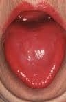 What Is A Fiery Red, Smooth, Burning Tongue?
A fiery red, smooth, burning tongue is an alteration in tongue tissue that is characteristic of advanced niacin deficiency.1
Q: How does niacin deficiency cause the tongue to be red and sore?
A: Niacin is an essential B vitamin that is required for a healthy tongue and by all body cells as well. Deficiency first shows in tissues with rapid cellular turnover, such as mucosal cells of the tongue.
When absorbed from the small intestines, niacin (the form in food) becomes the active form niacinamide. Niacinamide is converted by the body into co-enzymes which are present in all cells. These enzymes function in oxidation-reduction reactions essential for release of energy from carbohydrates, fats, and proteins and are needed as components for more than 200 enzymes involved in metabolism.
In addition to producing energy, niacinamide is essential for healthy skin and the mucosal lining of the digestive tract, normal functioning of the brain and nervous system, and production of steroid hormones from adrenal glands and hormones from sex glands.

A pale, smooth, burning tongue is an alteration in tongue tissue characteristic of iron deficiency. The tongue is also swollen.1
Additionally, the sore tongue surface may be invaded by candida yeast which takes advantage of the sore tissue.
Iron deficiency itself increases susceptibility to infection.
Q: What is iron deficiency?
A: Iron deficiency results when the level within cells is too low to meet metabolic needs of the body for this mineral.
Deficiency is characterized by impaired red blood cell formation, free-radical disposal, oxygenation of cells, immune response to infection, enzyme activity, cognitive performance, digestion, nail structure, and fetal health.2
Iron is an essential mineral that is required for normal body function. Almost two-thirds of iron in the body is found in hemoglobin, the protein in red blood cells that carries oxygen to tissues. Smaller amounts of iron are found in myoglobin, a protein that helps supply oxygen to muscle, and in enzymes that assist biochemical reactions.
Iron is also found in proteins that store iron for future needs and that transport iron in blood. Iron stores are regulated by intestinal iron absorption.3

C ollagenous colitis is a disease of the large intestine (colon) that is characterized by microscopic inflammation of the surface mucosal lining and an abnormally thickened collagen band of tissue that develops wthin the lining of the colon.
The thicker than normal layer of collagen of at least 10 µm (reference value: 2–7 µm) can vary in different locations. Inflammation occurs with increased numbers of lymphocytes (white blood cells) and plasma cells and epithelial (surface cell) damage. These changes can only be seen under microscopic examination of multiple biopsied tissue samples taken during a colonoscopy procedure.
Q: What is collagen?
A: Collagen is a strong, fibrous protein found in connective tissue of the colon and many other tissues such as tendons. The normal basement membrane in the bowel consists mainly of collagen type IV, laminin, and fibronectin. The increased collagen band observed in collagenous colitis consists basically of collagen type I and III, which are the subtypes produced by repair functions, indicating a reactive origin to some irritant or drug.1
The biopsies should preferably be taken from the ascending colon, since the pathological hallmarks may be absent in the descending colon, and in the normally occurring thicker collagen layer in the rectosigmoid region.1 Inflammation of the ileum (last segment of the small intestine next to colon) is common.2
Endoscopy and radiological (x-ray) examinations are usually normal.3
Autoimmune disorders are frequently seen in adult patients with collagenous colitis.4 In the study below by Koskela et al. concomittent autoimmune diseases were present in 53% of patients with collagenous colitis.5
Importantly, the finding of collagenous colitis in patients with autoimmune diseases may reflect the treatment with NSAIDs (non-steroidal anti-inflammatory drugs), such as Ibuprofin and aspirin, PPIs (proton pump inhibitors), and other drugs. However, if secondary forms of collagenous colitis are not taken into consideration, underlying, treatable diseases may be overlooked, while only the gastrointestinal symptoms are treated symptomatically or with budesonide (a steroid).6
Treatment with budesonide steroid is efficacious irrespective of bile acid malabsorption.7
Budesonide at a mean dose of 4.5 mg/day maintained clinical remission for at least 1 year in the majority of patients with collagenous colitis and preserved health-related quality of life without safety concerns. Treatment extension with low-dose budesonide beyond 1 year may be beneficial given the high relapse rate after budesonide discontinuation.8
See below for nutritional deficiency problems caused by steroid usage and steps to be taken for correction.

Riboflavin, also called vitamin B2, is fundamentally required for the breakdown of carbohydrates, amino acids, and fats in metabolism, enables oxygen to be used by cells, and acts as a component of more than a dozen enzymes involved in energy production and use.
Some enzymes needing riboflavin also require magnesium to properly function.
In other activities, riboflavin is required for healthy cardiovascular and nervous systems, eye health, and supports antioxidant protection. Functions are more fully described below.
Urinary excretion of riboflavin cannot be detected when vitamin intake is below the required levels. On the other hand, when intake exceeds saturation in the body, the vitamin and/or its metabolites are actively excreted into urine to prevent excessive toxicity of the vitamins.1
There is no specific name for riboflavin deficiency disease.
 What Is Prolonged Prothrombin Time?
Prolonged prothrombin time (PT) is a laboratory blood test result showing that blood clots too slowly which makes the patient subject to abnormal bleeding.
Q: What does the prothrombin time (PT) test measure?
A: The prothrombin test measures the clotting ability of blood protein factors I, II (prothrombin), V, VII, and X. If any of these factors are too low, it takes longer than normal for the blood to clot. Prothrombin is a vitamin K dependent factor meaning a deficiency of vitamin K will cause low prothrombin.
Blood is drawn into a blood collection tube with a light blue stopper which has a buffering additive. Tubes must be completely filled.

Chronic lesions of the oral mucosa are disorders of the mouth that are characterized by soreness and sores of the soft mucosal lining of the mouth.
 What Is Primary Biliary Cirrhosis?
P rimary biliary cirrhosis (PBC) is a biliary tract disease characterized by chronic cholestasis (build-up of bile) and gradual destruction of bile ducts within the liver, called intrahepatic bile ducts, caused by chronic inflammation.
Primary biliary cirrhosis comes under the umbrella term autoimune liver disease in which the end result is immune-mediated hepatocellular (liver cell) or hepatobiliary (bile duct) injury.1
Q: What is the end result of destruction of bile ducts?
A: The end result of destruction of bile ducts is liver damage.
Injured liver tissue from chronic inflammation and the buildup of bile leads to cirrhosis, a condition in which the liver slowly deteriorates and malfunctions.
Scar tissue replaces healthy liver tissue, partially blocking the flow of blood through the liver. Scarring also impairs the liver’s normal ability to control infections, remove bacteria and toxins from the blood, process nutrients, hormones, and drugs, make proteins that regulate blood clotting, produce bile, and effectively replace its own cells when they become damaged.2
The liver is the largest organ within the body. It lies mostly in the upper part of the abdomen on the right side just under the diaphragm. About 70% of liver tissue is made up of cube shaped cells called hepatocytes that do the main work of the liver. Other cells (epithelial) form structure and are arranged in single layers around blood vessels, sinusoids, and bile ducts.
Bile ducts carry bile, a greenish brown liquid made by the liver to the gall bladder for storage until needed to aid in the digestion and absorption of fat and fat-soluble vitamins A, D, E, and K from the small intestine. Bile emulsifies fat eaten in the diet so that the pancreatic enzyme called lypase can break it down into its fatty acid and glycerol components which can then be absorbed into the body.
Bile also carries away waste products produced by normal metabolism and toxic substances that are removed by the liver for eventual elimination in stool. Bile is continually made by the liver from phospholipids, bile acids, cholesterol, and aging blood cells it removes from circulation. As such, bile must continually flow out of the liver to prevent build-up in the liver.
There is no cure for primary biliary cirrhosis.
What Is Vitamin B12 Deficiency Anemia? Vitamin B12 deficiency anemia is a megaloblastic anemia that is characterized by defective DNA synthesis of red blood cells due to a lack of vitamin B12. Vitamin B12 is essential…

Angina pectoris, or simply angina, is a coronary syndrome characterized by an oppressive substernal pain (pain under breastbone) or pressure brought on by exertion and relieved by rest that results from failure of coronary arteries to deliver adequate oxygen to heart tissue due to ischemic heart disease.
Q: Why do coronary arteries fail to deliver adequate oxygen to heart tissue?
A: Coronary arteries are the blood vessels that serve the heart. In angina, these vessels fail to deliver adequate oxygen to heart tissue because they are narrowed or blocked by fatty buildups, called atherosclerotic plaques or by a blood clot which impair their ability to carry adequate blood that carries the oxygen. Diseased coronary arteries cannot deliver adequate oxygenated blood pumped by the heart to its own muscle cells.
The heart is a muscular organ that is working all the time without rest, so it needs a constant supply of oxygen. When heart muscle has to work harder, it needs more oxygen. Lack of oxygen causes pain which makes the affected person stop activity and rest.
Angina can be stable or unstable. Unstable angina is much more serious and can be life-threatening.