Erosions Resulting From Esophagitis Can Be A Cause Of Gastrointestinal Bleeding.
What Is Occult Gastrointestinal Bleeding?
Occult gastrointestinal bleeding is characterized by unseen or minute quantities of blood in stool. The origin of bleeding is from mucosa that lines the inside of the digestive tract at a site that must be found by medical testing and procedures to look at the lining.
Q: What tests and procedures are performed to locate the bleeding?
A: The simplest test to discover blood that cannot be seen is the stool test. This consists of a card with 3 spaces for 3 separate stool collection over 3 separate days. A tiny smear of stool is placed on a test card space on each of three days. Then the card is examined for a reaction that indicates the existence of blood in the stool.
If the stool test is positive, the origin of bleeding must be located. This search requires certain procedures that look at the mucosal lining directly by a gastroenterologist, usually under light sedation.
Gastroscopy procedure can visualize the upper gastrointestinal tract, which includes the esophagus and stomach.
Endoscopy procedure can also visualize the upper small intestine. However, endoscopy procedure is limited in that it cannot visualize the middle small intestine (jejunum).
Colonoscopy can visualize the end of the small intestine and colon all the way to the rectum.
Capsule endoscopy procedure. If gastroendoscopy and colonoscopy fail to discover the source of bleeding, the physician may administer a capsule endoscopy which visualizes the entire gastrointestinal tract. In this procedure, the patient swallows a capsule size camera (encased) which takes constant pictures over the course of a day until expelled through the rectum. Of course, the patient must be able to swallow the large size capsule.
What Is Occult Gastrointestinal Bleeding In Celiac Disease and/or Gluten Sensitivity?
This photo taken during laparoscopy shows the gall bladder (small white organ in middle) surrounded by yellow fat. Liver (dark red organ) is overlying.
What Is Impaired Gall Bladder Motility?
I mpaired gall bladder motility means the gall bladder is slow to empty or is dysfunctional.
The functional disorder of the gallbladder is caused initially either by metabolic abnormalities or by an alteration in its muscular ability to contract (motility dysfunction).
The diagnostic criteria based on symptoms of motility dysfunction of the gallbladder are episodes of moderate to severe steady pain located in the epigastrium and right upper abdominal quadrant that last at least 30 minutes.
Gallbladder motility disorder is suspected after gallstones and other structural abnormalities have been excluded.1
Q: What does the gallbladder do?
A: The gallbladder is a small pouch-like organ about the size of a pear that receives bile produced by the liver and stores it until needed during digestion. It lies just under the liver.
Bile is a complex fluid containing water, electrolytes and many organic molecules including bile acids, cholesterol, phospholipids and bilirubin. Bile acids are critical for digestion and absorption of fats and fat-soluble vitamins in the small intestine. Many waste products, including bilirubin, are eliminated from the body by secretion into bile and elimination in feces.2
Before a meal, the gallbladder is usually full of bile. In response to fat in the diet, the gallbladder squeezes stored bile into the small intestine through a series of ducts. When emptied after meals, the gallbladder is flat.
What Is Impaired Gall Bladder Motility In Celiac Disease and/or Gluten Sensitivity?
Sources:
Behar J, Corazziari E, Guelrud M, Hogan W, Sherman S, Toouli J. Functional gallbladder and sphincter of oddi disorders. Gastroenterology. 2006 Apr;130(5):1498-509. [↩]
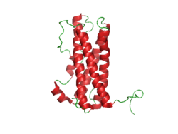
Prolactin Structure. Courtesy Boris TM at Wikipedia.
What Is Hyperprolactinemia?
H yperprolactinemia is an excess secretion of prolactin hormone causing an elevated blood level that is characterized by its effects on reproduction: in females causing amenorrhea and in males causing reduced sexual potency.
Q: What is prolactin?
A: Prolactin is a hormone produced by the the anterior pituitary gland that is transported by the bloodstream to stimulate androgen hormone production in male and estrogen hormone production in female sex glands. However, too much prolactin inhibits hormone production of estrogen in women and testosterone in men.
In pregnant females upon delivery of a baby, prolactin stimulates breast development and milk production for the purpose of feeding the infant. Sucking by the infant stimulates the release of prolactin which keeps the milk supply adequate.
Prolactin also promotes autoimmune mechanisms. Prolactin interferes specifically with B cell tolerance induction, enhances proliferative response to antigens and mitogens and increases the production of immune globulins, cytokines and autoantibodies.1
Other conditions that can stimulate prolactin release include nutritional deficiencies, thyroid stimulating hormone (TSH) produced by the pituitary gland, stress, exercise, and anterior pituitary tumors.
What Is An Elevated Prolactin Level In Celiac Disease and/or Gluten Sensitivity?
Sources:
Shelly S, Boaz M, Orbach H. Prolactin and autoimmunity. Autoimmun Rev. 2012 May;11(6-7):A465-70. doi: 10.1016/j.autrev.2011.11.009. [↩]
What Is Chronic Or Recurrent Abdominal Pain? A bdominal pain is characterized by pain anywhere in the abdominal cavity between the chest and groin. This region is often referred to as the belly. Abdominal pain,…
F olic acid deficiency anemia, also called folate deficiency anemia, is a macrocytic anemia characterized by defective DNA synthesis of red blood cells that results from a lack of folate in the body.
Q: How does folate deficiency cause anemia?
A: Folates are a family of B vitamins and folic acid is an active form.
Folate is required for the formation of both red and white blood cells in the bone marrow and for their maturation.
Also, folate serves as a carrier in the formation of heme, which contains iron, and is the non-protein part of the hemoglobin molecule.1
Red blood cells provide oxygen to body tissues. When there are not enough red blood cells or when they cannot properly carry oxygen, the condition is called anemia. In folic acid deficiency anemia, the red blood cells are abnormally large. Such cells are called macrocytes (macro size cells). They are also called megaloblasts (mega size cells) as seen in the bone marrow where they are produced. This is why this macrocytic anemia is also called megaloblastic anemia.2
Tests that may be done to determine folate adequacy are complete blood count (CBC), red blood cell folate level, methylmalonic acid level, and homocysteine level. Folic acid deficiency anemia shows a decrease in red blood cell folate and/or serum folate levels and normal plasma methylmalonic acid level with elevated homocysteine blood level. These levels distinguish folic acid deficiency anemia from vitamin B12 deficiency anemia.3
What Is Folate Deficiency Anemia In Celiac Disease and/or Gluten Sensitivity?
Section of small bowel surgically removed for adenocarcinoma that grew through the wall. By: CDC/ Dr. Edwin P. Ewing, Jr.
What Is Adenocarcinoma Of Small Intestine?
A denocarcinomas are malignant tumors, or cancer, of the small bowel arising out of glandular tissue. They fall in the category of rare neoplasm, comprising only 3% of all gastrointestinal malignancies.
Primary adenocarcinoma is the most common histological (cell) subtype constituting 35–50% of cases.1
Q: What does adenocarcinoma look like?
A: Adenocarcinoma may manifest as strictures, nodules, excavating masses, or annular lesions.2
What Is Adenocarcinoma Of Small Intestine In Celiac Disease and/or Gluten Sensitivity?
Sources:
Benhammane H, El M’rabet FZ, Serhouchni KI, El yousfi M, Charif I, Toughray I, et al. Small Bowel Adenocarcinoma Complicating Coeliac Disease: A Report of Three Cases and the Literature Review. Case Rep Oncol Med. 2012; 2012: 935183. Published online 2012 December 1. doi: 10.1155/2012/935183 [↩]
Ramachandran I, Sinha R, Rajesh A, Verma R. Multidetector row CT of small bowel tumors. Clinical Radiology. 2007; 62:607-614. [↩]
A ngina pectoris, or simply angina, is a coronary syndrome characterized by an oppressive substernal pain (pain under breastbone) or pressure brought on by exertion and relieved by rest that results from failure of coronary arteries to deliver adequate oxygen to heart tissue due to ischemic heart disease.
Q: Why do coronary arteries fail to deliver adequate oxygen to heart tissue?
A: Coronary arteries are the blood vessels that serve the heart. In angina, these vessels fail to deliver adequate oxygen to heart tissue because they are narrowed or blocked by fatty buildups, called atherosclerotic plaques or by a blood clot which impair their ability to carry adequate blood that carries the oxygen. Diseased coronary arteries cannot deliver adequate oxygenated blood pumped by the heart to its own muscle cells.
The heart is a muscular organ that is working all the time without rest, so it needs a constant supply of oxygen. When heart muscle has to work harder, it needs more oxygen. Lack of oxygen causes pain which makes the affected person stop activity and rest.
Angina can be stable or unstable. Unstable angina is much more serious and can be life-threatening.
Stable angina produces predictable pain and responds to rest and/or medication. It is less serious than unstable angina but can be very painful or uncomfortable. Anything that makes the heart muscle need more oxygen can cause an angina attack in someone with heart disease, including: smoking, cold weather, exercise, emotional stress, obesity, and large meals. Other causes of angina include: abnormal heart rhythms (usually ones that cause the heart to beat quickly), anemia, coronary artery spasm, heart failure, heart valve disease, and hyperthyroidism (overactive thyroid).1
Unstable angina produces unpredictable pain that may occur at rest, lasting more than 20 minutes. It is more severe than stable angina and less responsive to medication. Atherosclerosis is by far the most common cause of unstable angina. Oxidized low-density lipoprotein, so-called bad cholesterol, and oxysterols play an important role in atherogenesis, the development of atherosclerosis. Coronary arteries that are narrowed by atherosclerotic plaques can rupture causing injury to the coronary blood vessel resulting in blood clotting which blocks the flow of blood to the heart muscle. Blood clots may form, partially dissolve, and later form again and angina can occur each time a clot blocks blood flow in an artery. People with unstable angina are at increased risk of having a heart attack.2
What Is Angina In Celiac Disease and/or Gluten Sensitivity?
What Is Carbohydrate Malabsorption? C arbohydrate malabsorption is a digestive disorder characterized by the inability to properly digest and absorb carbohydrates within the small intestine to supply needed energy to the body. Q: What carbohydrates should be…
Figure on right shows how atherosclerosis impedes blood flow through coronary arteries while blood clots block blood flow. Courtesy Google.
What Is Coronary Artery Disease (CAD)?
C oronary artery disease (CAD), also called ischemic heart disease, is a gradual narrowing of medium and large arteries of the heart by fatty buildups, called atherosclerotic plaques.
It is characterized by slowly developing interference with blood flow to heart tissue itself, resulting in oppressive chest pain called angina and, ultimately, thrombosis (clot) causing heart attack.
The heart is a muscular organ that is working all the time, so it needs a constant supply of oxygen. Oxygen is brought to the working heart tissue by the coronary arteries with each beat of the heart. When heart muscle has to work harder, it needs more oxygen delivered to itself. Lack of oxygen causes pain.
In fact, failure of diseased coronary arteries to deliver adequate oxygen to heart tissue is the most common cause of angina pectoris – substernal pain (under breastbone) or pressure brought on by exertion and relieved by rest.
Thrombosis, or clot formation, occurs when blood cells within a narrowed artery can no longer get through. Trapped, blood cells pile up and block the artery thus triggering a cascade of events called heart attack. Coronary arteries that are narrowed by atherosclerotic plaques can rupture causing injury to the coronary blood vessel resulting in blood clotting which blocks the flow of blood to the heart muscle. Blood clots may form, partially dissolve, and later form again and angina can occur each time a clot blocks blood flow in an artery.1
Q: How does coronary artery disease develop?
A:Coronary artery disease slowly develops from this combination of events:
Dysfunction of epithelial cells that line the inside of arteries cause the vessels to stiffen, and subsequently
Accumulation of lipid (fat) in smooth muscle cells beneath the inside lining of arteries and in foam cells cause buildup of fatty deposits on the inside walls progressing to fibrous plaque formation.
Oxidized low-density lipoprotein (oxLDL), so-called bad cholesterol, and oxysterols play important roles in the development of atherosclerosis. OxLDL triggers the immune system to produce autoantibodies against oxLDL that are detectable in serum. These antibodies are called anti-oxLDL. Anti-oxLDL antibody and oxysterol concentrations are associated with coronary artery stenosis. Oxidative stress may be greatly increased in unstable angina.2 and Chronic inflammation in the general population is a major risk factor for ischemic heart disease.
The pathophysiology of atherosclerosis is, clearly, different in women when compared to the men. The women have a higher risk of blood coagulability making them at high risk for the blood clot formation. In a large number of women endothelial dysfunction, small vessel size and diffuse atherosclerosis have been identified as causes of ischemia without evidence of blockade in the coronary arteries.3
Also, atherosclerotic plaque in women is less fibrotic and contains more lipid filled foam cells, implying greater potential for reversibility but also potentially greater vulnerability for plaque rupture and thrombosis.4
Who is Affected in the General Population?
Coronary artery disease remains the leading cause of death in developed countries despite significant progress in primary prevention and treatment strategies.
It is the leading cause of death in women, as well as an important cause of disability.
Older patients are at particularly high risk of poor outcomes following acute coronary syndrome.5
What Is Coronary Artery Disease In Celiac Disease and/or Gluten Sensitivity?
Ischemic heart disease is the leading cause of death in the United States, making cardiovascular risk assessments and potential interventions or treatments imperative for patients with celiac disease.6
Yasunobu Y, Hayashi K, Shingu T, Yamagata T, Kajiyama G, Kambe M. Coronary atherosclerosis and oxidative stress as reflected by autoantibodies against oxidized low-density lipoprotein and oxysterosis. Atherosclerosis. Apr 2001;155(2):445-53. [↩]
Kunadian V, Ford GA, Bawamia B, Qiu W, Manson JE. Vitamin D deficiency and coronary artery disease: A review of the evidence. Am Heart J. 2014 Mar;167(3):283-291. doi: 10.1016/j.ahj.2013.11.012. Epub 2013 Dec 19. [↩]
Kunadian V, Ford GA, Bawamia B, Qiu W, Manson JE. Vitamin D deficiency and coronary artery disease: A review of the evidence. Am Heart J. 2014 Mar;167(3):283-291. doi: 10.1016/j.ahj.2013.11.012. Epub 2013 Dec 19. [↩]
Kunadian V, Ford GA, Bawamia B, Qiu W, Manson JE. Vitamin D deficiency and coronary artery disease: A review of the evidence. Am Heart J. 2014 Mar;167(3):283-291. doi: 10.1016/j.ahj.2013.11.012. [↩]
Robinson BL, Davis SC, Vess J, Lebel, J. Primary care management of celiac disease. Nurse Practitioner. February 2015: Vol 40 – Issue 2; 28–34. [↩]
C hronic diarrhea is an intestinal motility disorder characterized by 1) alteration in stool formation causing loose to fluid movements and quantity of movements with or without abdominal pain and 2) interference with normal carbohydrate salvage by the intestinal microbe population.
The severity of diarrhea is determined by the frequency and quantity of fluid lost.
Q: What is carbohydrate salvage by microbes in the colon?
A: Carbohydrate salvage in the colon is the necessary process whereby billions of microbes normally present in the colon work to breakdown undigestible carbohydrate foodstuffs such as fiber that continually arrive from the small intestine.
The microbial action releases energy to the body that would otherwise be lost with defecation, generates short-chain fatty acids, and stimulates sodium and fluid absorption. In this important process, butyrate and proprionate are produced which nourish the colonocytes (cells that line the colon), and acetate is produced for the liver.
While diarrhea may be a common symptom of small bowel mucosal disease, the consequent malabsorption can lead to substantial malnutrition and nutrient deficiencies. The small intestine, unlike the colon, has been relatively inaccessible, and systematic evaluation is often necessary to identify and treat small intestinal mucosal diseases that lead to diarrhea. All patients with severe diarrhea or diarrhea associated with features suggestive of malabsorption may have a disease of the small intestinal mucosa that requires careful evaluation and targeted management.1
What Is Chronic Diarrhea In Celiac Disease and/or Gluten Sensitivity?
Sources:
Murray JA1, Rubio-Tapia A. Diarrhoea due to small bowel diseases. Best Pract Res Clin Gastroenterol. 2012 Oct;26(5):581-600. doi: 10.1016/j.bpg.2012.11.013. [↩]