L aryngospasm is an acute disorder of the larynx that is characterized by life-threatening spasms of the muscles of the larynx located in the throat.
Laryngospasm is a rare feature of hypocalcemia (low blood calcium level) characterized by severe alteration in nerve conduction and muscle contraction.
What Is Laryngospasm In Celiac Disease and/or Gluten Sensitivity?
Anatomy of Upper Airway and Throat. Courtesy Wikipedia.org
What Is Post-Cricoid Carcinoma?
P ost-cricoid carcinoma is a rare malignancy arising in the hypopharynx.
Q: What is the hypopharynx?
A: The hypopharynx is the lower portion of the pharynx that opens into the larynx. It is comprised of the postcricoid region, pyriform sinus, and posterior hypopharyngeal wall.
Tumors rarely appear in the posterior pharyngeal wall or postcricoid region without also involving the pyriform sinus. The lack of anatomic barriers between these sites and a propensity for these tumors to develop in the pyriform sinus and spread outward account for this phenomenon.
Overall prognosis for these tumors is poor. Because of the rich organic lymphatic and vascular networks, aggressive growth and early cervical metastases (compared with cancers at other head and neck sites) characterize these cancers.
A study investigating the clinical course of 21 patients with post-cricoid carcinoma found the 5-year cause-specific survival rate was 52%. All the patients who eventually died did so within 3 years of the treatment. Seven patients had distant metastases, representing a higher frequency as compared with that of patients with recurrence of the primary focus and cervical lymph node metastasis. All of these patients who had been treated by surgery died of the cancer.1
Unfortunately, hypopharyngeal cancers are discovered at a later stage than other head and neck cancers. The delay in diagnosis from symptom onset averages 10 months.2
What Is Post-Cricoid Carcinoma In Celiac Disease and/or Gluten Sensitivity?
Sources:
Asakage T, Nakao K, Ebihara Y, Fujishiro Y, Watanabe K. A clinical study of post-cricoid carcinoma. Acta Otolaryngol Suppl. 2007 Dec;(559):118-22. doi: 10.1080/03655230701599354. [↩]
What Is Dysphagia? D ysphagia is a functional upper digestive disorder characterized by difficulty in swallowing or food getting stuck and swallowing that takes longer than 10 seconds to complete. Q: What determines the symptoms?…
Bristol Stool Chart Showing Normal and Abnormal Stool.
What Is Chronic Constipation Alternating With Diarrhea?
Chronic constipation alternating with diarrhea is an intestinal motility disorder, or irregularity, characterized by alteration in stool formation, consistency, and evacuation which results in a bowel movement that consists of some hard or balled stool along with some loose stool that can cause leakage.
Q: How do irregular movement patterns develop in the colon?
A: The colon produces irregular movements as a result of problems that originate in the colon (large intestine) itself and/or the small intestine which then affects function of the colon.
Here are listed the many types of problems or diseases that cause these abnormal bowel movements:
Disorders that adversely affect the colon, an organ which must propel stool, remove excess water, absorb electrolytes, ferment undigested food material that passes into it, and produce nutrients from the fermentation process:
Poor diet that does not contain adequate nutrition, fiber, probiotics, prebiotics, and water to form normal stool.
Diet that contains irritating, toxic or allergenic food that cause spasms.
Diseases that inflame the mucosa lining such as collagenous colitis, altering the proper absorption of water and electrolytes.
Diseases that damage and swell the colon walls, such as Crohn’s disease, ulcerative colitis, and diverticulitis.
Diseases that obstruct the lumen or passageway so that stool passes with difficulty.
Diseases that hamper normal peristalsis (muscle action), such as irritable bowel syndrome (IBS), diabetes and thyroid disease.
Disorders that adversely affect the small intestine, an organ which must digest and absorb nutrients needed by the body while passing unabsorbed food material to the colon:
Diet that conatins too much fat, sugar or artifical sweeteners, causing diarrhea.
Disorders that result in malabsorption, such as gluten enteropathy, milk enteropathy, steatorrhea (fat malabsorption), lactose intolerance, sucrose intolerance, maltose intolerance, and bacterial overgrowth, passing abnormal amounts of undigested food material to the colon where it is fermented producing excessive gas, diarrhea and spasm.
Disorders that impair peristalsis, such as active celiac disease, diabetes, scleroderma, and thyroid disease.
Tumors like cancer and lymphoma impair regular passage of material to colon.
Drugs that impair peristalsis, such as iron supplements, aluminum containing antacids, narcotics, some anti-depressants, some anti-seizure, and some diuretics.
What Is Chronic Constipation Alternating With Diarrhea In Celiac Disease and/or Gluten Sensitivity?
Figure 1: Normal Liver Lobule. Courtesy almostadoctor.com
What Is Autoimmune Cholangitis?
A utoimmune cholangitis is a rare inflammatory self-targeted disease of the biliary tract produced by high levels of immunoglobulin G (IgG) antibodies and antinuclear antibodies (ANA).
Autoimmune cholangitis is characterized by progressive inflammation and scarring that leads to narrowing and destruction of the bile ducts and development of biliary cirrhosis of the liver.
Autoimmune cholangitis is also termed anti-mitochondrial antibody-negative primary biliary cirrhosis because there is no production of anti-mitochondrial antibodies.
Q: What happens when bile ducts can no longer transport bile out of the liver?
A:Bile that cannot be removed from the liver by the biliary tract backs up and damages the liver, causing cirrhosis. In Figure 1, bile back up would directly damage the hepatic (liver) cells seen here as grey “blocks.”
Bile is a greenish brown liquid continually made by the liver cells (hepatocytes) from phospholipids salt, cholesterol, and aging blood cells that it removes from circulation.
Bile also carries away waste products produced by normal metabolism and toxic substances that are removed by the liver for eventual elimination in stool.As such, bile must continually flow out of the liver to prevent build-up in the liver.
Bile from the hepatic cells drains into small tubes called canniculi and then into larger bile ducts to be carried out of the liver to the gall bladder for storage until needed to aid in the digestion and absorption of fat from the small intestine. Bile emulsifies fat eaten in the diet so that the pancreatic enzyme called lypase can break it down into its fatty acid and glycerol components.
Unfortunately, autoimmune cholangitis is difficult to diagnose because it mimics many other diseases such as primary sclerosing cholangitis, sarcoidosis, cancer and lymphoma.
What Is Autoimmune Cholangitis In Celiac Disease and/or Gluten Sensitivity?
Erosions Resulting From Esophagitis Can Be A Cause Of Gastrointestinal Bleeding.
What Is Occult Gastrointestinal Bleeding?
Occult gastrointestinal bleeding is characterized by unseen or minute quantities of blood in stool. The origin of bleeding is from mucosa that lines the inside of the digestive tract at a site that must be found by medical testing and procedures to look at the lining.
Q: What tests and procedures are performed to locate the bleeding?
A: The simplest test to discover blood that cannot be seen is the stool test. This consists of a card with 3 spaces for 3 separate stool collection over 3 separate days. A tiny smear of stool is placed on a test card space on each of three days. Then the card is examined for a reaction that indicates the existence of blood in the stool.
If the stool test is positive, the origin of bleeding must be located. This search requires certain procedures that look at the mucosal lining directly by a gastroenterologist, usually under light sedation.
Gastroscopy procedure can visualize the upper gastrointestinal tract, which includes the esophagus and stomach.
Endoscopy procedure can also visualize the upper small intestine. However, endoscopy procedure is limited in that it cannot visualize the middle small intestine (jejunum).
Colonoscopy can visualize the end of the small intestine and colon all the way to the rectum.
Capsule endoscopy procedure. If gastroendoscopy and colonoscopy fail to discover the source of bleeding, the physician may administer a capsule endoscopy which visualizes the entire gastrointestinal tract. In this procedure, the patient swallows a capsule size camera (encased) which takes constant pictures over the course of a day until expelled through the rectum. Of course, the patient must be able to swallow the large size capsule.
What Is Occult Gastrointestinal Bleeding In Celiac Disease and/or Gluten Sensitivity?
This photo taken during laparoscopy shows the gall bladder (small white organ in middle) surrounded by yellow fat. Liver (dark red organ) is overlying.
What Is Impaired Gall Bladder Motility?
I mpaired gall bladder motility means the gall bladder is slow to empty or is dysfunctional.
The functional disorder of the gallbladder is caused initially either by metabolic abnormalities or by an alteration in its muscular ability to contract (motility dysfunction).
The diagnostic criteria based on symptoms of motility dysfunction of the gallbladder are episodes of moderate to severe steady pain located in the epigastrium and right upper abdominal quadrant that last at least 30 minutes.
Gallbladder motility disorder is suspected after gallstones and other structural abnormalities have been excluded.1
Q: What does the gallbladder do?
A: The gallbladder is a small pouch-like organ about the size of a pear that receives bile produced by the liver and stores it until needed during digestion. It lies just under the liver.
Bile is a complex fluid containing water, electrolytes and many organic molecules including bile acids, cholesterol, phospholipids and bilirubin. Bile acids are critical for digestion and absorption of fats and fat-soluble vitamins in the small intestine. Many waste products, including bilirubin, are eliminated from the body by secretion into bile and elimination in feces.2
Before a meal, the gallbladder is usually full of bile. In response to fat in the diet, the gallbladder squeezes stored bile into the small intestine through a series of ducts. When emptied after meals, the gallbladder is flat.
What Is Impaired Gall Bladder Motility In Celiac Disease and/or Gluten Sensitivity?
Sources:
Behar J, Corazziari E, Guelrud M, Hogan W, Sherman S, Toouli J. Functional gallbladder and sphincter of oddi disorders. Gastroenterology. 2006 Apr;130(5):1498-509. [↩]
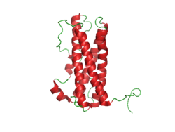
Prolactin Structure. Courtesy Boris TM at Wikipedia.
What Is Hyperprolactinemia?
H yperprolactinemia is an excess secretion of prolactin hormone causing an elevated blood level that is characterized by its effects on reproduction: in females causing amenorrhea and in males causing reduced sexual potency.
Q: What is prolactin?
A: Prolactin is a hormone produced by the the anterior pituitary gland that is transported by the bloodstream to stimulate androgen hormone production in male and estrogen hormone production in female sex glands. However, too much prolactin inhibits hormone production of estrogen in women and testosterone in men.
In pregnant females upon delivery of a baby, prolactin stimulates breast development and milk production for the purpose of feeding the infant. Sucking by the infant stimulates the release of prolactin which keeps the milk supply adequate.
Prolactin also promotes autoimmune mechanisms. Prolactin interferes specifically with B cell tolerance induction, enhances proliferative response to antigens and mitogens and increases the production of immune globulins, cytokines and autoantibodies.1
Other conditions that can stimulate prolactin release include nutritional deficiencies, thyroid stimulating hormone (TSH) produced by the pituitary gland, stress, exercise, and anterior pituitary tumors.
What Is An Elevated Prolactin Level In Celiac Disease and/or Gluten Sensitivity?
Sources:
Shelly S, Boaz M, Orbach H. Prolactin and autoimmunity. Autoimmun Rev. 2012 May;11(6-7):A465-70. doi: 10.1016/j.autrev.2011.11.009. [↩]
What Is Chronic Or Recurrent Abdominal Pain? A bdominal pain is characterized by pain anywhere in the abdominal cavity between the chest and groin. This region is often referred to as the belly. Abdominal pain,…
F olic acid deficiency anemia, also called folate deficiency anemia, is a macrocytic anemia characterized by defective DNA synthesis of red blood cells that results from a lack of folate in the body.
Q: How does folate deficiency cause anemia?
A: Folates are a family of B vitamins and folic acid is an active form.
Folate is required for the formation of both red and white blood cells in the bone marrow and for their maturation.
Also, folate serves as a carrier in the formation of heme, which contains iron, and is the non-protein part of the hemoglobin molecule.1
Red blood cells provide oxygen to body tissues. When there are not enough red blood cells or when they cannot properly carry oxygen, the condition is called anemia. In folic acid deficiency anemia, the red blood cells are abnormally large. Such cells are called macrocytes (macro size cells). They are also called megaloblasts (mega size cells) as seen in the bone marrow where they are produced. This is why this macrocytic anemia is also called megaloblastic anemia.2
Tests that may be done to determine folate adequacy are complete blood count (CBC), red blood cell folate level, methylmalonic acid level, and homocysteine level. Folic acid deficiency anemia shows a decrease in red blood cell folate and/or serum folate levels and normal plasma methylmalonic acid level with elevated homocysteine blood level. These levels distinguish folic acid deficiency anemia from vitamin B12 deficiency anemia.3
What Is Folate Deficiency Anemia In Celiac Disease and/or Gluten Sensitivity?
 What Is Laryngospasm?