Skip to content

Dysgeusia (Impaired Taste)

DyspareuniaWhat Is Autoimmune Thyroiditis (Hypothyroidism)?

A utoimmune thyroiditis, also called Hashimoto’s thyroiditis or Hashimoto’s Disease, is an autoimmune destruction of thyroid tissue characterized by insufficient thyroid hormone circulating in the body that causes formation of a goiter (enlarged thyroid gland) and hypothyroidism.

Hypothroidism refers to the condition of markedly reduced secretion of thyroid hormone. There are other causes of hypothyroidism besides Hashimoto’s thyroiditis.

Hashimoto’s thyroiditis is often associated with other autoimmune diseases such as celiac disease.

In Hashimoto’s thyroiditis, a profusion of antibodies are produced, which build up in the blood. Left untreated so that much of the thyroid gland is destroyed, this condition may progress to the very serious and life-threatening condition called myxedema.

Note: In myxedema, protein, electrolytes, and water abnormally accumulate in between cells which produce firm, inelastic puffy skin that is cool, dry, rough, scaly, and may appear yellow; in some people, areas such as the ankles become crusty with a look of tree bark. Many systemic changes develop shown by significant slowing of mental and physical functions. Please see below.

Q: What thyroid tissue is targeted for destruction?

A: In Hashimoto’s thyroiditis, high levels of autoantibodies target thyroglobulin and thyroid peroxidase, leading to inflammation and destruction of the thyroid gland. The resulting fibrosis or scarring of the gland results in lack of thyroid hormone production.

The thyroid gland consists of a large number of closed vesicles that contain a homogenous substance called colloid, which contains the thyroglobulin. Thyroglobulin is an iodine-containing protein secreted by the thyroid gland and stored within its colloid, from which the thyroid hormones thyroxine (T4) and triiodothyroinine (T3) are derived.1

T3 is the active hormone and is made from T4. Thyroid hormones affect metabolism, brain development, breathing, heart and nervous system functions, body temperature, muscle strength, skin dryness, menstrual cycles, weight, and cholesterol levels.

Thyroid hormone production is regulated by thyroid-stimulating hormone (TSH), which is made by the pituitary gland in the brain. Normally, when thyroid hormone levels in the blood are low, the pituitary releases more TSH. When thyroid hormone levels are high, the pituitary decreases TSH production.

Hashimoto’s disease, with or without the development of hypothyroidism, is treated with synthetic thyroxine, which is man-made T4. Health care providers prefer to use synthetic T4, such as Synthroid® (Levothyroxine), rather than synthetic T3, because T4 stays in the body longer, ensuring a steady supply of thyroid hormone throughout the day. The thyroid preparations made with animal thyroid are not considered as consistent as synthetic thyroid.2

What Is Autoimmune Thyroiditis In Celiac Disease and/or Gluten Sensitivity?

Sources:
  1. Taber’s Cyclopedic Medical Dictionary. 19th ed. F.A. Davis Company. Philadelphia, PA. []
  2. National Endocrine and Metabolic Diseases Information Service. []

Impotence

This White Spot Is A Feature of Zinc Deficiency.

What Are White Spots And White Bands In Nails?

W hite spots in the nails is a feature of zinc deficiency and white bands signify protein deficiency characterized by abnormal appearance anywhere in one or more nails.

White spots and bands can appear separately or together in nails. There may be just one mark or many marks depending on the level of deficiency.

Q: What part of the nail is affected by zinc and/or protein deficiency?

A: Zinc and protein deficiencies affect the nail plate.

The nail plate is the hard keratin cover of the finger tip and toe tip, simply called “the nail.” The nail plate is produced by the living nail tissue at its base. Here is an illustration of the parts that make up a fingernail:

Nail Anatomy. Nail Anatomy. A. Nail plate; B. lunula; C. root; D. sinus; E. matrix; F. nail bed; G. hyponychium; H. free margin. Courtesy Wikipedia.org.

A. Nail plate; B. lunula; C. root; D. sinus; E. matrix; F. nail bed; G. hyponychium; H. free margin. Courtesy Wikipedia.

What Are White Spots And White Bands In Nails In Celiac Disease and/or Gluten Sensitivity?

Bone Pain

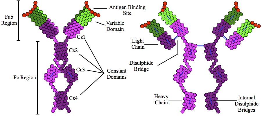
This is a depiction of immunoglobulin E (IgE) antibody which is elevated in allergic reactions.

What Is Allergic Rhinitis?

A llergic rhinitis is an immune disorder characterized by inflammation of the nasal mucosa by an IgE antibody reaction to an allergen.

An allergen is something that triggers an allergic immune response.

Q: What is the immune response?

A: Implicated in the response is an increase in T gamma-delta intraepithelial lymphocytes (IELs), which is a subset of pro-inflammatory T-cells located in the respiratory mucosa.  Lymphocytes are white blood cells.

When a person with allergic rhinitis breathes in an allergen such as pollen or dust, the body releases chemicals, including histamine that cause allergy symptoms. For example, hay fever involves an allergic reaction to pollen. A similar reaction occurs with allergy to mold, animal dander, dust, and other allergens that are breathed in.

What Is Allergic Rhinitis In Celiac Disease and/or Gluten Sensitivity?

Infertility In Males

Cutaneous Vasculitis. Courtesy quizlet.com

What Is Cutaneous Vasculitis?

Cutaneous vasculitis is a vascular disease of small blood vessels serving the skin that is characterized by segmental (spotty) inflammation of affected vessel walls resulting in their necrosis (destruction). Scarring is seen in the affected skin that lies over the destroyed blood vessels.

Skin appearance varies from small flat, purplish spots to raised purplish areas over damaged vessels.

In most cases, cutaneous vasculitis results from deposits of toxic immune complexes in the affected vessel walls that is caused by autoimmune activity targeting small blood vessels.1

Cutaneous vasculitis can also result from infection, drug reactions, or malignancies.

Who is Affected in the general Population? Cutaneous vasculitis affects all ages.

The overall annual incidence of cutaneous vasculitis was 38.6 persons per million in 1998.2

What Is Cutaneous Vasculitis In Celiac Disease and/or Gluten Sensitivity?

Sources:
  1. Abenavoli L, Proietti I, Leggio L, Ferrulli A, Vonghia L, Capizzi R, Rotoli M, Amerio PL, Gasbarrini G, Addolorato G. Cutaneous manifestations in celiac disease. World J Gastroenterol. 2006 Feb 14;12(6):843-52. []
  2. Watts RA, Jolliffe VA, Grattan CE, Elliott J, Lockwood M, Scott DG. Cutaneous vasculitis in a defined population–clinical and epidemiological associations. J Rheumatol. 1998 May;25(5):920-4. []

Osteitis Fibrosa Cystica 

Deep Vein Thrombosis in the Right Leg with Swelling and Redness. Coutesy wikipedia.
Deep Vein Thrombosis in the Right Leg with Swelling and Redness. Courtesy wikipedia.

What Is Antiphospholipid Syndrome?

Antiphospholipid syndrome (APS) is an autoimmune disease and a blood clotting disorder characterized by these clinical and laboratory criteria:

Clinical criteria – recurrent vascular thrombosis (clots in veins/arteries) from hypercoagulability (abnormal excessive clotting) and/or recurrent complications of pregnancy that include loss of the fetus  (miscarriage) and pre-eclampsia or eclampsia.

Laboratory criteria – persistently elevated anticardiolipin, anti–beta-2 glycoprotein I, and/or lupus anti-coagulant antibodies in blood.

In antiphospholipid syndrome autoantibodies are produced by the body and directed against negatively charged phospholipids that are found in the outer layer of cell membranes and platelets. B2-glycoprotein-I (a protein in blood plasma) has been found as a major target antigen for antiphospholipid antibodies.

Q: Are phospholipids important in the body?

A: Yes.  Phospholipid molecules are an essential part of cell membranes. They form a barrier around cells that protect the cell, allow movement of oxygen in and carbon dioxide out of the cell, and regulate other small molecules through the cell wall. Because phospholipids are widespread in the body, this disorder can produce a large variety of symptoms and affect many organs.

One severe effect of APS is the development of a blood clot in a vein deep in the arm or leg, called deep vein thrombosis (DVT). DVT can cause pain, swelling, redness, or increased warmth in the affected limb. Deep vein clots can break off, travel to the lungs, and cause pulmonary embolism.1 Pulmonary embolism is a medical emergency.

Treatment is with anticoagulant medications and blood monitoring.

What Is Antiphospholipid Syndrome In Celiac Disease and/or Gluten Sensitivity?

Sources:
  1. http://www.nhlbi.nih.gov/health/health-topics/topics/ebc/signs.html []

Osteomalacia

Drawing shows changes in airways during asthma attack. wikipedia

What Is Asthma?

Asthma is a chronic immune respiratory condition characterized by narrowing and inflammation of the lung airways (large bronchi, bronchial tubes and small bronchioles) in response to an allergen as the trigger or stimulus. As such, asthma occurs in episodes and does not result in progressive loss of pulmonary function.

During an asthma attack, airways constrict, trapping air so lungs become overinflated.  Normally, bronchial airways bring air to millions of air sacs that are attached to the ends of bronchioles. Air sacs, called alveoli, are only one cell thick to allow for rapid exchange of gases.

That is, oxygen from air breathed into the sacs moves into the bloodsteam and carbon dioxide is released from the bloodstream to air that is breathed out of air sacs.

The outer walls of bronchioles are made up of muscles which, in the process of breathing, normally contract on expiration to help expel air and then relax. During an asthma attack, these muscles abnormally constrict, impairing airflow into and out of the alveoli. This is called bronchospasm.

Common  allergens that cause inflammation include airborne dust mite feces, mold, and pollen and foods such as wheat, cow’s milk, eggs, and peanuts. Non-allergenic triggers include exercise, air pollution, smoking, and viral respiratory infection.

Q: What effect does inflammation have on the lungs?

A: Inflammation causes local tissue edema or swelling of the bronchioles and mucus formation. Inflammation with increased mucus secretions and edema narrows the airways that connect to alveoli which makes breathing difficult.  Two things happen:

  1. Inflammation  impairs exchange of gases in alveoli, resulting in lack of sufficient oxygen (O2) for body cell functions, called hypoxia, and build-up of carbon dioxide (CO2) in blood, called CO2 retention.
  2. Inflammation narrows passageways because of swelling, which reduces the movement of air to and from the alveoli through the airways, and this puts stress on the right side of the heart.

Treatment is aimed at controlling bronchospasm and reducing inflammation. Untreated asthma can be disabling and life threatening.

What Is Asthma In Celiac Disease and/or Gluten Sensitivity?

Common Variable Immunodeficiency 

Folic acid deficiency anemia gluten celiac disease symptom
Red Blood Cells.

What Is Folic Acid Or Folate Deficiency Anemia?

F olic acid deficiency anemia, also called folate deficiency anemia, is a macrocytic anemia characterized by defective DNA synthesis of red blood cells that results from a lack of folate in the body.

Q: How does folate deficiency cause anemia?

A: Folates are a family of B vitamins and folic acid is an active form.

Folate is required for the formation of both red and white blood cells in the bone marrow and for their maturation.

Also, folate serves as a carrier in the formation of heme, which contains iron, and is the non-protein part of the hemoglobin molecule.1

Red blood cells provide oxygen to body tissues. When there are not enough red blood cells or when they cannot properly carry oxygen, the condition is called anemia. In folic acid deficiency anemia, the red blood cells are abnormally large. Such cells are called macrocytes (macro size cells). They are also called megaloblasts (mega size cells) as seen in the bone marrow where they are produced. This is why this macrocytic anemia is also called megaloblastic anemia.2

Tests that may be done to determine folate adequacy are complete blood count (CBC), red blood cell folate level, methylmalonic acid level, and homocysteine level. Folic acid deficiency anemia shows a decrease in red blood cell folate and/or serum folate levels and normal plasma methylmalonic acid level with elevated homocysteine blood level. These levels distinguish folic acid deficiency anemia from vitamin B12 deficiency anemia.3

What Is Folate Deficiency Anemia In Celiac Disease and/or Gluten Sensitivity?

Sources:
  1. Kathleen Mahan and Sylvia Escott-Stump, ed. Krause’s Food, Nutrition & Diet Therapy, 10th Edition. Philadelphia, PA. USA: W.B. Saunders Company, 2000. []
  2. http://www.ncbi.nlm.nih.gov/pubmedhealth/PMH0001578/ []
  3. Mark Beers and Robert Berkow. The Merck Manual, 17th Edition. Whitehouse Station, N.J. USA: Merck Research Laboratories, 1999. []

Ataxia, Gluten

This photo taken during laparoscopy shows the gall bladder (small white organ in middle) surrounded by yellow fat. Liver (dark red organ) is overlying.

What Is Impaired Gall Bladder Motility?

I mpaired gall bladder motility means the gall bladder is slow to empty or is dysfunctional.

The functional disorder of the gallbladder is caused initially either by metabolic abnormalities or by an alteration in its muscular ability to contract (motility dysfunction).

The diagnostic criteria based on symptoms of motility dysfunction of the gallbladder are episodes of moderate to severe steady pain located in the epigastrium and right upper abdominal quadrant that last at least 30 minutes.

Gallbladder motility disorder is suspected after gallstones and other structural abnormalities have been excluded.1

Q: What does the gallbladder do?

A: The gallbladder is a small pouch-like organ about the size of a pear that receives bile produced by the liver and stores it until needed during digestion. It lies just under the liver.

Bile is a complex fluid containing water, electrolytes and many organic molecules including bile acids, cholesterol, phospholipids and bilirubin. Bile  acids are critical for digestion and absorption of fats and fat-soluble vitamins in the small intestine.  Many waste products, including bilirubin, are eliminated from the body by secretion into bile and elimination in feces.2

Before a meal, the gallbladder is usually full of bile. In response to fat in the diet, the gallbladder squeezes stored bile into the small intestine through a series of ducts. When emptied after meals, the gallbladder is flat.

What Is Impaired Gall Bladder Motility In Celiac Disease and/or Gluten Sensitivity?

Sources:
  1. Behar J, Corazziari E, Guelrud M, Hogan W, Sherman S, Toouli J. Functional gallbladder and sphincter of oddi disorders. Gastroenterology. 2006 Apr;130(5):1498-509. []
  2. http://www.vivo.colostate.edu/hbooks/pathphys/digestion/liver/bile.html []

Hair, Fine With Rough Texture

Inflamed Aorta. Courtesy Quizlet.com

What Is Aortic Vasculitis?

A ortic vasculitis is an inflammatory disease of the aorta that causes dilation of the aorta wall with narrowing of the inside passageway and results in widespread impairment of blood flow to tissues served by the aorta.

Q: What is the result of inflammatory disease of the aorta?

A: The aorta is the largest artery in the body. It extends from the left ventricle (lower chamber) of the heart and carries blood out of the heart with each beat to rest of the body. Narrowing of the aorta’s inside diameter due to swelling from inflammation causes elevated blood pressure and enlarged heart because of back pressure of blood unable to be fully pumped out of the heart with each beat. The heart enlarges because the difficulty of pumping blood out makes it work harder than normal.

Vasculitis that causes midaortic syndrome is a variety of aortic coarctation (narrowing) located in the lower thoracic aorta, the abdominal aorta or both, involving the intestinal and renal vessels (kidney). It usually presents with arterial hypertension.1

What Is Aortic Vasculitis  In Celiac Disease and/or Gluten Sensitivity?

Sources:
  1. Massel D. n-3 polyunsaturated fatty acids reduced mortality and morbidity after recent myocardial infarction. Therapeutics. Jan-Feb 2000:6 []

Brain Atrophy

What Is Chronic Or Recurrent Abdominal Pain? A bdominal pain is characterized by pain anywhere in the abdominal cavity between the chest and groin. This region is often referred to as the belly. Abdominal pain,…