Skip to content

Pancreatic Insufficiency

Model of coagulation factors found in blood.

What Are Low Coagulation Factors?

C oagulation factors II, VII, IX, X found in blood are essential for normal blood clotting.  Low coagulation factors on blood assay indicate an altered secondary coagulation disorder that is characterized by impaired clot formation.

Each coagulation factor must be present in sufficient quantity in order for normal clotting to occur, but the level required is different for each factor. Results are frequently reported as a percentage with 100% being normal. For example, a factor VIII that is 30% would be considered abnormally low.1

The production of the coagulation factors II, VII, IX, and X requires vitamin K without which the factors will be low.

Q: What happens when coagulation factors II, VII, IX, and X are low?

A: When any of the blood clotting factors are lacking or not working properly, the blood tests prothrombin (PT) and partial thromboplastin time (PTT) will be abnormally prolonged. Prothrombin and partial thromboplastin time measure the time it takes for blood to clot.  When you bleed, the body launches a series of activities that help the blood clot. This is called the coagulation cascade. There are three pathways to this event. These tests looks at coagulation factors, found in two of these pathways.2

What Are Low Coagulation Factors In Celiac Disease and/or Gluten Sensitivity?

Sources:
  1. http://labtestsonline.org/understanding/analytes/coagulation-factors/tab/test []
  2. http://www.nlm.nih.gov/medlineplus/ency/article/003653.htm []

Lymphoma, B-Cell Non-Hodgkin’s

e3343bf0a689b5f974a852fa2e84a718What Is Laryngospasm?

L aryngospasm is an acute disorder of the larynx that is characterized by life-threatening spasms of the muscles of the larynx located in the throat.

Laryngospasm is a rare feature of hypocalcemia (low blood calcium level) characterized by severe alteration in nerve conduction and muscle contraction.

What Is Laryngospasm In Celiac Disease and/or Gluten Sensitivity?

Steatorrhea

A 3-D model of homocysteine.

What Is Elevated Homocysteine?

E levated homocysteine in blood, called hyperhomocysteinemia, indicates an abnormal blood level of this transient amino acid.

Q: How does the level of homocysteine become abnormal?

A: In metabolism, homocysteine is briefly formed in the breakdown of the amino acid methionine. It is normally converted to cystathione and then to the amino acid cysteine by means of an enzyme that requires vitamin B6.

In the reverse, conversion of homocysteine to methionine requires an enzyme dependent on adequate folic acid and vitamin B12 levels.

Insufficient methionine levels and/or inefficiency in this process results in elevated homocysteine plasma levels that are toxic to blood vessels.

Folic acid, vitamin B12 and vitamin B6 are involved in the metabolic removal of homocysteine, but folic acid deficit occurs the most often.1

What Is Elevated Homocysteine In Celiac Disease and/or Gluten Sensitivity?

Sources:
  1. Lim PO, Tzemos N, Farquharson CA, et al. Reversible hypertension following coeliac disease treatment: the role of moderate hyperhomocysteinaemia and vascular endothelial dysfunction. Journal of Human Hypertension. Jun 2002;16(6):411-5. []

Celiac Disease, Refractory

Anatomy of Upper Airway and Throat. Courtesy Wikipedia.org

What Is Post-Cricoid Carcinoma?

P ost-cricoid carcinoma is a rare malignancy arising in the hypopharynx.

Q: What is the hypopharynx?

A: The hypopharynx is the lower portion of the pharynx that opens into the larynx. It is comprised of the postcricoid region, pyriform sinus, and posterior hypopharyngeal wall.

Tumors rarely appear in the posterior pharyngeal wall or postcricoid region without also involving the pyriform sinus. The lack of anatomic barriers between these sites and a propensity for these tumors to develop in the pyriform sinus and spread outward account for this phenomenon.

Overall prognosis for these tumors is poor. Because of the rich organic lymphatic and vascular networks, aggressive growth and early cervical metastases (compared with cancers at other head and neck sites) characterize these cancers.

A study investigating the clinical course of 21 patients with post-cricoid carcinoma found the 5-year cause-specific survival rate was 52%. All the patients who eventually died did so within 3 years of the treatment. Seven patients had distant metastases, representing a higher frequency as compared with that of patients with recurrence of the primary focus and cervical lymph node metastasis. All of these patients who had been treated by surgery died of the cancer.1

Unfortunately, hypopharyngeal cancers are discovered at a later stage than other head and neck cancers. The delay in diagnosis from symptom onset averages 10 months.2

What Is Post-Cricoid Carcinoma In Celiac Disease and/or Gluten Sensitivity?

Sources:
  1. Asakage T, Nakao K, Ebihara Y, Fujishiro Y, Watanabe K. A clinical study of post-cricoid carcinoma. Acta Otolaryngol Suppl. 2007 Dec;(559):118-22. doi: 10.1080/03655230701599354. []
  2. http://emedicine.medscape.com/article/848359-overview []

Primary Hyperparathyroidism

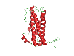
Prolactin Structure. Courtesy Boris TM at Wikipedia.

What Is Hyperprolactinemia?

H yperprolactinemia is an excess secretion of prolactin hormone causing an elevated blood level that is characterized by its effects on reproduction: in females causing  amenorrhea and in males causing reduced sexual potency.

Q: What is prolactin?

A: Prolactin is a hormone produced by the the anterior pituitary gland that is transported by the bloodstream to stimulate androgen hormone production in male and estrogen hormone production in female sex glands. However, too much prolactin inhibits hormone production of estrogen in women and testosterone in men.

In pregnant females upon delivery of a baby, prolactin stimulates breast development and milk production for the purpose of feeding the infant. Sucking by the infant stimulates the release of prolactin which keeps the milk supply adequate.

Prolactin also promotes autoimmune mechanisms. Prolactin interferes specifically with B cell tolerance induction, enhances proliferative response to antigens and mitogens and increases the production of immune globulins, cytokines and autoantibodies.1

Other conditions that can stimulate prolactin release include nutritional deficiencies, thyroid stimulating hormone (TSH) produced by the pituitary gland, stress, exercise, and anterior pituitary tumors.

What Is An Elevated Prolactin Level In Celiac Disease and/or Gluten Sensitivity?

Sources:
  1. Shelly S, Boaz M, Orbach H. Prolactin and autoimmunity. Autoimmun Rev. 2012 May;11(6-7):A465-70. doi: 10.1016/j.autrev.2011.11.009. []

Lymphomas, Extraintestinal

What Is Dysphagia? D ysphagia is a functional upper digestive disorder characterized by difficulty in swallowing or food getting stuck and swallowing that takes longer than 10 seconds to complete. Q: What determines the symptoms?… 

Hypoparathyroidism, Idiopathic

hypocalcemia celiac disease gluten symptomWhat Is Hypocalcemia?

H ypocalcemia, or low plasma calcium, means the level of calcium in blood is too low to meet metabolic needs of the body for calcium.

Low blood calcium is characterized by bone and tooth demineralization (loss of calcium causing weak teeth and fragile bones), and these impaired functions: nerve conduction, muscle contraction, blood clotting, blood pressure regulation, glycogen to glucose conversion, many hormone actions, many enzyme activities, and acetylcholine production.

Q: Where is calcium found in the body?

A: Calcium is the most abundant mineral in the body, with 99% residing in bones and teeth where it constitutes 40% of skeletal bone weight along with 45% phosphorus. As a component of bone (hard tissue), calcium fulfills a structural role to maintain body size and act as attachments for musculoskeletal tissues. The remaining 1% of calcium is present in blood and soft tissues.

Calcium levels in the blood are maintained within very strict limits by dietary intake, hormonal regulation by the parathyroid gland and a rapidly exchangeable pool in bone tissue.

What Is Hypocalcemia In Celiac Disease and/or Gluten Sensitivity?

Hypokalemic Rhabdomyolysis in Celiac Disease and Dermatitis Herpetiformis

mucosal web in plummer-vinson syndromeWhat Is Plummer-Vinson Syndrome Affecting the Esophagus?

P lummer-Vinson syndrome is a manifestation of severe, long-term, iron deficiency anemia that is characterized by post-cricoid esophageal webs and dysphagia.

Q: What are esophageal webs?

A: Esophageal webs are one or more thin horizontal membranes consisting of squamous epithelium (cells that line the surface of the esophagus) and submucosa. They usually protrude from the anterior (front) wall, extending laterally across the inside esophagus but not to the posterior (rear) wall, which means that they rarely encircle the lumen.1

Dysphagia, or difficulty swallowing, from these webs is commonly painless and intermittent or progressive and may cause obstruction.

Webs can be detected by barium swallow X-ray, but the best way for demonstration is videofluoroscopy and by upper gastrointestinal endoscopy. They appear smooth, thin, and gray with eccentric or central lumen space. The webs typically occur in the upper part of the esophagus and may be missed and accidentally ruptured unless the endoscope is introduced under direct visualization.1

Iron deficiency is believed to decrease the contraction amplitude or force of the esophageal muscle resulting in motility impairment. Slower transit times have been recorded at the proximal and middle parts of the esophagus of Plummer-Vinson syndrome patients compared to healthy volunteers.2 Transit time is how fast ingested food and fluids travel through the esophagus.

Gude et al, report that iron replacement does not necessarily reverse the dysphagia in all the cases of Plummer-Vinson syndrome and that close monitoring of the web is mandated to watch for malignant change.  In fact, 3 to 15 per cent of the patients with Plummer-Vinson syndrome, mostly women between 15 and 50 years of age, have been reported to develop esophageal or pharyngeal cancer.2

What Is Plummer-Vinson Syndrome Affecting the Esophagus In Celiac Disease and/or Gluten Sensitivity?

Sources:
  1. Novacek G. Plummer-Vinson syndrome. Orphanet J Rare Dis. 2006; 1: 36. Published online 2006 September 15. doi:  10.1186/1750-1172-1-36. [] []
  2. Gude D, Bansal DP, and Malu A. Revisiting Plummer Vinson Syndrome. Annals of Medical and Health Sciences Research. 2013 Jan-Mar;3(1):119-121. [] []

Allergic Rhinitis

hypomagnesemiaWhat Is Hypomagnesemia?

H ypomagnesemia means the level of magnesium in the bloodstream is too low to meet metabolic needs of the body for this mineral.

Q: What are the metabolic needs of the body for magnesium?

A: The metabolic needs of the body for magnesium are numerous which gives rise to very many distressing symptoms when this mineral is deficient.

A major function of magnesium is to stabilize the structure of an enzyme called adenosine triphosphate (ATP) within cells for the production of energy. In the brain, magnesium plays important roles in all the major metabolisms such as oxidation-reduction and regulation of ions (charged minerals).1

What Is Hypomagnesemia In Celiac Disease and/or Gluten Sensitivity?

Sources:
  1. Bourre JM. Effects of nutrients (in food) on the structure and function of the nervous system: update on dietary requirements for brain. Part 1: micronutrients. J Nutr Health Aging. 2006 Sep-Oct;10(5):377-85. []

Ataxia, Gait

Normal Liver Lobule. Courtesy almostadoctor.com
Figure 1: Normal Liver Lobule. Courtesy almostadoctor.com

What Is Autoimmune Cholangitis?

A utoimmune cholangitis is a rare inflammatory self-targeted disease of the biliary tract produced by high levels of immunoglobulin G (IgG) antibodies and antinuclear antibodies (ANA).

Autoimmune cholangitis is characterized by progressive inflammation and scarring that leads to narrowing and destruction of the bile ducts and development of biliary cirrhosis of the liver.

Autoimmune cholangitis is also termed anti-mitochondrial antibody-negative primary biliary cirrhosis because there is no production of anti-mitochondrial antibodies.

Q: What happens when bile ducts can no longer transport bile out of the liver?

A: Bile that cannot be removed from the liver by the biliary tract backs up and damages the liver, causing cirrhosis. In Figure 1, bile back up would directly damage the hepatic (liver) cells seen here as grey “blocks.” 

Bile is a greenish brown liquid continually made by the liver cells (hepatocytes) from phospholipids salt, cholesterol, and aging blood cells that it removes from circulation. 

Bile also carries away waste products produced by normal metabolism and toxic substances that are removed by the liver for eventual elimination in stool. As such, bile must continually flow out of the liver to prevent build-up in the liver. 

Bile from the hepatic cells drains into small tubes called canniculi and then into larger bile ducts to be carried out of the liver to the gall bladder for storage until needed to aid in the digestion and absorption of fat from the small intestine.   Bile emulsifies fat eaten in the diet so that the pancreatic enzyme called lypase can break it down into its fatty acid and glycerol components. 

Unfortunately, autoimmune cholangitis is difficult to diagnose because it mimics many other diseases such as primary sclerosing cholangitis, sarcoidosis, cancer and lymphoma.

What Is Autoimmune Cholangitis In Celiac Disease and/or Gluten Sensitivity?