Cutaneous vasculitis is a vascular disease of small blood vessels serving the skin that is characterized by segmental (spotty) inflammation of affected vessel walls resulting in their necrosis (destruction). Scarring is seen in the affected skin that lies over the destroyed blood vessels.
Skin appearance varies from small flat, purplish spots to raised purplish areas over damaged vessels.
In most cases, cutaneous vasculitis results from deposits of toxic immune complexes in the affected vessel walls that is caused by autoimmune activity targeting small blood vessels.1
Cutaneous vasculitis can also result from infection, drug reactions, or malignancies.
Who is Affected in the general Population? Cutaneous vasculitis affects all ages.
The overall annual incidence of cutaneous vasculitis was 38.6 persons per million in 1998.2
What Is Cutaneous Vasculitis In Celiac Disease and/or Gluten Sensitivity?
Sources:
Abenavoli L, Proietti I, Leggio L, Ferrulli A, Vonghia L, Capizzi R, Rotoli M, Amerio PL, Gasbarrini G, Addolorato G. Cutaneous manifestations in celiac disease. World J Gastroenterol. 2006 Feb 14;12(6):843-52. [↩]
Watts RA, Jolliffe VA, Grattan CE, Elliott J, Lockwood M, Scott DG. Cutaneous vasculitis in a defined population–clinical and epidemiological associations. J Rheumatol. 1998 May;25(5):920-4. [↩]
Deep Vein Thrombosis in the Right Leg with Swelling and Redness. Courtesy wikipedia.
What Is Antiphospholipid Syndrome?
Antiphospholipid syndrome (APS) is an autoimmune disease and a blood clotting disorder characterized by these clinical and laboratory criteria:
Clinical criteria – recurrent vascular thrombosis (clots in veins/arteries) from hypercoagulability (abnormal excessive clotting) and/or recurrent complications of pregnancy that include loss of the fetus (miscarriage) and pre-eclampsia or eclampsia.
Laboratory criteria – persistently elevated anticardiolipin, anti–beta-2 glycoprotein I, and/or lupus anti-coagulant antibodies in blood.
In antiphospholipid syndrome autoantibodies are produced by the body and directed against negatively charged phospholipids that are found in the outer layer of cell membranes and platelets. B2-glycoprotein-I (a protein in blood plasma) has been found as a major target antigen for antiphospholipid antibodies.
Q: Are phospholipids important in the body?
A: Yes. Phospholipid molecules are an essential part of cell membranes. They form a barrier around cells that protect the cell, allow movement of oxygen in and carbon dioxide out of the cell, and regulate other small molecules through the cell wall. Because phospholipids are widespread in the body, this disorder can produce a large variety of symptoms and affect many organs.
One severe effect of APS is the development of a blood clot in a vein deep in the arm or leg, called deep vein thrombosis (DVT). DVT can cause pain, swelling, redness, or increased warmth in the affected limb. Deep vein clots can break off, travel to the lungs, and cause pulmonary embolism.1 Pulmonary embolism is a medical emergency.
Treatment is with anticoagulant medications and blood monitoring.
What Is Antiphospholipid Syndrome In Celiac Disease and/or Gluten Sensitivity?
Drawing shows changes in airways during asthma attack. wikipedia
What Is Asthma?
Asthma is a chronic immune respiratory condition characterized by narrowing and inflammation of the lung airways (large bronchi, bronchial tubes and small bronchioles) in response to an allergen as the trigger or stimulus. As such, asthma occurs in episodes and does not result in progressive loss of pulmonary function.
During an asthma attack, airways constrict, trapping air so lungs become overinflated. Normally, bronchial airways bring air to millions of air sacs that are attached to the ends of bronchioles. Air sacs, called alveoli, are only one cell thick to allow for rapid exchange of gases.
That is, oxygen from air breathed into the sacs moves into the bloodsteam and carbon dioxide is released from the bloodstream to air that is breathed out of air sacs.
The outer walls of bronchioles are made up of muscles which, in the process of breathing, normally contract on expiration to help expel air and then relax. During an asthma attack, these muscles abnormally constrict, impairing airflow into and out of the alveoli. This is called bronchospasm.
Common allergens that cause inflammation include airborne dust mite feces, mold, and pollen and foods such as wheat, cow’s milk, eggs, and peanuts. Non-allergenic triggers include exercise, air pollution, smoking, and viral respiratory infection.
Q: What effect does inflammation have on the lungs?
A: Inflammation causes local tissue edema or swelling of the bronchioles and mucus formation. Inflammation with increased mucus secretions and edema narrows the airways that connect to alveoli which makes breathing difficult. Two things happen:
Inflammation impairs exchange of gases in alveoli, resulting in lack of sufficient oxygen (O2) for body cell functions, called hypoxia, and build-up of carbon dioxide (CO2) in blood, called CO2 retention.
Inflammation narrows passageways because of swelling, which reduces the movement of air to and from the alveoli through the airways, and this puts stress on the right side of the heart.
Treatment is aimed at controlling bronchospasm and reducing inflammation. Untreated asthma can be disabling and life threatening.
What Is Asthma In Celiac Disease and/or Gluten Sensitivity?
Each antibody binds to a specific antigen; an interaction similar to a lock and key. Courtesy Wikipedia.
What Are Autoimmune Disorders?
A utoimmune disorders refer to those conditions that involve an abnormal immune attack on the body’s own tissues perpetuated by the production of autoantibodies directed against the body, or “self.” Auto means self.
Q: Why does the immune system attack the body?
A: The exact answer is not yet known why the immune system turns against body tissue or “self.”
Normally, the immune system protects the body from harmful substances and pathogens and produces antibodies against the offending foreign substances, called antigens, to get rid of them. The immune system (humoral) thereafter remembers all antigens and is ready for the next encounter should it happen.
Production of autoimmune antibodies iscatastrophic because there is no turning off the readiness to attack a remembered threat (antigen) which is unfortunately “self.”
Yes, steroids and anti-inflammatory drugs can control symptoms, but nothing can undo the memory programmed into the immune system to produce autoantibodies. There is an enormous research effort ongoing for the answer.
Autoimmune disorders cover a wide range of diseases that may target only a particular organ, such as autoimmune hepatitis (liver), while others are systemic because the autoantibodies target a particular tissue that is part of more than one organ, such as scleroderma (connective tissue).
Autoimmune diseases as a group affect approximately 8.5% of people worldwide.
What Are Autoimmune Disorders In Celiac Disease and/or Gluten Sensitivity?
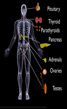
Endocrine glands targeted in polyglandular autoimmune syndrome.
What Are Autoimmune Polyglandular Syndromes?
A utoimmune polyglandular syndromes (APS) are rare clusterings of two or more endocrine and non-endocrine autoimmune disorders in the same affected person.
Polyglandular is somewhat of a misnomer since many of the manifestations of the diseases do not concern endocrine glands.1
Endocrine autoimmune disorders involve the abnormal production of autoantibodies that target and destroy the body’s own endocrine tissues, causing loss of essential hormone production by the targeted glands. Endocrine glands include the pituitary, thyroid, adrenal, parathyroid, islets of Langerhans (pancreas), testes in males, and ovaries in females.
First degree relatives (siblings of same parents, parents, children) have an increased incidence of latent, meaning not apparent, autoimmune pathology.2
Q: How many autoimmune polyglandular syndromes are described?
A:Three syndromes have been identified and they are all inherited: APS type-1, APS type-2, and APS type-3.
APS type-1 is a genetic mutation inherited in an autosomal recessive manner. A child with APS type-1 has inherited two mutated copies of a gene called the AIRE (autoimmune regulator) gene, which is on the long arm of 21st chromosome present in each cell.3 The parents, called carriers, are unaffected since they each have only a single copy of the AIRE mutated gene. Humans have a total of 23 pairs of chromosomes that contain genes inherited from each parent. Mutations in the genes cause disease.
Diagnosis criteria for autoimmune polyglandularsyndrome type-1 includes these three disorders:
Chronic candida infection (CMC), which usually develops first, typically attacks skin, but very commonly also nails, mouth, vagina, esophagus and intestine. CMC in APS type-1 patients is usually mild, and in most cases, it is chronic. It is found in 73–100 % of APS type-1 patients.
Hypoparathyroidism, causing loss of parathyroid function (hypoparathyreosis) is found in 76–93 % of APS type-1 patients.
Autommune Addison’s disease, also called autoimmune adrenalitis, is found in 72-100 % of APS type-1 patients. Still many of them die for unrecognized or late diagnosed autoimmune Addison’s disease, so regular follow-up for children in suspicion of APS type-1 (with CMC or/and hypoparathyroidism) is necessary.4
Other assocated disorders that may develop, but are not required for diagnosis, include: vitiligo, premature menopause, pernicious anemia, parathyroid gland failure, alopecia, and celiac disease. Thyroid disease rarely occurs.5
APS type-2 is linked to the inheritance of HLA antigens on chromosome 6 and appears to be autosomal dominant with incomplete penetrance. This suggests the contribution of environmental factors, such as bacterial and viral infections, medications, psychological factors, etc.6It does not have an identified mutation of the AIRE gene.
Diagnosis criteria for autoimmune polyglandular syndrome type-2 includes these two disorders:
Autommune Addison’s disease combined with
Autoimmune thyroid disease (thyroid atrophy, hypertrophic goiter related to Hashimoto’s thyroiditis, Graves’ disease, asymptomatic autoimmune thyroiditis)6 and/or type I diabetes mellitus. The conditions may occur in any order.
Polyglandular autoimmune syndrome type-2 is also known as Schmidt’s syndrome when adrenalitis (adrenal insufficiency) is associated with thyroiditis and Carpenter’s syndrome for adrenal insufficiency with hypoparathyreosis (impaired function of parathyroid glands).
Other disorders that may develop, but are not required for diagnosis, include: type 1 diabetes (50%), frequently gonadal failure or vitiligos, also celiac disease, autoimmune hepatitis, alopecia, pernicious anemia, and myasthenia gravis.7 Decades may arise between the onset of one disease and the onset of the second in the same patient.8
Therapy of APS type-2 consists of hormone replacement therapy for each separate condition, except that treatment for adrenal insufficiency must be given before thyroid therapy is started when the conditions occur together.9 Thyroxin replacement may induce life-threatening adrenal failure in a patient with untreated Addison’s disease. Thus, in case of doubt hydrocortisone should be given before the thyroxine administration is started.10
APS type-3 has a strong genetic background. Diagnosis criteria for autoimmune polyglandular syndrome type-3 involves these conditions:
Autoimmune thyroiditis that occurs with another organ-specific autoimmune disease, but not with autoimmune Addison’s disease, and
Other autoimmune diseases can include diabetes mellitus, pernicious anemia, vitiligo, alopecia, myasthenia gravis, celiac disease, and Sjögren’s syndrome. The most common APS type-3 combination is autoimmune disease of thyroid gland and pernicious anemia.11
Who is Affected in the General Population?
APS type-1 is usually apparent in childhood with the incidence of 1 in100,000 persons. It is more common among Finns (1 in 25,000), Sardinians (1 in 14,000), and Iranian Jews (1 in 6,500 to 1 in 9,000). The age of onset is usually early childhood, but new symptoms can develop throughout life. It affects both sexes equally.
APS type-2 has a peak onset in middle age, although the first signs usually develop between 20–30 years of age. Its prevalence is 1 in 20,000 persons. It is three times more frequent among women than men.12
APS type-3 is most frequent among middle-aged women.7
What Is Autoimmune Polyglandular Syndrome In Celiac Disease and/or Gluten Sensitivity?
Sources:
Wémeau JL, Proust-Lemoine E, Ryndak A, Vanhove L. Thyroid autoimmunity and polyglandular endocrine syndromes. Hormones (Athens). 2013 Jan-Mar;12(1):39-45. [↩]
Femiano P, Castaldo V, Iossa C. Complex family association in autoimmune polyendocrine syndrome. Minerva Pediatrica. Apr 2003;55(2):163-70. [↩]
Wémeau JL, Proust-Lemoine E, Ryndak A, Vanhove L. Thyroid autoimmunity and polyglandular endocrine syndromes. Hormones (Athens). 2013 Jan-Mar;12(1):39-45. [↩]
Wémeau JL, Proust-Lemoine E, Ryndak A, Vanhove L. Thyroid autoimmunity and polyglandular endocrine syndromes. Hormones (Athens). 2013 Jan-Mar;12(1):39-45. [↩]
Wémeau JL, Proust-Lemoine E, Ryndak A, Vanhove L. Thyroid autoimmunity and polyglandular endocrine syndromes. Hormones (Athens). 2013 Jan-Mar;12(1):39-45. [↩] [↩]
MAJERONI BA and PATEL P. Autoimmune Polyglandular Syndrome, Type II. Am Fam Physician. 2007 Mar 1;75(5):667-670. [↩]
Lipowsky C, Schorl-Schweikardt BA, Kehl O, Brändle M. 19-year-old patient with adrenal cortex insufficiency–only the tip of the iceberg. Polyendocrine autoimmune syndrome type II (Schmidt syndrome). Praxis (Bern 1994). 2008 Jan 23;97(2):77-81. [↩]
Van den Driessche A, Eenkhoorn V, Van Gaal L, De Block C. Type 1 diabetes and autoimmune polyglandular syndrome: a clinical review. Neth J Med. 2009 Dec;67(11):376-87. [↩]
I gA deficiency (IgAD) is an immunodeficiency disease characterized by lack of immunoglobulin A type antibody production, called IgA antibody, with no detectable levels in blood or secretions.
Q: What is an IgA antibody?
A: IgA is an antibody of the immune system that is secreted by plasma cells (specialized white blood cells) through epithelial cell linings of mucosal surfaces into mucosa secretions to protect the lining from microbe invasion.
In fact, immunoglobulin class A is the main protein of the mucosal immune system. This includes mucosa of the eye surface, digestive tract, respiratory tract, urinary tract, and genital tract.
Both major histocompatibility complex (MHC) and non-MHC genes contribute to susceptibility to the disease. The former genes appear to be located in different parts of the MHC region depending on the HLA haplotype. The latter show a marked overlap with genes associated with a variety of autoimmune disorders including Graves’ disease, systemic lupus erythematosus, type 1 diabetes and celiac disease, suggesting common pathophysiological mechanisms. The involvement of genes associated with autoimmunity may suggest that IgAD in itself is an autoimmune disease.1
IgA deficiency may progress into a common variable immunodeficiency (CVID).2
What Is IgA Deficiency (IgAD) In Celiac Disease and/or Gluten Sensitivity?
Sources:
Wang N, Hammarström L. IgA deficiency: what is new? Curr Opin Allergy Clin Immunol. 2012 Dec;12(6):602-8. doi: 10.1097/ACI.0b013e3283594219. [↩]
Binek A, Jarosz-Chobot P.Selective immunoglobulin A deficiency. Pediatr Endocrinol Diabetes Metab. 2012;18(2):76-8. [↩]
Testing the Eye for Tear Production (L) and Damage to Conjunctiva from Dryness (R).
What Is Sjögren’s Syndrome?
S jögren’s syndrome is a systemic inflammatory autoimmune disease with a chronic, progressive course that primarily attacks the lacrimal glands of the eye and the salivary glands of the mouth, which are exocrine glands. Exocrine glands secrete the substances they produce through a duct.
Sjögren’s syndrome is ordinarily characterized by dysfunction of the lacrimal glands to produce tears causing dry eye and the salivary glands to produce saliva causing dry mouth, but is not limited by or to these features.
Besides involvement of these exocrine glands, there may be involvement of other parts of the body, termed extraglandular, which may be more severe than eye or mouth features.
There is not yet agreement on classifying Sjögren’s syndrome. Primary and secondary are the two forms generally accepted.1 Both forms can cause mild to severe disease, called the spectrum:
Primary Sjögren syndrome. Disease occurs without involvement of other linked autoimmune disorders. In addition to the eyes and mouth, the nose, throat and skin may also be affected and joints, lungs, kidneys, blood vessels, digestive organs and nerves as well.2 Systemic manifestations (other than eyes and mouth) concern a third of patients, including lymphoma in 5% of the patients.3
Secondary Sjögren’s syndrome. Disease complicates other autoimmune disease such as systemic lupus erythematosus, rheumatoid arthritis, primary biliary cirrhosis, and celiac disease.
Diagnosis of Sjögren’s syndrome is made by most doctors based on Schimer’s test for tears and unstimulated whole salivary flow to assess objective eye and oral involvement, since these are the tests most physicians use in clinical practice.4 Specific antibody tests would be positive for anti-Ro (SSA)/anti-La (SSB) autoantibodies. Sjögren’s syndrome should also be considered when extraglandular manifestations such as vasculitis, polyneuropathy or arthritis occur, even when the patients do not complain of dry eyes and mouth.5
There is no cure for Sjögren’s syndrome. Treatment is aimed to diminish symptoms. For example, steroids and Ibupropen are used to decrease inflammation and pain in joints. Artificial tears and ointments are used for dry eye.
Most people who develop Sjogren’s syndrome are older than 40 years. Nine of ten people with Sjögren’s syndrome are women.2
What Is Sjögren’s Syndrome In Celiac Disease and/or Gluten Sensitivity?
Sources:
Huang YF, Cheng Q, Jiang CM, An S, Xiao L, Gou YC, Yu WJ, Lei L, Chen QM, Wang Y, Wang J. The immune factors involved in the pathogenesis, diagnosis, and treatment of Sjogren’s syndrome. Clin Dev Immunol. 2013;2013:160491. doi: 10.1155/2013/160491. Epub 2013 Jul 9. [↩]
Fazaa A, Bourcier T, Chatelus E, Sordet C, Theulin A, Sibilia J, Gottenberg JE. Classification criteria and treatment modalities in primary Sjögren’s syndrome. Expert Rev Clin Immunol. 2014 Apr;10(4):543-51. doi: 10.1586/1744666X.2014.897230. [↩]
Cornec D, Saraux A, Cochener B, Pers JO, Jousse-Joulin S, Renaudineau Y, Marhadour T, Devauchelle-Pensec V. Level of agreement between 2002 American-European Consensus Group and 2012 American College of Rheumatology classification criteria for Sjogren’s syndrome and reasons for discrepancies. Arthritis Res Ther. 2014 Mar 19;16(2):R74. [↩]
Witte T. Pathogenesis and diagnosis of Sjögren’s syndrome. Z Rheumatol. 2010 Feb;69(1):50-6. doi: 10.1007/s00393-009-0519-2. [↩]
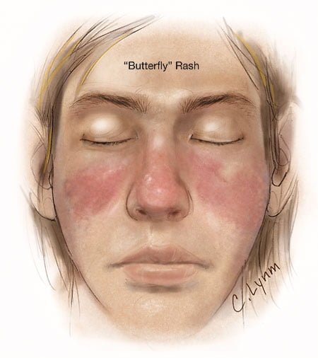
Image showing butterfly rash typical of SLE. Courtesy JAMA.
What Is Systemic Lupus Erythematosus?
S ystemic lupus erythematosus (SLE) is a chronic autoimmune inflammatory disease that is characterized by involvement of multiple organs due to the production of antibodies to components of the cell nucleus.1 SLE has an unpredictable course of acute flare-ups and remissions.
Severity depends on the extent of organs affected with skin and nail involvement, called discoid lupus, being the least serious and inflammmation of the kidney, called lupus nephritis, being the worst.
Nevetheless, a classic presentation is development of a rash over the cheeks and nose that resembles a butterfy with wings spread hence the name “butterfly rash.”
Symptoms are many and varied depending on the tissues affected and are often not specific, for example hair loss has a variety of causes. Symptoms can be confused by co-existence with other autoimmune disease such as Sjogren’s syndrome.
Systemic lupus erythematosus should be managed by a specialist. Symptoms can be controlled with steroid therapy, but this disease can be a cause of premature death mainly from active disease, organ failure (e.g., kidneys), infection, or cardiovascular disease from accelerated atherosclerosis.
Certain common medicines known to cause drug-induced lupus are:
Isoniazid
Hydralazine
Procainamide
Other less common drugs may also cause the condition. These may include:
Anti-seizure medications
Capoten
Chlorpromazine
Etanercept
Infliximab
Methyldopa
Minocycline
Penicillamine
Quinidine
Sulfasalazine
Symptoms tend to occur after taking the drug for at least 3 to 6 months.2
Although there is a strong familial aggregation, the disease is relatively uncommon and most cases are sporadic.1 According to the Center for Diseases (CDC), lupus most commonly affects women of childbearing age but also occurs in infants, children, adolescents, and men with peak occurrence between ages 15 and 40. Blacks (and possibly Hispanics, Asians, and Native Americans) are affected more than Whites.
What Is Systemic Lupus Erythematosus In Celiac Disease and/or Gluten Sensitivity?
D iffuse alopecia is characterized by abnormal hair loss or baldness.
Hair loss usually develops gradually and may be patchy or all over (diffuse). The average scalp contains about 100,000 hairs. Roughly 100 hairs are shed from the head every day.
Diffuse baldness not related to male pattern or heredity can be related to aging, nutritional deficiencies, some froms of dermatitis, radiation, endocrine disorders, especially thyroid hormone imbalance and diabetes, and undue stress.
Q: Can sudden stress cause hair loss?
A: A sudden physical or emotional stress may cause one-half to three-quarters of the hair throughout the scalp to shed. Other causes that need to be evaluated include use of common medications such as birth control pills, blood thinners, and anti-inflammatory pain drugs, and continued exposure to environmental chemicals such cleaning products.
What Is Diffuse Alopecia In Celiac Disease and/or Gluten Sensitivity?
F ine hair (lower diameter across the width) with rough texture is an abnormal hair shaft feature altered from the normal diameter and smooth quality of hair.
Q: What is the normal diameter and smooth quality of hair?
A: Although hair may appear to be a simple structure, it is actually a complex part of the anatomy whose biology is only partially understood. Hair grows from small organs (follicles) located within the complex microenvironment of the skin which has multiple layers of tissue, three glands whose secretions bathe hair, and multiple vascular systems.1
An individual hair is a thread-like shaft made up of cornified cells. It consists of the outermost layer, or cuticle, the cortex which is a horny component, and the medulla which is the central part.
Hairs receive nourishment from capillaries via the papilla at the base of their follicles (roots). Since the number of hair follicles are determined at birth, it is important to properly nourish them so they remain healthy.
What Is Fine Hair with Rough Texture In Celiac Disease and/or Gluten Sensitivity?
Sources:
Harkey MR. Anatomy and physiology of hair. Forensic Sci Int. 1993 Dec;63(1-3):9-18. [↩]