Selenium is a mineral that is required by the body in trace amounts for a healthy immune system, normal thyroid function, and antioxidant protection.
Selenium is absolutely required in the production of at least 30 selenoproteins in the body. First, selenium is joined to the amino acids cysteine as selenocysteine and to methionine as selenomethionine before being used as components for selenoproteins. Many selenoproteins are important antioxidant enzymes such as glutathione peroxidase.
Q: How does glutathione peroxidase work?
A: Glutathione peroxidase activity helps the recycling of vitamins C and E in optimizing the performance of the antioxidant system. The antioxidant properties of selenoproteins help prevent cellular damage from free radicals. Free radicals are natural by-products of oxygen metabolism that may contribute to the development of chronic diseases such as cancer and heart disease.
In the immune system, selenium stimulates immune properties of lymphocytes (white blood cells) by contributing to higher natural killer lymphocyte activity. Natural killer lymphocytes have the ability to destroy cancer cells and bacterial and viral agents.
Other selenoproteins help protect the thyroid gland from anti-oxidants and regulate thyroid function. Specifically, selenium plays an integral role in thyroid gland metabolism.1 Functions are more fully described below.
According to the Food and Agriculture Organization, United Nations, approximately 30 percent of tissue selenium is contained in the liver, 15 percent in kidney, 30 percent in muscle, and 10 percent in blood plasma.
What Is Selenium Deficiency In Celiac Disease and/or Gluten Sensitivity?
Sources:
Stazi AV, Trinti B. Selenium status and over-expression of interleukin-15 in celiac disease and autoimmune thyroid diseases. Ann Ist Super Sanita. 2010;46(4):389-99.DOI: 10.4415/ANN_10_04_06. [↩]
Figure on right shows how atherosclerosis impedes blood flow through coronary arteries while blood clots block blood flow. Courtesy Google.
What Is Coronary Artery Disease (CAD)?
Coronary artery disease (CAD), also called ischemic heart disease, is a gradual narrowing of medium and large arteries of the heart by fatty buildups, called atherosclerotic plaques.
It is characterized by slowly developing interference with blood flow to heart tissue itself, resulting in oppressive chest pain called angina and, ultimately, thrombosis (clot) causing heart attack.
The heart is a muscular organ that is working all the time, so it needs a constant supply of oxygen. Oxygen is brought to the working heart tissue by the coronary arteries with each beat of the heart. When heart muscle has to work harder, it needs more oxygen delivered to itself. Lack of oxygen causes pain.
In fact, failure of diseased coronary arteries to deliver adequate oxygen to heart tissue is the most common cause of angina pectoris – substernal pain (under breastbone) or pressure brought on by exertion and relieved by rest.
Thrombosis, or clot formation, occurs when blood cells within a narrowed artery can no longer get through. Trapped, blood cells pile up and block the artery thus triggering a cascade of events called heart attack. Coronary arteries that are narrowed by atherosclerotic plaques can rupture causing injury to the coronary blood vessel resulting in blood clotting which blocks the flow of blood to the heart muscle. Blood clots may form, partially dissolve, and later form again and angina can occur each time a clot blocks blood flow in an artery.1
Q: How does coronary artery disease develop?
A:Coronary artery disease slowly develops from this combination of events:
Dysfunction of epithelial cells that line the inside of arteries cause the vessels to stiffen, and subsequently
Accumulation of lipid (fat) in smooth muscle cells beneath the inside lining of arteries and in foam cells cause buildup of fatty deposits on the inside walls progressing to fibrous plaque formation.
Oxidized low-density lipoprotein (oxLDL), so-called bad cholesterol, and oxysterols play important roles in the development of atherosclerosis. OxLDL triggers the immune system to produce autoantibodies against oxLDL that are detectable in serum. These antibodies are called anti-oxLDL. Anti-oxLDL antibody and oxysterol concentrations are associated with coronary artery stenosis. Oxidative stress may be greatly increased in unstable angina.2 and Chronic inflammation in the general population is a major risk factor for ischemic heart disease.
The pathophysiology of atherosclerosis is, clearly, different in women when compared to the men. The women have a higher risk of blood coagulability making them at high risk for the blood clot formation. In a large number of women endothelial dysfunction, small vessel size and diffuse atherosclerosis have been identified as causes of ischemia without evidence of blockade in the coronary arteries.3
Also, atherosclerotic plaque in women is less fibrotic and contains more lipid filled foam cells, implying greater potential for reversibility but also potentially greater vulnerability for plaque rupture and thrombosis.4
Who is Affected in the General Population?
Coronary artery disease remains the leading cause of death in developed countries despite significant progress in primary prevention and treatment strategies.
It is the leading cause of death in women, as well as an important cause of disability.
Older patients are at particularly high risk of poor outcomes following acute coronary syndrome.5
What Is Coronary Artery Disease In Celiac Disease and/or Gluten Sensitivity?
Ischemic heart disease is the leading cause of death in the United States, making cardiovascular risk assessments and potential interventions or treatments imperative for patients with celiac disease.6
Yasunobu Y, Hayashi K, Shingu T, Yamagata T, Kajiyama G, Kambe M. Coronary atherosclerosis and oxidative stress as reflected by autoantibodies against oxidized low-density lipoprotein and oxysterosis. Atherosclerosis. Apr 2001;155(2):445-53. [↩]
Kunadian V, Ford GA, Bawamia B, Qiu W, Manson JE. Vitamin D deficiency and coronary artery disease: A review of the evidence. Am Heart J. 2014 Mar;167(3):283-291. doi: 10.1016/j.ahj.2013.11.012. Epub 2013 Dec 19. [↩]
Kunadian V, Ford GA, Bawamia B, Qiu W, Manson JE. Vitamin D deficiency and coronary artery disease: A review of the evidence. Am Heart J. 2014 Mar;167(3):283-291. doi: 10.1016/j.ahj.2013.11.012. Epub 2013 Dec 19. [↩]
Kunadian V, Ford GA, Bawamia B, Qiu W, Manson JE. Vitamin D deficiency and coronary artery disease: A review of the evidence. Am Heart J. 2014 Mar;167(3):283-291. doi: 10.1016/j.ahj.2013.11.012. [↩]
Robinson BL, Davis SC, Vess J, Lebel, J. Primary care management of celiac disease. Nurse Practitioner. February 2015: Vol 40 – Issue 2; 28–34. [↩]
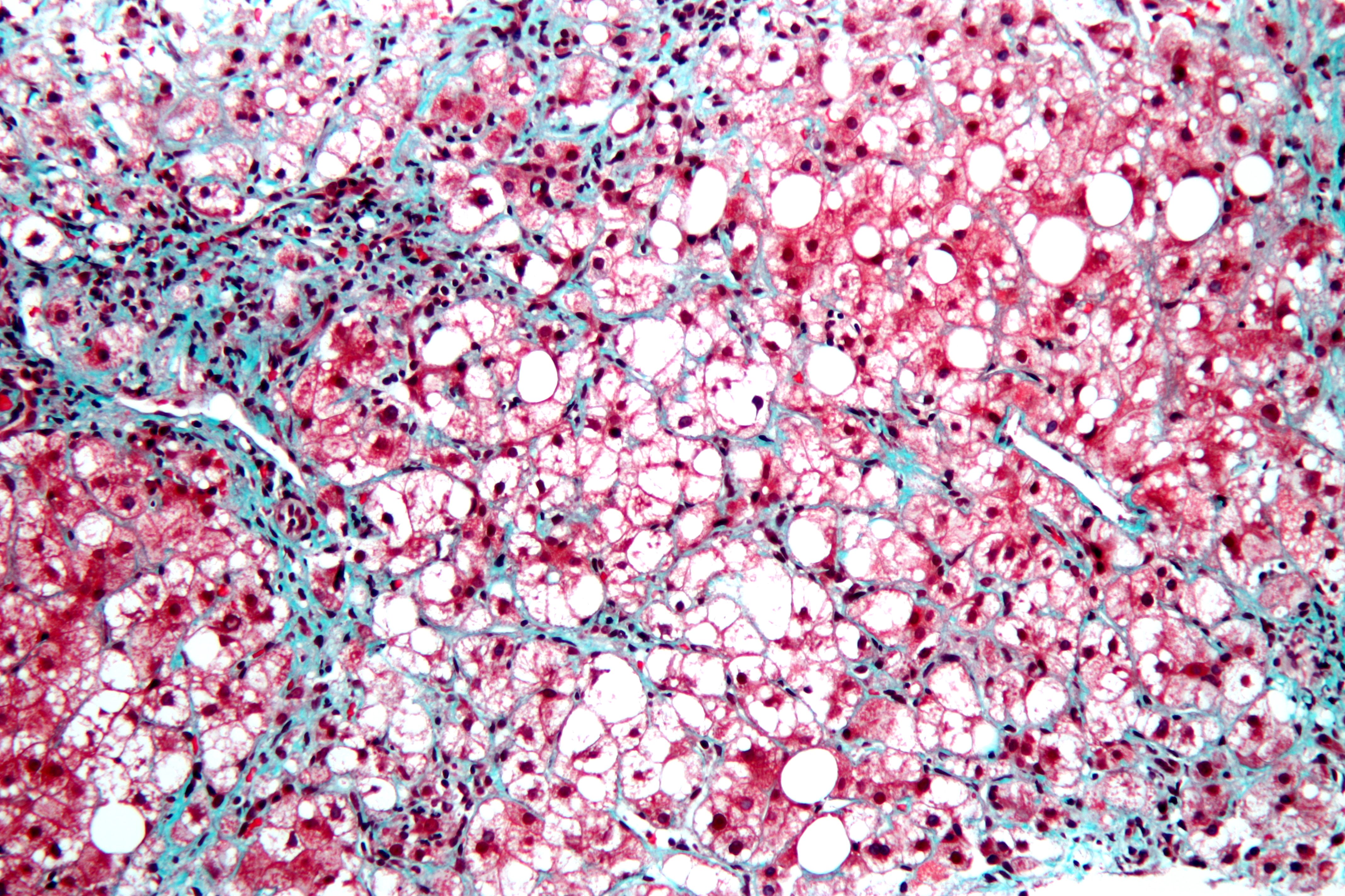
This is a stained liver biopsy sample showing advanced cellular changes in non-alcoholic fatty liver disease. Blue is fibrosis. White is fat accumulation in degenerated cells. Courtesy of Nephron’s work.
What Is Non-Alcoholic Fatty Liver Disease?
N on-alcoholic fatty liver is a non-inflammatory liver disorder characterized by degenerative changes in the liver caused by excessive accumulation of lipid (fat) in hepatocytes (liver cells) that is called free fatty acid-generated lipotoxicity.
Non-alcoholic fatty liver shows an increase in liver enzymes called transaminases.
Q: What are the enzymes that increase?
A: The transaminases that increase are ALT and AST. ALT is the abbreviation for alanine aminotransferase enzyme and AST is the abbreviation for aspartate aminotransferase enzyme. They are commonly measured in blood tests to determine liver function and when elevated indicate inflammation.
What Is Non-Alcoholic Fatty Liver In Celiac Disease and/or Gluten Sensitivity?
Dermatitis Herpetiformis On Forearm. Skin Is Darkened Where Old Blisters Healed.
What Is Dermatitis Herpetiformis?
D ermatitis herpetiformis (DH) is an autoimmune extremely itchy, painful bullous skin rash (blistering eruptions) arising from the underlying dermis layer of skin as a consequence of gluten sensitivity.
Dermatitis herpetiformis is characterized by multiple intensely itchy, red blisters appearing on the elbows which can extend down the forearm to the wrist and the knees. Less usual areas involve the back, buttocks, scalp, and abdomen.
Q: Do the blisters leave a mark when healed?
A: Crops of skin eruptions begin with itching or a burning sensation in reddened papules. There are grouped vesicles and tense blisters. The blister contents may be serous or bloody, with symmetrical distribution (eg, both knees or both elbows). Fluid filled elements rupture leaving denuded areas of sore skin and crust. Subsequently, there is residual hypopigmentation (a white area) or hyperpigmentation (dark area).1
Rupture of blisters begins relief from intense burning and itching.
Dermatitis Herpetiformis Eruptions On Knees. Notice White Areas Showing Loss of Pigmentation From Healed Blisters.
What Is Dermatitis Herpetiformis In Celiac Disease and/or Gluten Sensitivity?
Primary care providers should be aware of this skin condition, as they are more likely than a gastroenterologist to be confronted with this type of presentation of celiac disease.2
Sources:
Mendes FB, Hissa-Elian A, de Abreu MA, Gonçalves VS. Review: dermatitis herpetiformis. An Bras Dermatol. 2013 Jul-Aug;88(4):594-9. [↩]
Robinson BL, Davis SC, Vess J, Lebel, J. Primary care management of celiac disease. Autoimmune Disorders. Nurse Practitioner. February 2015: Vol 40 – Issue 2; 28–34. [↩]
E rythema nodosum is an inflammatory disorder involving the deep dermis layer of skin and subcutaneous fat septa that underlies the skin. It is characterized by eruptions of recurrent or persistent multiple painful, red nodules under the skin that leave a bruised appearance when healing and do not scar.
The lower legs are most affected, but sores can appear anywhere there is subcutaneous fat.
Q: How do the nodules develop in erythema nodosum?
A: The edges of nodules are poorly defined, and the nodules vary from 2-6 cm.
During the first week of eruption, nodules become tense, hard, and painful. During the second week, they change color from bright red to bluish or livid and may become soft, but do not ulcerate. As absorption progresses, the color gradually fades to a yellowish hue, resembling a bruise. This disappears in 1 or 2 weeks as the overlying skin sloughs off and is replaced.1
The eruptive phase of erythema nodosum begins with flulike symptoms of fever and generalized aching followed by a painful rash within 1-2 days. Aching legs and swelling ankles may occur and precede the eruption or appear during the eruptive phase and may persist for weeks.2
Currently, the most common cause of erythema nodosum is streptococcal infection in children and streptococcal infection and sarcoidosis in adults.3Most sores in infection-induced erythema nodosum heal within 7 weeks, but active disease may last up to 18 weeks.
In contrast, 30% of idiopathic erythema nodosum cases may last more than 6 months. Idiopathic means that the cause is not known.
What Is Erythema Nodosum In Celiac Disease and/or Gluten Sensitivity?
H angnail is a broken strip of epidermis (piece of skin) at root or lateral (side) edge of fingernail or toenail that causes sharp pain.
A hangnail develops because the skin around the nail is unhealthy due to inadequate nutrition. Injury from trauma including biting the skin and pushing back the cuticles or exposure to excessive detergents and water that remove protective oils promote the development of hangnail.
All ages and both sexes can be affected.
Q: Can a hangnail become infected?
A: Infection, called paronychia, may develop from invasion of sore skin by any of these pathogens: bacteria, fungus, or yeast (Candida). Infected skin is red, swollen, and painful. Topical ointment is required to treat the infection.1
What Is Hangnail In Celiac Disease and/or Gluten Sensitivity?
P ityriasis rubra pilaris (PRP) is a chronic generalized exfoliative dermatitis (sloughing skin) characterized by erythema (redness), scaling, dilated plugged hair follicles, and keratoderma (thickened skin) of the hands and feet that is often associated with anemia and low serum albumin.
It may manifest either as Type I classical adult onset PRP, Type II atypical adult (onset) PRP, or Type VI PRP (HIV-associated PRP pityriasis rubra pilaris) in contrast to classical juvenile (Type III) and circumscribed juvenile (Type IV) encountered among children.1
Q: Who is affected in the general population?
A: All ages are affected. Pityriasis rubra pilaris occurs all over the world but with racial variations – it is 1 in 5,000 in Great Britainand 1 in 50,000 in India.2
What Is Pityriasis Rubra Pilaris In Celiac Disease and/or Gluten Sensitivity?
Sources:
Sehgal VN, Srivastava G, Dogra S. Adult onset pityriasis rubra pilaris. Indian J Dermatol Venereol Leprol. 2008 Jul-Aug;74(4):311-21. [↩]
Sehgal VN, Srivastava G, Dogra S. Adult onset pityriasis rubra pilaris. Indian J Dermatol Venereol Leprol. 2008 Jul-Aug;74(4):311-21. [↩]
P rurigo nodularis is a chronic dermatitis characterized by hard, dry, deep seated, intensely itchy papules (small bumps like pimples) and/or nodules (large bumps) that erupt most commonly on the arms, legs, and back.
Papules and nodules vary in number and may become infected after picking or scratching.
Q: Does the itching go away?
A: New nodules develop from time to time, and existing nodules may remain itchy indefinitely, although some may regress spontaneously and leave scars. In most cases, the disease runs a very protracted course with exacerbations and remissions.1
Prurigo nodularis is an unusual disorder of unknown etiology, which is notoriously resistant to therapy. A variety of systemic conditions have been reported to be associated with prurigo nodularis. However, the mechanism by which these disorders may trigger prurigo nodularis is unknown.2
It has been shown to be associated with malnutriton and infection such as tonsillitis, which resolved after removal of tonsils.3
What Is Prurigo Nodularis In Celiac Disease and/or Gluten Sensitivity?
Sources:
Katotomichelakis M, Balatsouras DG, Bassioukas K, Kontogiannis N, Simopoulos K, Danielides V. Recurrent prurigo nodularis related to infected tonsils: a case report. J Med Case Rep. 2008 Jul 24;2:243. doi: 10.1186/1752-1947-2-243. [↩]
Lee MR, Shumack S. Prurigo nodularis: a review. Australas J Dermatol. 2005 Nov;46(4):211-18; quiz 219-20. [↩]
Katotomichelakis M, Balatsouras DG, Bassioukas K, Kontogiannis N, Simopoulos K, Danielides V. Recurrent prurigo nodularis related to infected tonsils: a case report. J Med Case Rep. 2008 Jul 24;2:243. doi: 10.1186/1752-1947-2-243. [↩]
P soriasis is a chronic, autommune relapsing skin disorder characterized by scaling, erythema (redness), and less commonly, pustulation.1
The body surface area affected and the degree to which psoriasis is a problem varies considerably among patients and over time.2 Often there are additional manifestations in the nails and in joints.3
Q: Are there different forms of psoriasis?
A: There are five forms of psoriasis. The lesions in all forms are itchy and red but vary in appearance and severity. Plaque psoriasis is the most common form observed in more than 80% of patients. Atypical forms include guttate, inverse, pustular, and erythrodermic psoriasis.4
Plaque psoriasis features thickened or raised red areas that have a distinct edge and are covered with silvery white buildup of flaky skin typically on elbows, knees, scalp and buttocks.
Gutate psoriasis appears as small, flat red patches with shiny buildup that are not usually painful, just itchy. There may be a few or many patches and they can group together.
Inverse psoriasis affects folds of skin, armpits and the groin area. Lesions are deep red with shiny buildup. It can be a thin red area along a crease line or involve, for example, the whole armpit.
Pustular psoriasis features an itchy, red base followed by blisters of white, non-infectious pus that appears glossy after a day or two and then sloughs in cycles. These areas may be limited to certain areas such as the hands and feet or be more widespread.
Erythrodermic psoriasis involves large areas of the body’s surface, inflaming normal skin and changing it into very red, raw looking flesh that is painful, swollen and itchy. This form requires extensive treatment, and complications can be life-threatening. Fortunately, this form of psoriasis is the least common.
Psoriasis in children has been reported to differ from that among adults being more frequently itchy and plaque lesions are relatively thinner, softer, and less scaly, face and flexural involvement is common and guttate type is the characteristic presentation.5
In children, psoriasis is a common skin disorder with about one third of all patients having onset of disease in the first or second decade of life. A chronic disfiguring skin disease, such as psoriasis, in childhood is likely to have profound emotional and psychological effects, and hence requires special attention.6
What Is Psoriasis In Celiac Disease and/or Gluten Sensitivity?
Sources:
Addolorato G, Parente A, de Lorenzi G, et al. Rapid regression of psoriasis in a coeliac patient after gluten-free diet. A case report and review of the literature. Digestion. 2003;68(1):9-12. [↩]
Stern, R. S., Nijsten, T., Feldman, S. R., Margolis, D. J. and Rolstad, T.
Psoriasis is common, carries a substantial burden even when not extensive, and is associated with widespread treatment dissatisfaction. J. Invest. Dermatol. Symp.. 2004 Mar;9(2):136-9.. [↩]
Weigle N, McBane S. Psoriasis. Am Fam Physician. 2013 May 1;87(9):626-33. [↩]
Weigle N, McBane S. Psoriasis. Am Fam Physician. 2013 May 1;87(9):626-33. [↩]
Dogra S, Kaur I. Childhood psoriasis. Indian J Dermatol Venereol Leprol. 2010 Jul-Aug;76(4):357-65. doi: 10.4103/0378-6323.66580. [↩]
Dogra S, Kaur I. Childhood psoriasis. Indian J Dermatol Venereol Leprol. 2010 Jul-Aug;76(4):357-65. doi: 10.4103/0378-6323.66580. [↩]
S cleroderma is a chronic skin manifestation of progressive systemic sclerosis characterized by generalized thickened, edematous skin firmly bound to subcutaneous tissue which causes limited movement.
Systemic sclerosis a connective tissue disease that involves destructive changes in the skin, blood vessels, muscles, and internal organs. The course can be mild or it can be fatal. Cardiopulmonary complications from fibrosis are the most common cause of death.
Gastrointestinal problems mainly due to fibrosis affect 50 to 90% of patients.1
Q: Is there a cure for scleroderma?
A: There is no cure for scleroderma. Treatment is aimed at improving symptoms.
Heartburn (acid reflux) can be treated with antacid drugs.
Scleroderma kidney disease can be treated with blood pressure medications called “angiotensin converting enzyme inhibitors” (ACE inhibitors). These can often effectively control kidney damage if started early and use of these drugs has been a major advance for treating scleroderma.
Muscle pain and weakness can be treated with anti-inflammatory drugs such as prednisone, intravenous immunoglobin (IVIg), and/or immunosuppressive medications. Physical therapy may be useful to maintain joint and skin flexibility.2
What Is Scleroderma In Celiac Disease and/or Gluten Sensitivity?
Sources:
Forbes A, Marie I. Gastrointestinal complications: the most frequent internal complications of systemic sclerosis. Rheumatology (Oxford). 2009 Jun;48 Suppl 3:iii36-9. doi: 10.1093/rheumatology/ ken485. [↩]
 What Is Selenium?



![Crohnie_sores_4[1]](https://glutenfreeworks.com/health/wp-content/uploads/sites/10/Crohnie_sores_41.jpg)


![Psoriasis_on_back[1]](https://glutenfreeworks.com/health/wp-content/uploads/sites/10/Psoriasis_on_back1.jpg)
