Skip to content

Increased Intestinal Permeability (Leaky Gut)

Close-up Slice of a Small Intestinal Villus Showing How Enterocytes Appear Tightly Lining the Entire Outside Surface Of A Villus. Courtesy Cleo Libonati

What Is Increased Intestinal Permeability?

Increased intestinal permeability is characterized by dysfunctional intestinal permeability (leakiness) allowing for the penetration of harmful entities from the gut into the bloodstream such as undigested proteins and microbes. The popular name is “leaky gut.”

Q: Why does intestinal permeability increase?

A: Intestinal permeability is an essential function of the small intestinal mucosal lining by which wanted substances such as properly digested foodstuffs are allowed to permeate through the lining to enter the body via the bloodstream and lymphatics. At the same time unwanted substances are kept out.

The mucosal lining is one cell thick and makes up the surface between the digested foodstuffs inside the hollow of the intestine and the underlying tissues.

The mucosal lining is covered by millions of microscopic finger-like structures called villi that project toward the inside of the intestine giving the appearance of a shag rug.

Each one, called a villus, contains a capillary bringing blood to absorb nutrients, a vein to take away nutrients, and a lacteal to absorb and take away digested fat. Its wall is made up of a single layer of tightly connecting cells, called enterocytes.

This single layer of cells separates the contents of our small intestine from the lamina propria (underlying tissues of the small intestine) and the rest of our body. Breaching of this single layer of cells by leakiness can expose lymphocytes (immune cells) located in the lamina propria to a myriad of microorganisms and food antigens, leading to immune reactions.1

To protect the body from unwanted substances, a gatekeeping barrier system operates to regulate the passage of nutrients, or permeation, through the surface mucosal lining. This system acts to seal the inside body from the gut.

The integrity of intestinal permeability is determined by interactions among several barrier components including the unstirred water layer, mucosal surface hydrophobicity, the surface mucous coat, and cell factors (especially tight junctions).

Tight junctions hold cells tightly together side-by-side to prevent unwanted substances from passing through the lining. Tight junctions are complex structures comprising over 50 proteins, such as the claudin proteins which are considered to be the structural backbone of tight junctions.

Tight junctions include a series of special proteins forming fibrils (springy like proteins) that cross the plasma membrane and interact with proteins in the adjoining cells. Tight junctions are regulated by the protein zonulin.2

If zonulin deregulates from the action of substances such as gliadin (gluten in wheat) and bacteria, the tight junction barrier fails which results in increased intestinal permeability. Dysfunction of the barrier system allows unwanted substances to enter the body where they are damaging to many tissues.

Tight junction dysfunction has been shown to be a part of certain autoimmune diseases such as celiac disease, type I diabetes mellitus, multiple sclerosis, and rheumatoid arthritis. Other diseases associated are cancer, allergies, and infections.3

Important gastrointestinal infections that cause leaky gut include rotavirus, parasites, pathogenic bacteria (escherichia coli, clostridium difficile), and mycotoxins produced by fungi found in stored grain and dried fruit.4

Fortunately, the presence of some commensal (friendly intestinal bacteria) and probiotic strains leads to an increase in tight junctions  proteins at the cell boundaries and in some cases prevents or reverses the adverse effects of pathogens, food and stress. Various dietary components are also known to regulate epithelial permeability by modifying expression and localization of  tight junctions proteins.2

What Is Increased Intestinal Permeability In Celiac Disease and/or Gluten Sensitivity?

Sources:
  1. Fahardi A, Banan A, Fields J, Keshavarzian A. Intestinal barrier: an interface between health and disease. Journal of Gastroenterology and Hepatology. 2003; 18: 479-497. []
  2. Ulluwishewa D, Anderson RC, McNabb WC, Moughan PJ, Wells JM, Roy NC. Regulation of tight junction permeability by intestinal bacteria and dietary components. J Nutr. 2011 May;141(5):769-76. doi: 10.3945/jn.110.135657. [] []
  3. Fasano A. Zonulin and Its Regulation of Intestinal Barrier Function: The Biological Door to Inflammation, Autoimmunity, and Cancer. Physiological Reviews. January 2011Vol. 91no. 151-175DOI: 10.1152/physrev.00003.2008 []
  4. Farhadi A, Banan A, Fields J, Keshavarzian A. Intestinal barrier: an interface between health and disease. Journal of Gastroenterology and Hepatology. 2003;18:479-91. []

Constipation, Chronic

Constipation in a young child as seen on X-ray. Lowest circle shows hard feces in the pelvis. Source, James Heilman, MD.

What Is Chronic Constipation?

Chronic constipation is an intestinal motility disorder characterized by abnormal stool formation, consistency, and evacuation.

Motility disorder means the normal rhythmic movement of intestinal muscles, called peristalsis, that moves food matter through the gut is hampered or dysfunctional.

Studies show that methane gas present in the colon induces constipation by delaying transit time, which is the time it takes for stool to pass through the colon.

Researchers investigating the relationship between methane and constipation found that methane positivity was detected in 75% of patients with slow transit, 44% of patients with normal transit and and 28% of the patients who were controls. However, methane positivity was not related with stool consistency.1

Other researchers investigating the total amount of methane produced found that there was significantly more methane production in patients with constipation (21.1 ppm vs. 6.1 ppm, respectively) than in controls without constipation.2

Q. How does methane get into the colon?

A. Methane is produced in the colon by intestinal methanogens (microbes) that metabolize hydrogen, one of the end products of normal anaerobic (meaning without oxygen) bacterial fermentation.  Fermentation of the undigested starchy part of carbohydrates produces hydrogen in the intestine which is the substrate (food) for methane production by intestinal methanogens.

Hydrogen and methane are excreted in the flatus and in breath giving the opportunity to indirectly measure their production using breath testing. Methane is detected in 30%-50% of the healthy adult population worldwide.3

Other common causes of constipation include not getting enough exercise, not drinking enough fluids, not eating enough fiber in the diet, not eating foods that supply microbes needed by the colon (probiotics), not eating foods that nourish the good microbe population (prebiotics) and supply minerals needed for healthy movement of stool, and food sensitivities. Too much cows milk is a common cause of stool that forms into balls.

Who is Affected in the General Population? Chronic constipation is a remarkably common and costly condition that can negatively impact the quality of life and result in a major social and economic burden. Based on the definition, either self-reported or using Rome criteria, chronic constipation can affect up to 27% of the population. There is strong evidence that constipation occurs more frequently in women.4

What Is Chronic Constipation In Celiac Disease and/or Gluten Sensitivity?

Sources:
  1. Triantafyllou K, Chang C, Pimentel M. Methanogens, Methane and Gastrointestinal Motility. J Neurogastroenterol Motil. 2014 Jan;20(1):31-40. Epub 2013 Dec 30. []
  2. Triantafyllou K, Chang C, Pimentel M. Methanogens, Methane and Gastrointestinal Motility. J Neurogastroenterol Motil. 2014 Jan;20(1):31-40. Epub 2013 Dec 30. []
  3. Triantafyllou K, Chang C, Pimentel M. Methanogens, Methane and Gastrointestinal Motility. J Neurogastroenterol Motil. 2014 Jan;20(1):31-40. Epub 2013 Dec 30. []
  4. Sanchez MI, Bercik P. Epidemiology and burden of chronic constipation. Can J Gastroenterol. 2011 Oct;25 Suppl B:11B-15B. []

Systemic Lupus Erythematosus 

bruiseWhat Is Hypoprothrombinemia?

H ypoprothrombinemia is a deficiency of prothrombin (clotting factor II) in the blood that is characterized by impaired hemostasis in response to trauma or a laceration.

Q: What is hemostasis and how is it altered by a deficiency of prothrombin?

A: Hemostasis encompasses the tightly regulated processes of blood clotting, platelet activation, and blood vessel repair.1

Prothrombin is a protein clotting factor present in blood that is involved in the first part of hemostasis, which is blood clotting or coagulation. Vitamin K is required for prothrombin production.

When a laceration or wound is sustained, prothrombin is converted to the enzyme thrombin. Thrombin in turn acts on fibrinogen to convert it to fibrin which then forms the framework of a clot to stop bleeding. Deficiency of prothrombin prevents this series of events and bleeding is not properly stopped.

After the clotting process of hemostasis would come the second part, platelet activation. Eventually, coagulation and platelet activation are switched off by blood-borne inhibitors.

What Is Hypoprothrombinemia In Celiac Disease and/or Gluten Sensitivity?

Sources:
  1. Versteeg HH, Heemskerk JWM, Levi M,  Reitsma PH. New Fundamentals in Hemostasis. Physiological Reviews Published 1 January 2013Vol. 93no. 327-358DOI: 10.1152/physrev.00016.2011 []

Candida Albicans Infection

Close-up shows inflammation and yellowish white patches of roof and back of the mouth caused by candidiasis. Uvula is greatly swollen, hanging near the level of the tongue. Courtesy: Wikipedia.
Close-up shows inflammation and infected patches on roof and back of the mouth caused by candidiasis. Uvula is swollen, hanging near the tongue. Courtesy: Wikimedia.

What Is Candida Albicans Infection?

Candida albicans infection, called candidosis or candidiasis, is an opportunistic invasion of mucous membrane or skin by candida albicans, an endogenous yeast found in 40 to 80% of normal human beings. A former name for this small, budding fungus is monilia albicans.

Opportunistic means that yeast living on mucosal and skin surfaces does not invade (infect) unless these tissues become unhealthy and therefore cannot protect themselves.

Q: How does candida albicans cause infection?

A: Candida albicans lives on the mucosal surfaces and skin in most people without causing infection (colonizes) because of our normal defenses against invasion. In fact, candida albicans is a very effective colonizer of humans. For example, Russell and Lay found that 47% of 1-month-old infants were orally colonized with candida albicans, and 49% were colonized with other fungi.

During growth within the intestinal tract, the organism senses pH (acidity), oxygen, carbon sources, and the presence of surfaces allowing it to optimize gene expression for a particular environment. With these mechanisms for sensing, candida albicans is able to efficiently colonize humans in infancy.1

Candida Infection Of The Esophagus on X-ray. Courtesy Radiology Assistant.nl
Candida Infection Of The Esophagus (White Area) On X-ray. Courtesy Radiology Assistant.nl

Lowered host defenses allow yeast already present on mucosal and skin surfaces to take advantage and can grow rapidly, becoming pathogenic (disease producing) so that infection results.

Infection is characterized by superficial, irregular white patches on mucosal surfaces and possible invasion of the bloodstream by a filamentous form (thread-like structures) that can rapidly develop.

Candida albicans is unique among oral pathogens in its ability to invade cornified layers of stratified squamous epithelium of the tongue, mouth surfaces, hard and soft palate, esophagus, and gut. Stratified squamous epithelium is the tough surface cells that ordinarily protect underlying tissues from damage or invasion by microbes.

Candida albicans is also capable of invading the lungs and causing pneumonia and septicemia, which is the spread of infection into the bloodstream.

Here is a time honored simple do-it-youself test for infection of the mouth or throat: First thing in the morning before brushing your teeth or eating, fill a small see through glass with water then gently spit onto the surface. If after an hour the spit remains on the water surface, it is unlikely you have candida in the mouth. If it grows legs downward, it indicates that yeast is growing. If the spit sinks to the bottom, you have this problem. Yeast in the mouth can quickly travel down the esophagus and into the gut.

Medical diagnosis. Difinitive diagnosis for the oral cavity is made by your clinician by swabbing the areas of your mouth and/or throat and viewing under a microscope for evidence of candida.

Infections of the esophagus and gut require inspection by gastroscopy or endoscopy procedure and the taking of samples to be examined under microscope. This examination also give the opportunity to rule out other problems. Barium swallow can show the extent of infection and any disfiguration of the esophagus that results.

What Is Candida Albicans Infection In Celiac Disease and/or Gluten Sensitivity?

Sources:
  1. Rosenbach A, Dignard D, Pierce JV, Whiteway M, Kumamoto CA. Adaptations of Candida albicans for growth in the mammalian intestinal tract. Eukaryot Cell. 2010 Jul;9(7):1075-86. doi: 10.1128/EC.00034-10. Epub 2010 Apr 30. []

Low Stomach Acid Production

low-stomach-acid1What Is Low Stomach Acid Production?

Low stomach acid production is a common disorder in celiac disease and dermatitis herpetiformis that is characterized by lack of sufficient hydrochloric acid needed 1) to properly digest food, which results in malnutrition and subsequent nutritional deficiencies, and 2) to destroy swallowed bacteria and other microbes, which may allow infections to develop.

It is also common in the general population, as well, affecting 50% of people age 60 years and about 80% by age 85 years.  Nevertheless, low stomach acid is not generally looked for as a cause of acute and chronic disorders that rob health with far-reaching effects.

Understanding Stomach Acid Production and Function

The stomach digests incoming food into a liquid state, thereby releasing nutrients so they can be absorbed by the small intestine. Powerful stomach muscles churn food and mix it with gastric juice, dissolving and breaking it down.

Gastric juice is produced by gastric glands located in the stomach lining. These numerous, microscopic glands produce about 3 liters of juice a day. Gastric juice is composed of a high concentration of

Crohn’s Disease

Endoscopic image of Crohn'sDisease showing deep ulceration in sigmoid colon.
Endoscopic Image of Crohn’s Disease Showing Deep Ulceration in the Sigmoid Colon.

What Is Crohn’s Disease?

Crohn’s disease is an inflammatory bowel disease characterized by patchy inflamed areas involving the full thickness of the intestinal wall that can occur anywhere in the intestinal tract, in addition to, mucosal disease.

In Crohn’s disease there is ongoing immune activation which produces inflammation and ulceration but the cause is not known and the severity varies among patients. At diagnosis of Crohn’s disease, factors predictive of subsequent 5-year aggressive disease are an age below 40 years, the presence of perianal disease, and the initial requirement for steroids.1

Dysbiosis is a factor that develops in and worsens Crohn’s disease and stress is a factor in both of these conditions. Psychological stress activates multiple physiological processes aimed at maintaining balance within the body. These physiological processes also have the capacity to influence the composition of microbial communities in the digestive tract, and research now indicates that exposure to stressful stimuli leads to gut microbiota dysbiosis.2

While the relative abundance of many different bacterial types can be altered during stressor exposure, findings in nonhuman primates and laboratory rodents, as well as humans, indicate that bacteria in the genus Lactobacillus are consistently reduced in the gut during stress.2

Q: Is there a cure for Crohn’s disease?

A: Presently, Crohn’s disease cannot be cured. This condition has a course of remissions, when symptoms subside, and flares, when symtpoms get worse. Treatment is aimed to reduce flares and promote remission.

What Is Crohn’s Disease In Celiac Disease and/or Gluten Sensitivity?

Sources:
  1. Beaugerie L, Seksik P, Nion-Larmurier I, Gendre JP, Cosnes J. Predictors of Crohn’s disease. Gastroenterology. 2006;130:650–656. []
  2. Galley JD, Bailey MT. Impact of stressor exposure on the interplay between commensal microbiota and host inflammation. Gut Microbes. 2014 May 1;5(3):390-396. Epub 2014 Apr 1. [] []

Dysbiosis (Intestinal)

What Is Intestinal Dysbiosis? Intestinal dysbiosis is an imbalance of the composition and quantity of microbe populations (called the microbiota), that naturally inhabit our human gut. Dysbiosis causes altered gut immunity, abnormal fermentation of undigested foodstuffs,… 

Colitis, Collagenous

Collagenous Colitis.
Microscopic Image Showing a Pink Collagen Band in Collagenous Colitis.

What Is Collagenous Colitis?

Collagenous colitis is a disease of the large intestine (colon) that is characterized by microscopic inflammation of the surface mucosal lining and an abnormally thickened collagen band of tissue that develops wthin the lining of the colon.

The thicker than normal layer of collagen of at least 10 µm (reference value: 2–7 µm) can vary in different locations. Inflammation occurs with increased numbers of lymphocytes (white blood cells) and plasma cells and epithelial (surface cell) damage. These changes can only be seen under microscopic examination of multiple biopsied tissue samples taken during a colonoscopy procedure.

Q: What is collagen?

A: Collagen is a strong, fibrous protein found in connective tissue of the colon and many other tissues such as tendons. The normal basement membrane in the bowel consists mainly of collagen type IV, laminin, and fibronectin. The increased collagen band observed in collagenous colitis consists basically of collagen type I and III, which are the subtypes produced by repair functions, indicating a reactive origin to some irritant or drug.1

The biopsies should preferably be taken from the ascending colon, since the pathological hallmarks may be absent in the descending colon, and in the normally occurring thicker collagen layer in the rectosigmoid region.1 Inflammation of the ileum (last segment of the small intestine next to colon) is common.2

Endoscopy and radiological (x-ray) examinations are usually normal.3

Autoimmune disorders are frequently seen in adult patients with collagenous colitis.4 In the study below by Koskela et al. concomittent autoimmune diseases were present in 53% of patients with collagenous colitis.5

Importantly, the finding of collagenous colitis in patients with autoimmune diseases may reflect the treatment with NSAIDs (non-steroidal anti-inflammatory drugs), such as Ibuprofin and aspirin, PPIs (proton pump inhibitors), and other drugs. However, if secondary forms of collagenous colitis are not taken into consideration, underlying, treatable diseases may be overlooked, while only the gastrointestinal symptoms are treated symptomatically or with budesonide (a steroid).6

Treatment with budesonide steroid is efficacious irrespective of bile acid malabsorption.7

Budesonide at a mean dose of 4.5 mg/day maintained clinical remission for at least 1 year in the majority of patients with collagenous colitis and preserved health-related quality of life without safety concerns. Treatment extension with low-dose budesonide beyond 1 year may be beneficial given the high relapse rate after budesonide discontinuation.8

See below for nutritional deficiency problems caused by steroid usage and steps to be taken for correction.

What Is Collagenous Colitis In Celiac Disease and/or Gluten Sensitivity?

Sources:
  1. Ohlsson B. New insights and challenges in microscopic colitis. Therap Adv Gastroenterol. 2015 Jan;8(1):37-47. doi: 10.1177/1756283X14550134. [] []
  2. Bjørnbak C, Engel PJ, Nielsen PL, Munck LK. Microscopic colitis: clinical findings, topography and persistence of histopathological subgroups. Aliment Pharmacol Ther. 2011 Nov;34(10):1225-34. doi: 10.1111/j.1365-2036.2011.04865.x. []
  3. Abdo AA, Urbanski SJ, Beck PL. Lymphotcytic and collagenous colitis: the emerging entity of microscopic colitis. An update on pathophysiology, diagnosis and management. Canadian Journal of Gastroenterology. Jul 2003;17(7):425-32. []
  4. Leung ST, Chandan VS, Murray JA, Wu TT. Collagenous gastritis: histopathologic features and association with other gastrointestinal diseases. Am J Surg Pathol. 2009 May;33(5):788-98. doi: 10.1097/PAS.0b013e318196a67f. []
  5. Koskela RM, Niemela SE, Karttunen TJ, Lehtola JK. Clinical characteristics of collagenous and lymphocytic colitis. Scandanavian Journal of Gastroenterology. Sep 2004;39(9):837-45. []
  6. Ohlsson B. New insights and challenges in microscopic colitis. Therap Adv Gastroenterol. 2015 Jan;8(1):37-47. doi: 10.1177/1756283X14550134. []
  7. Bjørnbak C, Engel PJ, Nielsen PL, Munck LK. Microscopic colitis: clinical findings, topography and persistence of histopathological subgroups. Aliment Pharmacol Ther. 2011 Nov;34(10):1225-34. doi: 10.1111/j.1365-2036.2011.04865.x. []
  8. Münch A, Bohr J, Miehlke S, et al. Low-dose budesonide for maintenance of clinical remission in collagenous colitis: a randomised, placebo-controlled, 12-month trial. Gut. 2014 Nov 25. pii: gutjnl-2014-308363. doi: 10.1136/gutjnl-2014-308363. []

Colitis, Lymphocytic

Microscopic Slide of Lymphocytic Colitis. Courtesy Quizlet.com
Microscopic  Slide of Biopsy Sample Showing Lymphocytic Colitis. Courtesy Quizlet.com

What Is Lymphocytic Colitis?

Lymphocytic colitis is a microscopic inflammation of the large intestinal mucosa with infiltration of lymphocytes (IELs)  that is characterized by non-bloody secretory diarrhea.

Secretory diarrhea describes bowel movements that consist of a large volume of liquid stool.

Q: What are IELs?

A: IELs is an abbreviation for intraepithelial lymphocytes, which are white blood cells that infiltrate within epithelial cells or between them. Epithelial cells form the surface mucosa of the large intestine also called the colon.

The histopathological criteria (biopsy) for lymphocytic colitis are a density of at least 20 IELs per 100 surface epithelial cells; chronic inflammatory infiltrate of mononuclear cells in the lamina propria; epithelial damage; and a subepithelial collagen layer of less than 10 µm. The increased collagen band consists basically of collagen type I and III, which are the subtypes produced by repair functions, indicating a reactive origin.1That is, the mucosa is reacting to some irritative substance.

Up to 10% of adults undergoing colonoscopy for investigation of chronic diarrhea and having visibily normal appearing mucosa may have lymphocytic colitis.2

Bile acid malabsorption has been shown to coexist in 60% of patients with lymphocytic colitis.1

Lymphocytic colitis (LC) is categorized as primary or secondary.  Primary LC is a clinical and histopathological disease of unknown cause. Secondary LC may develop as the result of iritating factors acting on the colon such as smoking or many medications.  In one study, the most common drug treatments as a percentage of the study group were corticosteroids (32.1%), proton pump inhibitors (26.0%), antidepressant drugs, specifically selective serotonin reuptake inhibitors (21.4%), angiotensin-converting enzyme inhibitors or angiotensin II receptor antagonists (18.3%), statins (17.6%), thyroid hormones (17.6%), and beta-blockers (16.0%).3

Secondary lymphocytic colitis is associated with several concomitant diseases including celiac disease. This is why lymphocytic changes must be interpreted with caution before considering them as a separate entity of autoimmune origin, instead of secondary reactions to ischemia and toxic stimulants. Efforts must be made to better classify and diagnose patients with real, primary lymphocytic colitis to avoid over-prescription of corticosteroids for treatment.3

What Is Lymphocytic Colitis In Celiac Disease and/or Gluten Sensitivity?

Sources:
  1. Ohlsson B. New insights and challenges in microscopic colitis. Therap Adv Gastroenterol. 2015 Jan;8(1):37-47. doi: 10.1177/1756283X14550134. [] []
  2. Abdo AA, Urbanski SJ, Beck PL. Lymphotcytic and collagenous colitis: the emerging entity of microscopic colitis. An update on pathophysiology, diagnosis and management. Canadian Journal of Gastroenterology. Jul 2003;17(7):425-32. []
  3. Roth B, Manjer J, Ohlsson B. Drug Target Insights. 2013 Aug 11;7:19-25. doi: 10.4137/DTI.S12109. [] []

Colitis, Ulcerative

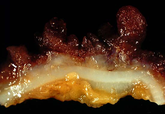
This photo was from a total colectomy done for clinically severe, intractable chronic ulcerative colitis. It shows a closer view of a longitudinal section through the colon wall. This demonstrates not only the angry red mucosa but also the tendency for the inflamed tissue to throw itself up into inflammatory pseudopolyps. Source: Ed Uthman, MD. Public domain.
This photo is from a total colectomy done for severe, intractable chronic ulcerative colitis. It shows a close view of a lengthwise section through the colon wall. This demonstrates not only the angry red mucosa, but also, the tendency for the inflamed tissue to throw itself up into inflammatory pseudopolyps.
Source: Ed Uthman, MD. Public domain.

What Is Ulcerative Colitis?

Ulcerative colitis is an inflammatory disorder of the colon characterized by continuous inflammation of the mucosa and submucosa usually with small ulcers, extending from the rectum and typically involving the distal colon, rectum, and anus and producing bloody diarrhea.

While the severity of ulcerative colitis varies among patients, iron deficiency anemia often develops due to blood loss especially when there are many bloody bowel movements in a day.

The onset of ulcerative colitis is most commonly in young adulhood.

Q: Is this disease painful?

A: Yes with the passage of stool.

Psychological stress and subsequent dysbiosis exacerbate ulcerative colitis.

Psychological stress activates multiple physiological processes aimed at maintaining balance within the body. These physiological processes also have the capacity to influence the composition of microbial communities in the digestive tract, and research now indicates that exposure to stressful stimuli leads to gut microbiota dysbiosis.1

While the relative abundance of many different bacterial types can be altered during stressor exposure, findings in nonhuman primates and laboratory rodents, as well as humans, indicate that bacteria in the genus Lactobacillus are consistently reduced in the gut during stress.2

Presently, ulcerative colitis cannot be cured. This condition has a course of remissions, when symptoms subside, and flares, when symtpoms get worse. Treatment is aimed to reduce flares and promote remission. In all cases, correction of dysbiosis improves the condition.

Ulcerative colitis is associated with increased incidence of cancer of the colon.3

What Is Ulcerative Colitis In Celiac Disease and/or Gluten Sensitivity?

Sources:
  1. Galley JD, Bailey MT. Impact of stressor exposure on the interplay between commensal microbiota and host inflammation. Gut Microbes. 2014 May 1;5(3):390-396. Epub 2014 Apr 1. []
  2. Galley JD, Bailey MT. Impact of stressor exposure on the interplay between commensal microbiota and host inflammation. Gut Microbes. 2014 May 1;5(3):390-396. Epub 2014 Apr 1. []
  3. Taber’s Cyclopedic Medical Dictionary. F. A. Davis. Philadelphia, PA []