Skip to content

Cancer Of The Pharynx 

Areas of the Pharynx Where Cancer May Occur. Courtesy Wikimedia.

What Is Cancer Of The Pharynx?

Cancer of the pharynx is a malignant growth of stratified squamous cells that line the pharynx, or throat.

Q: What are stratified squamous cells that line the pharynx?

A: Stratified squamous cells are thin, flat epithelial cells consisting of several layers forming the surface mucosa that protects underlying layers of the pharynx. These cells lie very close together and have no blood vessels.

The pharynx is the upper passageway of the throat where swallowing food and fluids from the mouth first pass on the way to the esophagus and breathing air passes from the nose and mouth to and from the lungs.

The pharynx provides for speech production, adequate taste reception, and proper hearing through equalization of air pressure in the eustachian tubes.

The pharynx has three areas and pharyngeal cancer can occur in any of them: the nasopharynx behind the nose, the oropharynx behind the mouth, and the hypopharynx or laryngopharynx, just above the larynx. Tonsils, adenoids, and other lymph tissue lie at the back of the throat.

Unfortunately, cancers of the pharynx tend not to be detected until late in their course when symptoms become evident. This makes the prognosis poor. See symptom below.

What Is Cancer Of The Pharynx In Celiac Disease and/or Gluten Sensitivity?

Adenocarcinoma Of Small Intestine (Cancer)

Section of small bowel surgically removed for adenocarcinoma that grew through the wall. By: CDC/ Dr. Edwin P. Ewing, Jr.

What Is Adenocarcinoma Of Small Intestine?

Adenocarcinomas are malignant tumors, or cancer, of the small bowel arising out of glandular tissue. They fall in the category of rare neoplasm, comprising only 3% of all gastrointestinal malignancies.

Primary adenocarcinoma is the most common histological (cell) subtype constituting 35–50% of cases.1 

Q: What does adenocarcinoma look like?

A: Adenocarcinoma may manifest as strictures, nodules, excavating masses, or annular lesions.2

What Is Adenocarcinoma Of Small Intestine In Celiac Disease and/or Gluten Sensitivity?

Sources:
  1. Benhammane H, El M’rabet FZ, Serhouchni KI, El yousfi M, Charif I, Toughray I, et al. Small Bowel Adenocarcinoma Complicating Coeliac Disease: A Report of Three Cases and the Literature Review. Case Rep Oncol Med. 2012; 2012: 935183. Published online 2012 December 1. doi: 10.1155/2012/935183 []
  2. Ramachandran I, Sinha R, Rajesh A, Verma R. Multidetector row CT of small bowel tumors.  Clinical Radiology. 2007; 62:607-614. []

Tongue – Beefy Red, Smooth, Burning 

tongue in B12 deficiencyWhat Is Beefy Red, Smooth, Burning Tongue?

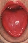
Beefy, red, smooth, burning tongue is an inflammatory alteration in tongue tissue characteristic of vitamin B12 deficiency.

Inflammation makes the tongue red and sore which, when untreated, progresses to atrophied papillae (shrunken taste buds) that makes the tongue smooth.

Q: Why does vitamin B12 alter the tongue?

A: Vitamin B12 is essential for the metabolism of all cells, especially for those of the digestive tract of which the tongue is an organ.1

These easily seen changes to tongue tissue are an important diagnostic feature of deficient vitamin B12, which causes other devastating unseen damage, because the discomfort generally causes the individual to seek medical care.

What Is Beefy Red, Smooth, Burning Tongue In Celiac Disease and/or Gluten Sensitivity?

Sources:
  1. Krause’s Food, Nutrition, & Diet Therapy. 10th Edition. Kathleen Mahan, Sylvia Escott-Stump. 2000. W.B. Saunders Company. []

Tongue – Fiery Red, Smooth, Burning

niacin deficiencyWhat Is A Fiery Red, Smooth, Burning Tongue?

A fiery red, smooth, burning tongue is an alteration in tongue tissue that is characteristic of advanced niacin deficiency.1

Q: How does niacin deficiency cause the tongue to be red and sore?

A: Niacin is an essential B vitamin that is required for a healthy tongue and by all body cells as well. Deficiency first shows in tissues with rapid cellular turnover, such as mucosal cells of the tongue.

When absorbed from the small intestines, niacin (the form in food) becomes the active form niacinamide. Niacinamide is converted by the body into co-enzymes which are present in all cells. These enzymes function in oxidation-reduction reactions essential for release of energy from carbohydrates, fats, and proteins and are needed as components for more than 200 enzymes involved in metabolism.

In addition to producing energy, niacinamide is essential for healthy skin and the mucosal lining of the digestive tract, normal functioning of the brain and nervous system, and production of steroid hormones from adrenal glands and hormones from sex glands.

What Is A Fiery Red, Smooth, Burning Tongue In Celiac Disease and/or Gluten Sensitivity?

Sources:
  1. Krause’s Food, Nutrition, & Diet Therapy. 10th Edition. Kathleen Mahan, Sylvia Escott-Stump. 2000. W.B. Saunders Company. []

Tongue – Pale, Smooth, Burning

Swollen Tongue Causing Tooth Indentations. GFW
Swollen Tongue Causing Tooth Indentations. Notice the Accompanying Denuded Area Due to Riboflavin Deficiency and Mild Candida Overgrowth. GFW

What Is A Pale, Smooth, Burning Tongue?

A pale, smooth, burning tongue is an alteration in tongue tissue characteristic of iron deficiency. The tongue is also swollen.1

Additionally, the sore tongue surface may be invaded by candida yeast which takes advantage of the sore tissue.

Iron deficiency itself increases susceptibility to infection.

Q: What is iron deficiency?

A: Iron deficiency results when the level within cells is too low to meet metabolic needs of the body for this mineral.

Deficiency is characterized by impaired red blood cell formation, free-radical disposal, oxygenation of cells, immune response to infection, enzyme activity, cognitive performance, digestion, nail structure, and fetal health.2

Iron is an essential mineral that is required for normal body function. Almost two-thirds of iron in the body is found in hemoglobin, the protein in red blood cells that carries oxygen to tissues. Smaller amounts of iron are found in myoglobin, a protein that helps supply oxygen to muscle, and in enzymes that assist biochemical reactions.

Iron is also found in proteins that store iron for future needs and that transport iron in blood. Iron stores are regulated by intestinal iron absorption.3

What Is A Pale, Smooth, Burning Tongue In Celiac Disease and/or Gluten Sensitivity?

Sources:
  1. Krause’s Food, Nutrition, & Diet Therapy. 10th Edition. Kathleen Mahan, Sylvia Escott-Stump. 2000. W.B. Saunders Company. []
  2. Kathleen Mahan and Sylvia Escott-Stump, ed. Krause’s Food, Nutrition & Diet Therapy, 10th Edition. Philadelphia, PA. USA: W.B. Saunders Company, 2000. []
  3. http://ods.od.nih.gov/factsheets/Iron-HealthProfessional accessed 12/11/12 []

Vomiting

VomitingWhat Is Vomiting?

Vomiting is the regurgitation of swallowed food back out of the mouth.

What Is Vomiting In Celiac Disease and/or Gluten Sensitivity?

Small Bowel Intussusception

Invagination_Schema[1]What Is Small Bowel Intussusception?

Small bowel intussusception is a bowel derangement that is characterized by the slipping of one section of intestine into another, leading to bowel obstruction.

Acute bowel intussusception is a rare manifestation in adults, which mainly involves the small intestine.1

What Is Small Bowel Intussusception In Celiac Disease and/or Gluten Sensitivity?

Sources:
  1. Grados A, Bernard F, Coquet-Reinier B, Rossi P, Bagneres D, Demoux AL, Marciano S, Frances Y, Granel B. Acute bowel intussusception revealing celiac disease a new case and literature review. Rev Med Interne. 2011 Oct;32(10):628-32. doi: 10.1016/j.revmed.2011.03.334 []

Sucrose Intolerance and Sucrosemia 

Sweet Bon Bons: Courtesy Freepik.
Sugary Bon Bons: Courtesy Freepik.

What Is Sucrose Intolerance And Sucrosemia?

Sucrose intolerance is the inability to digest sucrose, a widely available sugar, while sucrosemia is the abnormal presence of sucrose in the bloodstream.

Q: Why cannot the body digest sucrose?

A: Sucrose, such as cane or beet sugar, is a double molecule sugar which must first be digested before being absorbed from the gut into the bloodstream. That is, sucrose must be split into its component single molecules of fructose and glucose, which are then properly absorbed.

The inability to properly digest sucrose results directly from low production and activity of sucrase in the small intestine. Sucrase is the specific enzyme that splits or digests sucrose.

Undigested sucrose does not remain idle. Its presence acts osmotically to draw water from the body into the intestine, causing watery diarrhea.

Meanwhile, microbiota (normal bacteria) in the colon eagerly ferment the abnormally present sucrose that arrives from the small intestine. Fermentation generates short-chain fatty acids and hydrogen gas, which results in bloating pain.1

In sucrosemia, sucrose molecules abnormaly pass through an unhealthy small intestinal lining and enter the bloodstream where there presence is abnormal. Sucrose in the blood is filtered out by the kidneys and excreted in urine.

Positive response to a breath hydrogen test (BHT), involving 1 – 3 hours of time post ingestion of sucrose test dose, signifies malabsorption in the small intestine and fermentation in the colon. If BHT is positive before 60 minutes, the result implies bacteria is abnormally present in the small intestine, causing fermentation there. Endoscopy is used to measure sucrase activity in tissue samples.

What Is Sucrose Intolerance And Sucrosemia In Celiac Disease and/or Gluten Sensitivity?

Sources:
  1. Krause’s Food, Nutrition, & Diet Therapy. 10th Edition. Kathleen Mahan, Sylvia Escott-Stump. 2000. W.B. Saunders Company. []

Food Allergy – IgE and Non IgE

Baby with Allergic Reaction to Peanuts. GFW
Baby with Allergic Reaction to Peanuts. GFW photo.

What Is Food Allergy?

Food allergy is an abnormal immune response to food proteins that may damage the small intestinal  lining and produce malabsorption of food. The reaction is usually delayed which makes it difficult to identify the offending food that is causing symptoms.

Q: How does food allergy develop?

A: The gastrointestinal tract serves not only to digest and absorb foodstuffs but also to protect the body from unwanted substances. When allergic food substances are eaten, the immune response that is triggered in the gut produces inflammation with symptoms such as pain, vomiting and loose bowels.

Inflammation causes swelling of the gut lining that can interfere with the passage of nutrients through it to the body which results in malabsorption. Malabsorption deprives the body of nutrients needed for normal function.

Symptoms other than digestive may involve skin rashes, hives, and respiratory difficulties that can be distressing and life-threatening.

What Is Food Allergy In Celiac Disease and/or Gluten Sensitivity?

Plummer-Vinson Syndrome Affecting Esophagus

mucosal web in plummer-vinson syndromeWhat Is Plummer-Vinson Syndrome Affecting the Esophagus?

Plummer-Vinson syndrome is a manifestation of severe, long-term, iron deficiency anemia that is characterized by post-cricoid esophageal webs and dysphagia.

Q: What are esophageal webs?

A: Esophageal webs are one or more thin horizontal membranes consisting of squamous epithelium (cells that line the surface of the esophagus) and submucosa. They usually protrude from the anterior (front) wall, extending laterally across the inside esophagus but not to the posterior (rear) wall, which means that they rarely encircle the lumen.1

Dysphagia, or difficulty swallowing, from these webs is commonly painless and intermittent or progressive and may cause obstruction.

Webs can be detected by barium swallow X-ray, but the best way for demonstration is videofluoroscopy and by upper gastrointestinal endoscopy. They appear smooth, thin, and gray with eccentric or central lumen space. The webs typically occur in the upper part of the esophagus and may be missed and accidentally ruptured unless the endoscope is introduced under direct visualization.1

Iron deficiency is believed to decrease the contraction amplitude or force of the esophageal muscle resulting in motility impairment. Slower transit times have been recorded at the proximal and middle parts of the esophagus of Plummer-Vinson syndrome patients compared to healthy volunteers.2 Transit time is how fast ingested food and fluids travel through the esophagus.

Gude et al, report that iron replacement does not necessarily reverse the dysphagia in all the cases of Plummer-Vinson syndrome and that close monitoring of the web is mandated to watch for malignant change.  In fact, 3 to 15 per cent of the patients with Plummer-Vinson syndrome, mostly women between 15 and 50 years of age, have been reported to develop esophageal or pharyngeal cancer.2

What Is Plummer-Vinson Syndrome Affecting the Esophagus In Celiac Disease and/or Gluten Sensitivity?

Sources:
  1. Novacek G. Plummer-Vinson syndrome. Orphanet J Rare Dis. 2006; 1: 36. Published online 2006 September 15. doi:  10.1186/1750-1172-1-36. [] []
  2. Gude D, Bansal DP, and Malu A. Revisiting Plummer Vinson Syndrome. Annals of Medical and Health Sciences Research. 2013 Jan-Mar;3(1):119-121. [] []