Skip to content

Appendectomy: definition

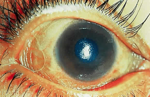
Slit lamp view of cataract in human eye. Courttesy Wikimedia
Slit lamp view of cataract in human eye. Courtesy Wikimedia

What Are Cataracts?

C ataract is a clouding of the normally clear lens in an affected eye characterized by blurred vision and progressive blindness due to loss of the len’s ability to focus light rays on the retina. Cataracts can occur in either or both eyes.

Q: How does a cataract form?

A: The lens is a transparent, colorless, oval-like structure of the eye made of mostly water and protein. The protein is arranged in a precise way that keeps the lens clear and lets light coming through the pupil to pass through it to reach the retina where it is recorded. Once an image reaches the retina, it is changed into nerve signals that are sent to the brain.1

In each eye, the lens is enclosed in a capsule that is held in place directly behind the pupil by the ciliary body and the suspensory ligaments. The lens consists primarily of lens fibers that at the periphery are soft, forming the cortis lentis, and in the center are of a harder consistency, forming the nucleus lentis. Beneath the capsule on the front surface is a layer of cells, the lens epithelium. The shape of the lens is changed by the ciliary muscle to focus light rays onto the retina.2

A cataract begins to form when some of the protein clumps together and starts to cloud a small area of the lens. Over time, the cataract may grow larger and cloud more of the lens, making it harder to see.3

A cataract is diagnosed by an ophthalmologist, who is a medical doctor specializing in the treatment of eye conditions. The eye examination involves viewing the anterior (front) of the eye by means of a slit lamp microscope. This instrument allows detailed observation of the lens and its supporting structures.

What Are Cataracts In Celiac Disease and/or Gluten Sensitivity?

Sources:
  1. Tabers Cyclopedic Medical Dictionary. 19th ed. FA Davis Company, Philadelphia, PA. []
  2. Tabers Cyclopedic Medical Dictionary.C 19th ed. FA Davis Company, Philadelphia, PA. []
  3. http://www.nei.nih.gov/health/cataract/cataract_facts []

Anus: definition

Dry Eye Due to Riboflavin Deficiency. GFW

What Is Chronic Dry Eye Syndrome?

C hronic dry eye syndrome is an inflammatory condition of the conjunctiva with inadequate tear production or distribution over the eye surface. It is characterized by surface damage to the conjunctiva, giving the appearance of redness.

This complex condition involves the lacrimal glands, eyelids, and tear film, as well as a variety of eye surface tissues, including epithelial, inflammatory, immune, and goblet cells.1

Q: What is the conjunctiva?

A: The conjunctiva is the mucous membrane that lines the inside of the eyelids and covers the eyeball surface, also called the white of eye. The conjunctiva has a rich blood supply that can quickly bring microbe fighting cells to prevent invasion and overcome infection.

While infection and inflammation go hand-in-hand, they are not the cause of, but may result from, chronic dry eyes.

Dry eye syndrome develops from poor health of the conjunctiva that is associated with inadequate nutrition and poor quality of the tears that steadily lubricate the eye surface. These tears are called the tear film and are not the same as tears produced when crying or that result from irritating substances such as onions. Blinking spreads the tears that protect the surface from drying and flushes away unwanted matter.

The tear film is made up of three layers: mucous layer produced by goblet cells in the conjunctiva, aqueous (water) layer produced by the lacrimal glands, and lipid (fat) layer produced by the meibomian glands in the eyelids. Dysfunction of any of these layers impairs the the tear film. Without an adequate tear film the conjunctiva becomes dry and inflamed.

When the meibomian glands fail to produce sufficient lipid that is clear and fluid but rather cloudy and thick, the condition is called meibomian gland dysfunction (MGD). The main cause of MGD is hyperkeratinization (thickening of the glands) and its related pathogenesis (for example, ductal dilatation and acinar atrophy). Other disorders such as atopy, pemphigoid acne, rosacea, and seborrhea are related to MGD and may result in a chronic inflammation of the eye surface.2

MGD is “the most underrecognized, underappreciated and undertreated disease in ophthalmic care. It is so common as to be taken as ‘normal’ in many clinical practices,” according to Joseph Tauber, MD, an anterior segment subspecialist and refractive surgeon in Kansas City, Mo.3

Untreated dry eye progresses to xerophthalmia, a condition of extreme drying and thickening of the conjunctiva characterized by hazy, dry cornea. Other causes of dry eye, beside celiac disease, include certain systemic conditions such as Sjogren’s syndrome, an autoimmune disease.

What Is Chronic Dry Eye Syndrome In Celiac Disease and/or Gluten Sensitivity?

Sources:
  1. Liu A, Ji J. Omega-3 essential fatty acids therapy for dry eye syndrome: a meta-analysis of randomized controlled studies. Med Sci Monit. 2014 Sep 6;20:1583-9. doi: 10.12659/MSM.891364. []
  2. Oleñik A, Jiménez-Alfaro I, Alejandre-Alba N, Mahillo-Fernández I. A randomized, double-masked study to evaluate the effect of omega-3 fatty acids supplementation in meibomian gland dysfunction. Clin Interv Aging. 2013;8:1133-8. doi: 10.2147/CIA.S48955. Epub 2013 Aug 30. []
  3. Rethinking Meibomian Gland Dysfunction: How to Spot It, Stage It and Treat It. Linda Roach, PhD. OPHTHALMIC PEARLS. []

Apoptosis: definition

blurred visionWhat Is Unexplained Blurred Vision?

U nexplained blurred vision is unclear or distorted vision that is not caused by allergy, poor refraction (eye test), medication side effects, metabolic disorders, or defects in any part of the eyeball, optic nerve, or in the occipital area of the brain resulting from such problems as calcifications, aneurysm, or tumors.

What Is Unexplained Blurred Vision In Celiac Disease and/or Gluten Sensitivity?

Barium Enema X Ray: definition

X-ray showing osteitis fibrosa cystica in long bones. Courtesy Wikimedia.

What Is Osteitis Fibrosa Cystica?

O steitis fibrosa cystica is a metabolic bone disease characterized by decalcification and softening of bones with bone cyst formation and bone tumors developing from effects of chronic vitamin D deficiency causing hyperparathyroidism and parathyroid adenoma (tumor).

Hyperparathyroidism is a sustained elevated parathyroid hormone level in the blood which draws calcium from bone tissue.

Q: How does vitamin D deficiency cause hyperparathyroidism?

A: Vitamin D is required to absorb calcium from the food we eat. Deficiency of vitamin D  in turn causes deficiency of calcium to develop. Without adequate calcium for essential body functions, the pituitary gland stimulates the parathyroid glands to produce parathyroid hormone for the purpose of getting calcium from bone tissue which is less essential to life.

Unfortunately, the bones become depleted of calcium and become soft and disfigured.

What Is Osteitis Fibrosa Cystica In Celiac Disease and/or Gluten Sensitivity?

Appendix: definition

 Keratomalacia in a severely malnourished 10-month-old Indonesian boy.  (Sommer A, Sugana T: Corneal xerophthalmia and keratomalacia. Arch Ophthalmol 100:404, 1982)
Keratomalacia in a severely malnourished 10-month-old Indonesian boy.1

What Is Keratomalacia?

K eratomalacia is a severe non-inflammatory eye condition characterized by a hazy, dry cornea that becomes softened and denuded (loss of protective layer), which when liquified is then irreversibly destroying the eyeball and causing blindness.

The conjunctiva and lacrimal glands (tear producing) are also affected resulting in ulceration of the eye surface with, or without, Bitot’s spots (foamy patches) because these tissues also require adequate vitamin A to function normally.

The conjunctiva is the mucous membrane that lines the inside of the eyelids and covers the eyeball surface, also called the white of eye.

Q: What does the term keratomalcia mean?

A: In the term keratomalcia, kerato refers to the cornea and malacia means softening of the tissues.

What Is Keratomalacia In Celiac Disease and/or Gluten Sensitivity?

Sources:
  1. Sommer A, Sugana T: Corneal xerophthalmia and keratomalacia. Arch Ophthalmol 100:404, 1982 []

Ascending Colon: definition

NightblindnessWhat Is Night Blindness?

N ight blindness is a visual defect caused by lack of rhodopsin in the rods of the retina located in the back of the eyeball due to inadequate vitamin A.

Q: How does inadequate vitamin A cause a lack of rhodopsin in the eyeball?

A: Rhodopsin is a visual pigment that requires vitamin A as a component.

Night blindness is characterized by decreased ability to see at night or in the dark because rhodopsin is a chemical required for the retina to distinquish light and dark. It is an early feature of vitamin A deficiency and also decreased content of oxygen in the blood such as anemia.

With increasing vitamin A deficiency, damage to the rods and to the eyeball itself occurs because these tissues also require vitamin A for structure and function.

Night blindness can be brought on by poor diet, gastroenteritis and fever due to increased requirements for vitamin A if stores in the liver are low, malabsorption, and systemic disease that interferes with storage in the liver or transport in the blood.

Breastfeeding infants of mothers who are vitamin A deficient are at high risk since they will not have adequate liver stores and cannot get enough from mother’s milk.

In teenagers it can be the beginning symptoms of retinitis pigmentosa, a group of retinal degenerations that primarily affect the rod photoreceptors.1

What Is Night Blindness In Celiac Disease and/or Gluten Sensitivity?

Sources:
  1. Gordon K. Klintworth, MD, PhD. The Eye Pathologist.com []

Astrovirus: definition

Drawing of eyeball to illustrate uveitis. Courtesy National Eye Institute
Drawing of Eye to Illustrate Uvea Tract. Courtesy
National Eye Institute

What Is Non-Infectious Uveitis?

N on-infectious uveitis, as opposed to that which is caused by an infection, is an inflammatory eye disorder characterized mainly by swelling of the uveal tract structures in the anterior (front) of the eye.  

Inflammation can also affect the lens, retina, optic nerve, and vitreous, producing reduced vision or blindness if left untreated. 

Q: What are structures of the uveal tract?

A: Structures of the uveal tract are composed of the iris, the ciliary body, and the choroid: 

  • The iris is the colored circle at the front of the eye. It defines eye color, secretes nutrients to keep the lens healthy, and controls the amount of light that enters the eye by adjusting the size of the pupil (opening).
  • The ciliary body is located between the iris and the choroid. It helps the eye focus by controlling the shape of the lens and it provides nutrients to keep the lens healthy.
  • The choroid is a thin, spongy network of blood vessels, which primarily provides nutrients to the retina in the back of the eye.1

Uveitis may be caused by problems or diseases occurring in the eye itself or it can be part of an inflammatory disease affecting other parts of the body. The uveal tract has a rich supply of blood vessels that contain immune cells to fight microbial invasion. These immune cells are lymphocytes, phagocytes, and plasma cells.

The uvea can be attacked by autoimmune antibodies produced in autoimmune diseases that affect similar tissues in other parts of the body such as psoriasis, Behcet’s syndrome, multiple sclerosis, sarcoidosis, Vogt Koyanagi Harada’s disease, and celiac disease.

Uveitis can last for a short (acute) or a long (chronic) time. The severest forms of uveitis reoccur many times.

What Is Non-Infectious Uveitis In Celiac Disease and/or Gluten Sensitivity?

Sources:
  1. http://www.nei.nih.gov/health/uveitis/uveitis.asp []

Ascites: definition

Opthalmoplegia. Courtesy bmj.com
Opthalmoplegia. Courtesy BMJ from lookfordiagnosis.com

What Is Ocular Myopathy?

O cular myopathy is an impairment of musculature of the eyeball that is characterized by abnormalities of eye movement that make initiating and maintaining eye movement and eye contact difficult.

What Is Ocular Myopathy In Celiac Disease and/or Gluten Sensitivity?

Asymptomatic: definition

Marked conjunctival and corneal xerosis. The entire bulbar conjunctiva is dry and thickened and almost skinlike. (Sommer A, Sugana T, Djunaedi E, Green R: Vitamin A-responsive panocular xerophthalmia in a healthy adult. Arch Ophthalmol 96:1630, 1978)

What Is Xerophthalmia?

X erophthalmia is a serious condition of extreme drying and thickening of the conjunctiva following chronic inflammation and failure to make tears, characterized by hazy, dry cornea.

Vitamin A is obtained from the diet and is required in the cornea for maintaining epithelial (surface cell) health.1

Xerophthalmia is most often associated with malnutrition involving chronic, severe vitamin A deficiency and may develop from chronic liver disease, intestinal disorders such as celiac disease, pancreatitis, and extensive bowel surgery that cause malabsorption.

It may also develop in autoimmune diseases such as Sjogrens syndrome.

Q: What is the prognosis for xerophthalmia?

A: Xerophthalmia has the potential to rapidly advance to corneal necrosis (keratomalacia) and blindness.

What Is Xerophthalmia In Celiac Disease and/or Gluten Sensitivity?

Sources:
  1. Alwitry A. Vitamin A deficiency in coeliac disease. Br J Ophthalmol. 2000 September; 84(9): 1075. doi:  10.1136/bjo.84.9.1075e []

Atonic Colon: definition

Courtesy encyclopedia.lubopitko-bg.com

What Is Loss of Smell?

L oss of smell is a disorder that is characterized by impaired olfactory sense, or olfaction.

Partial or complete loss of smell can result from malnutrition, chronic rhinitis, polyps, chronic sinusitis, medications, old age, head trauma, Alzheimer’s disease and Parkinson’s disease.

Q: How do we smell things?

A: The sense of smell is complex. It involves specialized olfactory cells that are chemoreceptors, that is, they detect odor molecules in the air we breathe in through our nose and transmit the information to the olfactory area of our brain, called the olfactory cortex, by way of the olfactory tract.   Olfactory cells are located in the top of the nasal cavity.

The ability to smell strongly contributes to the ability to taste, so that individuals with loss of smell have great difficulty in perceiving the taste of food. The result is loss of the appetite for and pleasure from food.

What Is Loss of Smell In Celiac Disease and/or Gluten Sensitivity?