Skip to content

Antacids: definition

schizophrenia gluten
PET scan showing schizophrenia. Courtesy Wikimedia.

What Is Schizophrenia?

S chizophrenia is a thought disorder characterized by psychotic symptoms and inappropriate and bizarre behavior.

Schizophrenia involves dysfunction in any of these areas:

  • Cognition which are thinking skills that include intellect, language, mathematical ability, memory, perception, reasoning, judgment, learning, and imagination.
  • Interpersonal relationships.
  • Ability to work or attend education.
  • Ability to take care of physical and financial needs.

Q: What are psychotic symptoms?

A: In psychosis the symptoms vary but include loss of contact with reality shown by hallucinations, delusions, disorganized speech, and disorientation. Some may be depressed or manic. Some may be withdrawn and mute while others may become violent if frustrated or thwarted.

In hallucinations, affected persons see what is not real such as a burgler in the process of robbing them but which is not really happening or hear voices that are not real.

In delusions, affected persons may express strange ideas, claim to read minds or that they are a famous person living or dead.

Early-onset schizophrenia with onset before age 18 is defined by the same diagnostic criteria as in adults because it is continuous with adult schizophrenia. The onset before age 12 years comprises less than 1% of adult cases, and onset from age 12–18 years constitutes about 12–33% of all adult cases of schizophrenia.1

What Is Schizophrenia In Celiac Disease and/or Gluten Sensitivity?

Sources:
  1. Pagsberg AK, Tarp S, Glintborg D, Stenstrøm AD, Fink-Jensen A, Correll CU, Christensen R. Antipsychotic treatment for children and adolescents with schizophrenia spectrum disorders: protocol for a network meta-analysis of randomised trials. BMJ Open. 2014 Oct 10;4(10):e005708. doi: 10.1136/bmjopen-2014-005708. []

Antiemetics: definition

Autopsy View of Lung Abcess.
Autopsy View Of Lung Cavity.

What Is Abscess Formation Causing Lung Cavities?

A bscess formation and subsequent lung cavities that develop from necrosis (death) of affected lung tissue constitute severe life-threatening respiratory disease  of the lung.

Q: What is the difference between lung abscess and lung cavity?

A: Lung abscess is characterized by localized pus formation in the lung that develops from uncontrolled infection from encapsulated bacteria such as streptococcus pneumoniae and klebsiella. The capsules that surround bacteria protect them from phagocytosis by macrophages and neutrophils, white blood cells that are specialized for this normal defensive action. Phagocytosis is a seek and destroy mission.

Lung cavities are holes that form from dead tissue during the destructive abscess development, which involves significant inflammation and edema of the area. Lung cavities often cause death of the patient. The outlook is very poor.

What Is Abscess Formation Causing Lung Cavities In Celiac Disease and/or Gluten Sensitivity?

Anticholinergics: definition

Bronchiectasis Image. Courtesy Quizlet.com

What Is Bronchiectasis?

B ronchiectasis is a pulmonary disorder characterized by chronic dilation of bronchi (main airways) and bronchioles (small airways) with chronic inflammation and swelling, increasing the risk of infection.

In bronchiectasis, mucus produced to trap foreign substances, such as dust and bacteria, builds-up because the ability of airways to move it out is impaired. Normally, hair-like cilia that line airways beat mucus out of the lungs. Loss of this action increases mucus in the airways which increases risk of pneumonia.

The anatomy of airways is likened to a branching tree. The trachea, or windpipe, like the trunk of a tree, branches into two primary bronchi that connect it to the lungs. One bronchus connects to the left lung and the other to the right lung. Each bronchus further divides into smaller bronchi that connect to the lobes. Lobes are large subdivisions of lung tissue: two of the left lung and three of the right lung.

Lobe bronchi divide into smaller bronchial tubes ending in small bronchioles. The ends of bronchioles are surrounded with and attached to tiny alveoli which are air sacs that look like clusters of grapes.

Q: What effect does inflammation have on the lungs?

A: Alveoli are only one cell thick, where oxygen is obtained by the bloodsteam from air breathed into them and carbon dioxide is released from the bloodstream to air that is breathed out of air sacs through the pulmonary tree. Two things happen:

  1. Inflammation  impairs exchange of gases in alveoli, resulting in lack of sufficient oxygen (O2) for body cell functions, called hypoxia, and build-up of carbon dioxide (CO2) in blood, called CO2 retention.
  2. Inflammation narrows passageways, which reduces the movement of air to and from the alveoli, and this puts stress on the right side of the heart.

Bronchiectasis can affect one or both lungs or one segment or multiple segments of a lung. Treatment is with antibiotics, chest physiotherapy and good nutrition.

What Is Bronchiectasis In Celiac Disease and/or Gluten Sensitivity?

Antidiarrheals: definition

bronchoalveolitisWhat Is Lymphocytic Bronchoalveolitis?

L ymphocytic bronchoalveolitis is a pulmonary disorder characterized by lymphocytic airway inflammation of the bronchi (main passageways branching from the windpipe), bronchioles (small passageways), and alveoli (air sacs) at the end.

The chronic, productive cough that develops and worsens with progression is due to increased mucus secretion in the large and small airways.

If infection develops in the bronchioles, it is call bronchopneumonia. If infection develops in the alveoli, it is called interstitial pneumonia. The most common organism causing infection is staphylcoccus aureus.

Q: What effect does inflammation have on the lungs?

A: Alveoli are air sacs, each only one cell thick, where oxygen is obtained by the bloodsteam from air breathed into them and carbon dioxide is released from the bloodstream to air that is breathed out of air sacs. Two things happen:

  1. Inflammation  impairs exchange of gases in alveoli, resulting in lack of sufficient oxygen (O2) for body cell functions, called hypoxia, and build-up of carbon dioxide (CO2) in blood, called CO2 retention.
  2. Inflammation narrows passageways, which reduces the movement of air to and from the alveoli, and this puts stress on the right side of the heart.

Lymphocytes, which are a type of white blood cell, can be collected and assessed by a procedure called bronchoalveolar lavage to determine the degree of involvement.

What Is Lymphocytic Bronchoalveolitis In Celiac Disease and/or Gluten Sensitivity?

Antispasmodics: definition

Pneumococcus bacteria
Pneumococcal Bacteria Occuring in Pairs.

What Is Pneumoccocal Septicemia?

P neumococcal septicemia (sepsis) is a life-threatening syndrome characterized by a cascade of systemic (body-wide) inflammatory responses to lung infection caused by bacterial infection spread to the bloodstream.

It is fatal in about 40% of cases, showing inadequate blood flow to internal organs. About 400,000 people die of sepsis each year in the United States.

Q: What is a pneumococcus?

A: A pneumococcus is an oval shaped , encapsulated, non-spore forming, gram positive bacterium of the genus Streptococcus. These bacteria usually occur in pairs.1

Infection can be prevented with pneumococcal vaccination.

What Is Pneumococcal Septicemia In Celiac Disease and/or Gluten Sensitivity?

Sources:
  1. Taber’s Encyclopedic Medical Dictionary. 10th ed., Phila. []

Antigen, Immunogen Type: definition

Pulmonary Hemosiderosis. Courtesy Quizlet.com
Lung from a Patient who Had Pulmonary Hemosiderosis. Courtesy Quizlet.com

What Is Idiopathic Pulmonary Hemosiderosis?

P ulmonary hemosiderosis (IPH) is a rare and severe pulmonary syndrome characterized by a triad of recurrent episodes of alveolar hemorrhage (bleeding into air sacs), hemoptysis (coughing blood), and iron deficiency anemia.

Q: What is the prognosis for pulmonary hemosiderosis?

A: Untreated pulmonary hemosiderosis ends in death. Idiopathic is a term that means the cause of a condition is not known.

What Is Pulmonary Hemosiderosis In Celiac Disease and/or Gluten Sensitivity?

Antibody: definition

Image shows
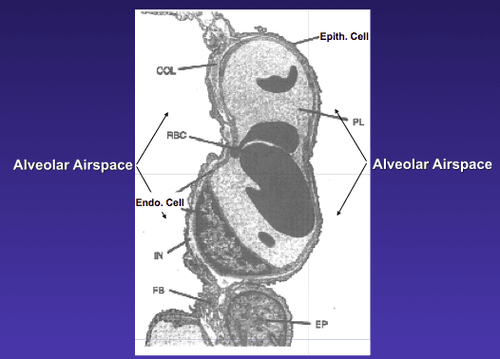
Image Showing  Blood-Air Barrier in the Lung. Courtesy quizlet.com

What Is Increased Pulmonary Permeability?

I ncreased pulmonary permeability is a mucosal defect in the lung characterized by alteration of the normally tight epithelial  blood-air barrier in the lung apparently caused by inflammation.

Q: What is the blood-air barrier in the lung?

A: This blood-air barrier in the lung consists of the alveolar epithelium (surface cells of the alveoli), the underlying capillary endothelium (surface cells of the capillaries), their basement membranes and the interstitial space between the cell layers.

Alveoli are also called “air sacs.” The exchange of oxygen breathed into air sacs from air on inspiration and carbon dioxide breathed out from capillary blood on expiration occurs between  air sacs and capillaries.

The capillary endothelium prevents proteins in blood from leaking (permeating) into the air sacs while allowing water and small molecules to pass. This is why breath has moisture in it.

Little is known about the interactions between the alveolar and the blood compartment.1

What Is Increased Pulmonary Permeability In Celiac Disease and/or Gluten Sensitivity?

Sources:
  1. Neuhaus W, Samwer F, Kunzmann S, Muellenbach RM, Wirth M, Speer CP, Roewer N, Förster CY. Lung endothelial cells strengthen, but brain endothelial cells weaken barrier properties of a human alveolar epithelium cell culture model. Differentiation. 2012 Nov;84(4):294-304. doi: 10.1016/j.diff.2012.08.006. Epub 2012 Sep 27. []

Antibody, Immunoglobulin G: definition

Chest x-ray showing tuberculosis. Courtesy wikipedia.
Chest x-ray showing tuberculosis with consolidation of left lung (white area). Courtesy wikipedia.

What Is Non-Response to Tuberculosis Treatment?

N on-response to treatment for tuberculosis means that proper medical treatment failed to control active disease. Tuberculosis is an infection that may be dormant or active.

Q: What happens in active tuberculosis?

A: Tuberculosis is an infectious disease caused by a bacteria called mycobacterium tuberculosis. It is characterized by chronic bacterial infection most commonly affecting lungs that develops in stages.

Active tuberculosis  produces inflammation and formation of tubercles, necrosis, abcess, fibrosis, and calcification. Calcification is the body’s action to encapsulate the bacterial invasion. Active tuberculosis is life-threatening and may result in death.

About one third of the world’s population is infected with tuberculosis bacteria. In 2012 the number reached a staggering 8.6 million people. Of these, 1.3 million people died from tuberulosis.  About 95% of tuberulosis deaths occur in low- and middle-income countries and it is among the top three causes of death among women aged 15 to 44.1

People with weakened immune systems have a much greater risk of falling ill from tuberculosis. For example, a person living with HIV is about 20 to 30 times more likely to develop active tuberculosis.2

What Is Non-Response To Tuberculosis Treatment In Celiac Disease and/or Gluten Sensitivity?

Sources:
  1. http://www.who.int/features/factfiles/tb_facts/en/index.html []
  2. http://www.who.int/features/factfiles/tuberculosis/en/ []

Antibody, Immunoglobulin A: definition

Bacteria that causes tuberculosis. Courtesy Wikimedia.

What Is Increased Susceptibility To Tuberculosis?

T uberculosis (TB), is an infectious disease caused by a bacteria called mycobacterium tuberculosis. It is characterized by chronic bacterial infection most commonly affecting lungs that develops in stages.

Increased susceptibility to tuberculosis menas the person’s defense mechanisms against developing infection are inadequate. Tuberculosis may be dormant or active.

Q: What happens in active tuberculosis?

A: Active tuberculosis  produces inflammation and formation of tubercles, necrosis (death of tissues), abcess, fibrosis, and calcification. Calcification is the body’s action to encapsulate the bacterial invasion. Active tuberculosis is life-threatening and may result in death.

About one third of the world’s population is infected with tuberculosis bacteria. In 2012 the number reached a staggering 8.6 million people. Of these, 1.3 million people died from tuberulosis.  About 95% of tuberulosis deaths occur in low- and middle-income countries and it is among the top three causes of death among women aged 15 to 44.1

People with weakened immune systems have a much greater risk of falling ill from tuberculosis. For example, a person living with HIV is about 20 to 30 times more likely to develop active tuberculosis.2 The combination of tuberculosis, HIV coinfection, and malnutrition has been commonly termed as “triple trouble.”3

What Is Increased Susceptibility To Tuberculosis In Celiac Disease and/or Gluten Sensitivity?

Sources:
  1. http://www.who.int/features/factfiles/tb_facts/en/index.html []
  2. http://www.who.int/features/factfiles/tuberculosis/en/ []
  3. Steinbrenner H, Al-Quraishy S, Dkhil MA, Wunderlich F, Sies H. Dietary selenium in adjuvant therapy of viral and bacterial infections. Adv Nutr. 2015 Jan 15;6(1):73-82. doi: 10.3945/an.114.007575. Print 2015 Jan. []

Antibody, Immunoglobulin E: definition

Classic Bitot's spot in a 29-year-old man that shrunk dramatically with vitamin A therapy. (Sommer A: Nutritional Blindness: Xerophthalmia and Keratomalacia. New York, Oxford University Press, 1982)
Classic Bitot’s spot in a 29-year-old man that shrunk dramatically with vitamin A therapy.*

What Are Bitot’s Spots?

B itot’s spots are superficial foamy patches that develop on the exposed bulbar conjunctiva (white of the eye) as a manifestation of advanced vitamin A deficiency.

This painless eye disorder is reversible only with vitamin A therapy. It is named after Charles Bitot, who first described it.

Q: What makes up these foamy alterations of the conjunctiva?

A: Bitot’s spots are composed of epithelial debris (dead surface cells) and secretions.1

Bitot’s spots may develop in malnutrition, reduced intake including alcoholism, medication adverse effect, old age, low stomach acid, and disease causing vitamin A deficiency such as celiac disease, Crohn’s disease, pancreatic insufficiency, and short bowel syndrome.  Other conditions associated with vitamin A deficiency may include disordered transport (Abetalipoprotenemia, a genetic disorder) and reduced liver storage caused by liver disease.

What Are Bitot’s Spots In Celiac Disease and/or Gluten Sensitivity?

Sources:
  1. Sadowski B, Rohrback JM, Steuhl KP, Weidle EG, Castrillon-Obendorfer WL. Corneal manifestations in Vitamin A deficiency. Klinische Monatsblatter fur Augenheilkunde. Aug 1994;205(2)76-85. []