Skip to content

Insomnia

Sweet Bon Bons: Courtesy Freepik.
Sugary Bon Bons: Courtesy Freepik.

What Is Sucrose Intolerance And Sucrosemia?

S ucrose intolerance is the inability to digest sucrose, a widely available sugar, while sucrosemia is the abnormal presence of sucrose in the bloodstream.

Q: Why cannot the body digest sucrose?

A: Sucrose, such as cane or beet sugar, is a double molecule sugar which must first be digested before being absorbed from the gut into the bloodstream. That is, sucrose must be split into its component single molecules of fructose and glucose, which are then properly absorbed.

The inability to properly digest sucrose results directly from low production and activity of sucrase in the small intestine. Sucrase is the specific enzyme that splits or digests sucrose.

Undigested sucrose does not remain idle. Its presence acts osmotically to draw water from the body into the intestine, causing watery diarrhea.

Meanwhile, microbiota (normal bacteria) in the colon eagerly ferment the abnormally present sucrose that arrives from the small intestine. Fermentation generates short-chain fatty acids and hydrogen gas, which results in bloating pain.1

In sucrosemia, sucrose molecules abnormaly pass through an unhealthy small intestinal lining and enter the bloodstream where there presence is abnormal. Sucrose in the blood is filtered out by the kidneys and excreted in urine.

Positive response to a breath hydrogen test (BHT), involving 1 – 3 hours of time post ingestion of sucrose test dose, signifies malabsorption in the small intestine and fermentation in the colon. If BHT is positive before 60 minutes, the result implies bacteria is abnormally present in the small intestine, causing fermentation there. Endoscopy is used to measure sucrase activity in tissue samples.

What Is Sucrose Intolerance And Sucrosemia In Celiac Disease and/or Gluten Sensitivity?

Sources:
  1. Krause’s Food, Nutrition, & Diet Therapy. 10th Edition. Kathleen Mahan, Sylvia Escott-Stump. 2000. W.B. Saunders Company. []

Bone Fractures 

Proptosis and lid retraction are features of  Grave's disease, or hyperthyroidism. Courtesy of Wikimedia.
Proptosis and Lid Retraction are Features of Grave’s Disease, or Hyperthyroidism.

What Is Grave’s Disease (Hyperthyroidism)?

G rave’s disease, or hyperthyroidism, is an autoimmune thyroid disease characterized by diffuse nontender goiter, elevated thyroxine hormone levels (T4, T3), suppressed thyroid stimulating hormone (TSH), and presence of thyroid receptor antibodies in the blood.

The autoantibodies involved are anti-thyroid peroxidase and anti-thyroglobulin antibodies. They bind to the thyroid stimulating hormone receptors, causing thyroid stimulation. These antibodies are detected by blood tests.

Q: What happens to the thyroid gland in Grave’s disease?

A: The thyroid gland is located in the front of the neck. This butterfly shaped gland consists of a large number of closed vesicles that contain a homogenous substance called colloid, which contains the thyroglobulin. Thyroglobulin is an iodine-containing protein secreted by the thyroid gland and stored within its colloid, from which the thyroid hormones thyroxine (T4) and triiodothyroinine (T3) are derived.1

Thyroxine molecule, chemical structure. Thyroid gland hormone that plays a role in energy metabolism regulation. It is a iodine containing derivative of thyrosine. Atoms are represented as spheres with conventional color coding: hydrogen (white), carbon (grey), oxygen (red), nitrogen (blue), iodine (purple).
Thyroxine molecule. Atoms are represented as spheres with conventional color coding: hydrogen (white), carbon (grey), oxygen (red), nitrogen (blue), iodine (purple).

T3 is the active hormone and is made from T4. Thyroid hormones are released into the bloodstream as needed to control metabolism, brain development, breathing, heart and nervous system functions, body temperature, muscle strength, skin dryness, menstrual cycles, weight, and cholesterol levels.

Thyroid hormone production is regulated by thyroid-stimulating hormone (TSH), which is made by the pituitary gland in the brain. Normally, when thyroid hormone levels in the blood are low, the pituitary releases more TSH in response to stimulation by the nearby hypothalamus which is continually monitoring levels of thyroxin. When thyroid hormone levels are high, the pituitary decreases TSH production. So in Grave’s disease, release of TSH by the pituitary gland is suppressed by the hypothalamus because thyroid hormone is elevated. Goiter develops from growth stimulation by thyroid stimulating autoantibodies.

What Is Grave’s Disease In Celiac Disease and/or Gluten Sensitivity?

Sources:
  1. Taber’s Cyclopedic Medical Dictionary. 19th ed. F.A. Davis Company. Philadelphia, PA. []

Psoriasis

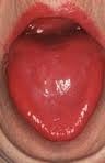
niacin deficiencyWhat Is A Fiery Red, Smooth, Burning Tongue?

A fiery red, smooth, burning tongue is an alteration in tongue tissue that is characteristic of advanced niacin deficiency.1

Q: How does niacin deficiency cause the tongue to be red and sore?

A: Niacin is an essential B vitamin that is required for a healthy tongue and by all body cells as well. Deficiency first shows in tissues with rapid cellular turnover, such as mucosal cells of the tongue.

When absorbed from the small intestines, niacin (the form in food) becomes the active form niacinamide. Niacinamide is converted by the body into co-enzymes which are present in all cells. These enzymes function in oxidation-reduction reactions essential for release of energy from carbohydrates, fats, and proteins and are needed as components for more than 200 enzymes involved in metabolism.

In addition to producing energy, niacinamide is essential for healthy skin and the mucosal lining of the digestive tract, normal functioning of the brain and nervous system, and production of steroid hormones from adrenal glands and hormones from sex glands.

What Is A Fiery Red, Smooth, Burning Tongue In Celiac Disease and/or Gluten Sensitivity?

Sources:
  1. Krause’s Food, Nutrition, & Diet Therapy. 10th Edition. Kathleen Mahan, Sylvia Escott-Stump. 2000. W.B. Saunders Company. []

Rickets

Seborrhea patches at the inner eyebrows. GFW

What Is Seborrhea Dermatitis?

S eborrhea dermatitis is a recurring inflammatory disorder of sebaceous glands characterized by scaly patches of skin, often with bumps.

Seborrhea dermatitis results from the body’s inflammatory reaction to invasion by pityrosporum yeast that naturally inhabits the scalp and skin.1 Inflammation is the normal response to tissue injury and germ invasion.

Pityrosporum is a yeast that is commonly present worldwide. Its development depends on various factors that predispose to pityriasis versicolor, a chronic and mild superficial yeast infection. These infections usually are asymptomatic without itching or pain and without cellular and/or antibody responses.2

Q: Why are the sebaceous glands particularly affected by this yeast?

A: Pityrosporum yeast is an organism that needs oil produced by sebaceous glands to grow. If conditions permit, this yeast invades the superficial layer of skin and hair shafts to reproduce, causing infection. Such conditions include weakened skin due to nutritional deficiencies, excessive build-up of oil on skin, and altered immunity due to systemic disease such as psoriasis.

In adults, areas of skin that are the most affected have the greatest number of sebaceous glands especially the scalp, back, underarms, and the face including the eyelids, eyebrows and side folds of the nose.

All ages are subject to seborrhea dermatitis, and males have a higher occurence than females.

What Is Seborrhea In Celiac Disease and/or Gluten Sensitivity?

Sources:
  1. Krause’s Food, Nutrition, & Diet Therapy. 10th Edition. Kathleen Mahan, Sylvia Escott-Stump. 2000. W.B. Saunders Company. []
  2. Zarei-Mahmoudabadi A, Zarrin.  M, Mehdinezhad F. Seborrheic dermatitis due to Malassezia species in Ahvaz, Iran. Iran J Microbiol. 2013 Sep;5(3):268-71. []

Delayed Puberty in Boys

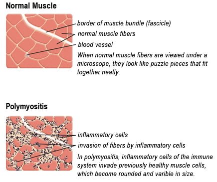
Drawing of Biopsy Showing Muscle Fibers Invaded by Immune Cells. Courtesy MDA.org

What Is Polymyositis?

P olymyositis is a body-wide connective tissue disease resulting from autoimmune attack of skeletal muscles that is characterized by inflammatory and degeneratory changes. The course is unpredictable being marked by spontaneous flare-ups and remissions.

Polymyositis can begin slowly or abruptly according to the factor that is triggering the onset such as infection, medications like phenytoin, and autoimmune disease.

Progressive muscle weakness starts in the proximal skeletal muscles (muscles closest to the trunk of the body).

Skeletal muscles, also called voluntary, are muscles that move the body as we want, such as walking and  lifting objects, as opposed to those we cannot voluntarily control, such as the muscles of digestion. 

Q: What are the degeneratory changes in skeletal muscles?

A: In polymyositis, degeneratory changes in skeletal muscles means that muscles are being destroyed (called necrosis), resulting in fibrosis, or scarring. When scar tissue takes the place of lost muscle tissue, it cannot act like muscle to contract and relax.   Muscle destruction is what causes muscle pain and weakness.

After the clinical work-up of exams and blood studies to determine muscle damage, the diagnosis of polymyositis is confirmed by muscle biopsy. See image at above left. The black dots are inflammatory cells. Edema (fluid) between cells caused by inflammation pushes muscle fibers apart.

There is no cure for polymyositis, but the symptoms can be treated.  Options include medication, physical therapy, exercise, heat therapy (including microwave and ultrasound), orthotics and assistive devices, and rest.  The standard treatment for polymyositis is a corticosteroid drug, given either in pill form or intravenously.  Immunosuppressant drugs, such as azathioprine and methotrexate, may reduce inflammation in people who do not respond well to prednisone. 

Periodic treatment using intravenous immunoglobulin can also improve recovery.  Other immunosuppressive agents used to treat the inflammation associated with polymyositis include cyclosporine A, cyclophosphamide, and tacrolimus.  Physical therapy is usually recommended to prevent muscle atrophy and to regain muscle strength and range of motion.1

Diagnosis is based on elevated muscles enzymes, increased urinary creatine level, and electromyograph abnormalities.

Polymyositis can affect people at any age. It is most common in adults between ages 50 and 70, and in children ages 5 to 15. It affects women twice as often as men and is more common in African Americans than Caucasians.2 The major causes of death from polymyositis are cancer and lung disease, including pneumonia.  The 5-year mortality rate can be as high as 1 in 5 patients.2

What Is Polymyositis In Celiac Disease and/or Gluten Sensitivity?

Sources:
  1. National Institute of Neurological Disorders and Stroke []
  2. http://www.nlm.nih.gov/medlineplus/ency/article/000428.htm [] []

Osteopenia In Childhood 

sclerodermaWhat Is Scleroderma?

S cleroderma is a chronic skin manifestation of progressive systemic sclerosis characterized by generalized thickened, edematous skin firmly bound to subcutaneous tissue which causes limited movement.

Systemic sclerosis a connective tissue disease that involves destructive changes in the skin, blood vessels, muscles, and internal organs. The course can be mild or it can be fatal. Cardiopulmonary complications from fibrosis are the most common cause of death.

Gastrointestinal problems mainly due to fibrosis affect 50 to 90% of patients.1

Q: Is there a cure for scleroderma?

A: There is no cure for scleroderma. Treatment is aimed at improving symptoms.

  • Heartburn (acid reflux) can be treated with antacid drugs.
  • Scleroderma kidney disease can be treated with blood pressure medications called “angiotensin converting enzyme inhibitors” (ACE inhibitors). These can often effectively control kidney damage if started early and use of these drugs has been a major advance for treating scleroderma.
  • Muscle pain and weakness can be treated with anti-inflammatory drugs such as prednisone, intravenous immunoglobin (IVIg), and/or immunosuppressive medications. Physical therapy may be useful to maintain joint and skin flexibility.2


What Is Scleroderma In Celiac Disease and/or Gluten Sensitivity?

Sources:
  1. Forbes A, Marie I. Gastrointestinal complications: the most frequent internal complications of systemic sclerosis. Rheumatology (Oxford). 2009 Jun;48 Suppl 3:iii36-9. doi: 10.1093/rheumatology/ ken485. []
  2. http://www.rheumatology.org []

Delayed Puberty In Girls 

sick woman on bed, symptom of cold, flu, insomnia, stress, headache, hangover, dizzinessWhat Is Hypokalemic Rhabdomyolysis?

H ypokalemic rhabdomyolysis is an acute and sometimes fatal disease due to its rapid progression of muscle destruction when untreated.

It is characterized by the accumulation of by-products of skeletal muscle destruction in the renal (kidney) tubules and producing acute kidney failure caused by rapid potassium loss.

This condition puts you in bed because the legs muscles cannot support the body and arms are too weak to move.

What Is Hypokalemic Rhabdomyolysis In Celiac Disease and/or Gluten Sensitivity and Dermatitis Herpetiformis?

Hypogonadism In Adult Males That Is Unexplained  

Nail Anatomy. A. Nail plate; B. lunula; C. root; D. sinus; E. matrix; F. nail bed; G. hyponychium; H. free margin. Courtesy Wikipedia.org
Nail Anatomy. A. Nail plate; B. lunula; C. root; D. sinus; E. matrix; F. nail bed; G. hyponychium; H. free margin. Courtesy Wikimedia.org

What Are Rounded Nails With Curved Ends?

R ounded, curved, dark and dry nails are a well known feature of vitamin B12 deficiency.

Q: How does vitamin B12 deficiency cause the nails to grow this way?

A: Vitamin B12 deficiency affects the nail plate. The nail plate is the hard keratin cover of the finger tip and toe tip, simply called “the nail.” The nail plate is produced by the living nail matrix at its base.

What Are Rounded Nails With Curved Ends In Celiac Disease and/or Gluten Sensitivity?

Impotence

This White Spot Is A Feature of Zinc Deficiency.

What Are White Spots And White Bands In Nails?

W hite spots in the nails is a feature of zinc deficiency and white bands signify protein deficiency characterized by abnormal appearance anywhere in one or more nails.

White spots and bands can appear separately or together in nails. There may be just one mark or many marks depending on the level of deficiency.

Q: What part of the nail is affected by zinc and/or protein deficiency?

A: Zinc and protein deficiencies affect the nail plate.

The nail plate is the hard keratin cover of the finger tip and toe tip, simply called “the nail.” The nail plate is produced by the living nail tissue at its base. Here is an illustration of the parts that make up a fingernail:

Nail Anatomy. Nail Anatomy. A. Nail plate; B. lunula; C. root; D. sinus; E. matrix; F. nail bed; G. hyponychium; H. free margin. Courtesy Wikipedia.org.

A. Nail plate; B. lunula; C. root; D. sinus; E. matrix; F. nail bed; G. hyponychium; H. free margin. Courtesy Wikipedia.

What Are White Spots And White Bands In Nails In Celiac Disease and/or Gluten Sensitivity?

Bone Pain

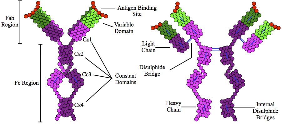
This is a depiction of immunoglobulin E (IgE) antibody which is elevated in allergic reactions.

What Is Allergic Rhinitis?

A llergic rhinitis is an immune disorder characterized by inflammation of the nasal mucosa by an IgE antibody reaction to an allergen.

An allergen is something that triggers an allergic immune response.

Q: What is the immune response?

A: Implicated in the response is an increase in T gamma-delta intraepithelial lymphocytes (IELs), which is a subset of pro-inflammatory T-cells located in the respiratory mucosa.  Lymphocytes are white blood cells.

When a person with allergic rhinitis breathes in an allergen such as pollen or dust, the body releases chemicals, including histamine that cause allergy symptoms. For example, hay fever involves an allergic reaction to pollen. A similar reaction occurs with allergy to mold, animal dander, dust, and other allergens that are breathed in.

What Is Allergic Rhinitis In Celiac Disease and/or Gluten Sensitivity?