Skip to content

Macroamylasemia

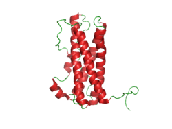
Depiction of pancreatic amylase molecule.

What Is Macroamylasemia?

[dropcap]M[/dropcap]acroamylasemia is an acquired enzyme disorder that causes elevated levels of the enzyme amylase (hyperamylasaemia) in the bloodstream. It is characterized by altered amylase molecules that have become abnormally bound with plasma proteins in the bloodstream, commonly IgG (immunoglobulin G) and/or IgA (immunoglobulin A).

The resulting molecule is too large to be properly filtered by the kidneys and excreted in the urine as would be normal unbound amylase, causing sustained elevation of amylase levels in the plasma.

With normal kidney function, a hyperamylasemia without an increase in urine amylase suggests the diagnosis of macroamylasemia, and is confirmed by identifying the macromolecular components.1

Amylase is a an enzyme produced by the pancreas and the parotid glands to digest starch in the diet. Comparatively small amounts are also produced by other organs.

Q: How do the pancreas and parotid glands deliver their amylase for digestion of starch?

A: The parotid glands deliver their amylase into the mouth. The action of amylase in the mouth starts the breakdown of starch as it is chewed and moistened with saliva.

After the starchy food is liquified in the stomach, the mass empties into the duodenum. Here pancreatic amylase is delivered through the common bile duct into the duodenum where it turns starch into maltose sugar. The next step to final digestion of starch is carried out by enzymes in the small intestinal lining that split maltose into its component sugar which is glucose.

Glucose is a simple sugar that can be absorbed into the bloodstream for use in the body. Humans cannot live without adequate glucose.

Distinquishing macroamylasemia from hyperamylasemia due to pancreatic disease is necessary to prevent needless treatment and investigation for pancreatitis.2

What Is Macroamylasemia In Celiac Disease and/or Gluten Sensitivity?

Sources:
  1. Torrent Vernetta A, Segarra Cantón O, Soler Palacín P, Segura Cardona RM, Infante Pina D. Macroamylasaemia in paediatrics. An Pediatr (Barc). 2008 Nov;69(5):439-41. []
  2. Isham CA, Ridgeway NA, Hedrick R, Cate JC 4th. Screening for macroamylase in a community hospital. Clin Chem. 1984 May;30(5):741-2. []

Macrolipasemia

Lipase Enzyme. Courtesy johnsonmatthey.
Lipase Enzyme. Courtesy Johnson Matthey Catalysts.

What Is Macrolipasemia?

[dropcap]M[/dropcap]acrolipasemia is a rare enzyme disorder characterized by altered molecules of lipase, a pancreatic enzyme needed to digest fats, that are abnormally bound with serum antibody proteins. These antibodies are commonly immunoglobulin G (IgG) and/or less likely immunoglobulin A (IgA).

Q: What happens when lipase is bound to immunoglobulins (IgA and/or IgG)?

A: The resulting molecule is too large to be filtered by the kidneys and excreted in the urine, consequently these abnormal molecules build up in the plasma causing sustained elevation of lipase levels called macrolipasemia.

Macrolipasemia occurs with or without macroamylasemia, which is the binding of immunoglobulin A and/or G to amylase, a pancreatic enzyme needed to digest starches.1

What Is Macrolipasemia In Celiac Disease and/or Gluten Sensitivity?

Sources:
  1. La Villa G, Pantaleo P, Tarquini R, Cirami L, Perfetto F, Mancuso F, Laffi G. Multiple immune disorders in unrecognized celiac disease: a case report. World J Gastroenterol. 2003;9(6):1377-1380, Available at: http://www.wjgnet.com/1007-9327/9/1377.asp. Accessed Jan 3, 2005. []

Hemochromatosis (Iron Overload Disease)

Hemochromatosis
Microscopic View of Hemochromatosis Stained Blue. Courtesy Wikimedia

What Is Hemochromatosis?

[dropcap]H[/dropcap]emochromatosis, also called iron overload liver disease, is a common inherited disease in the Caucasian population that is characterized by increased iron deposition within the tissues (overload) associated with injury to them.

Hemochromatosis is an autosomal recessive disease, meaning a pair of abnormal genes are inherited from each parent. Parents are unaffected because they carry only one gene.

Q: Where is iron deposited?

A: In hemochromatosis, more iron is absorbed from the small intestine than is needed by the body. Subsequently, because the body has no satisfactory means to release iron overload, excess iron is deposited in various organs such as the liver causing cirrhosis, joints causing arthritis, and the pancreas causing diabetes mellitus.1

Consumption of alcoholic drinks with food should be avoided because alcohol increases leaky gut and greatly increases the absorption of iron. On the flip side, eggs and foods containing calcium such as milk and cheese are beneficial for hemochromatosis because they impair the absorption of iron. Another iron inhibitor is food that contains phytic acid which includes the bran and outer layer of grains, seeds, nuts, peas, beans, and lentils.

Iron overloading, as measured by a random (non-fasting) elevated transferrin saturation value, is estimated to occur in 1 to 6 people per 100 in the United States according to the CDC (Center for Disease Control and Prevention). Too much iron increases the risk for metabolic syndrome, type 2 diabetes mellitus, cancer, liver disease, and osteoporosis.

Medical treatment to remove excess iron from the body uses phlebotomy that draws blood through a vein and/or certain injectable drugs (deferoxamine and deferairox) that bind to iron.

What Is Hemochromatosis In Celiac Disease and/or Gluten Sensitivity?

Sources:
  1. www.irondisorders.org []

Immune Thrombocytopenic Purpura

Photo of Skin Rash in ITP Courtesy Wikipedia.

What Is Immune Thrombocytopenic Purpura?

[dropcap]I[/dropcap]mmune thrombocytopenia purpura (ITP), formerly called idiopathic, is an immune mediated bleeding disorder characterized by destruction of circulating platelets (thrombocytes) by autoantibodies in the presence of normal bone marrow and without other conditions that cause bleeding problems including many medications.

Q: How do autoantibodies destroy platelets?

A: Autoantibodies bind to normal platelet surface antigens (targets) which forms a complex that is cleared from blood by the reticuloendothelial system. Consequently, this condition results in an abnormal decrease in the number of platelets circulating in blood plasma.

Platelets are cells that are needed to stop bleeding. A normal platelet count is in the range of 150,000 to 450,000. With ITP, the platelet count is less than 100,000. By the time significant bleeding occurs, the patient may have a platelet count of less than 10,000. The lower the platelet count, the greater the risk of bleeding. 

Because of inability of blood to properly clot due to lack of sufficient platelets, a distinctive purplish rash develops under the skin from blood leakage and an appearance of many small red spots of varying size can cover the skin. Depending on the number of platelets, there may be minimal to extensive bleeding in various parts of the body.  Bleeding in the brain is the worst complication, causing death in approximately 1% of affected children and 5% of affected adults.

Immune thrombocytopenic purpura is common in childhood, on average 50 cases per 1,000,000 per year with same incidence in boys as girls. It may follow a viral infection or immunization and is caused by an inappropriate response of the immune system to cross react against the body’s platelets.1

The incidence of immune thrombocytopenic purpura in adults is approximately 66 cases per 1,000,000 per year.  Older persons and persons with a previous history of hemorrhage are at increased risk of severe bleeding in adult ITP.  

Spontaneous remission occurs in most children (80%).  Remission in adults usually requires treatment. 

What Is Immune Thrombocytopenic Purpura In Celiac Disease and/or Gluten Sensitivity?

Sources:
  1. Dogan M, Sal E, Akbayram S, Peker E, Cesur Y, Oner AF. Concurrent celiac disease, idiopathic thrombocytopenic purpura and autoimmune thyroiditis. Clin Appl Thromb Hemost. 2011 Nov-Dec;17(6):E13-6. doi: 10.1177/1076029610378502. Epub 2010 Aug 3. []

Erythroblastopenia, Transient

Courtesy of slideshow.net

What Is Transient Erythroblastopenia?

[dropcap]T[/dropcap]ransient erythroblastopenia is a rare disorder of red blood cell formation characterized by brief, reversible disappearance of erythroblasts (red blood cell precursors) in the bone marrow of children.

Q: What do the red blood cells look like?

A: Circulating red blood cells appear normal so that the anemia which develops is a normocytic (normal cellular) anemia in contrast to microcytic (small cell) anemia in iron deficiency or macrocytic (large cell) anemia in vitamin B12 or folic acid deficiency.

Because new red blood cells are failing to mature, packed red blood cell transfusions may be necessary in severe anemia.

In adults, this disorder is called pure red blood cell anemia.

What Is Transient Erythroblastopenia In Celiac Disease and/or Gluten Sensitivity?

Hyperprolactinemia (Excess Prolactin Hormone)

Prolactin Structure. Courtesy Boris TM at Wikipedia.

What Is Hyperprolactinemia?

[dropcap]H[/dropcap]yperprolactinemia is an excess secretion of prolactin hormone causing an elevated blood level that is characterized by its effects on reproduction: in females causing  amenorrhea and in males causing reduced sexual potency.

Q: What is prolactin?

A: Prolactin is a hormone produced by the the anterior pituitary gland that is transported by the bloodstream to stimulate androgen hormone production in male and estrogen hormone production in female sex glands. However, too much prolactin inhibits hormone production of estrogen in women and testosterone in men.

In pregnant females upon delivery of a baby, prolactin stimulates breast development and milk production for the purpose of feeding the infant. Sucking by the infant stimulates the release of prolactin which keeps the milk supply adequate.

Prolactin also promotes autoimmune mechanisms. Prolactin interferes specifically with B cell tolerance induction, enhances proliferative response to antigens and mitogens and increases the production of immune globulins, cytokines and autoantibodies.1

Other conditions that can stimulate prolactin release include nutritional deficiencies, thyroid stimulating hormone (TSH) produced by the pituitary gland, stress, exercise, and anterior pituitary tumors.

What Is An Elevated Prolactin Level In Celiac Disease and/or Gluten Sensitivity?

Sources:
  1. Shelly S, Boaz M, Orbach H. Prolactin and autoimmunity. Autoimmun Rev. 2012 May;11(6-7):A465-70. doi: 10.1016/j.autrev.2011.11.009. []