Skip to content

DHA (Docosahexaenoic Acid) Deficiency

Smoked Salmon...Brain Food that's Good for the Eyes!
Salmon…Brain Food That’s Good for Your Eyes, Heart, Blood Vessels, Blood Sugar, Liver, Muscles, and Fights Inflammation!

What Is DHA?

[dropcap]D[/dropcap]ocosahexaenoic acid (DHA) is an essential omega-3 fatty acid that is abundant in the brain, being crucial in brain structure. As such DHA is a key component of neuronal membranes together with arachidonic acid (a major opposing omega-6 fatty acid), making up 15-20% of the brain’s dry mass.

This polyunsaturated fatty acid is obtained from fish sources of food.

In healthy human volunteers, positron emission tomography (PET) has shown that the normal human brain consumes 4.6 mg/day of DHA.1

DHA is particularly concentrated in highly active membranes such as nerve synapses (junctions) and photoreceptors in the eye (retina).

Q: How much DHA is in the retina?

A: DHA makes up more than 30% of the retina.2

In other roles, DHA is an  important building material for the eicosanoids, a large group of highly bioactive hormone-like substances including prostaglandins, leukotrienes, and thromboxanes that are involved in blood clotting, inflammation, and vasoconstriction.

DHA has been shown to increase insulin sensitivity as opposed to the opposite problem of insulin resistance, to improve muscle mass, and protect against non-alcoholic fatty liver disease.3

Egert et al. in a study of people aged 19 to 43 years with normal cholesterol showed that DHA intake significantly increased serum HDL (good) cholesterol. Also, DHA significantly decreased fasting serum triglycerides.4

What Is DHA Deficiency In Celiac Disease and/or Gluten Sensitivity?

Sources:
  1. Rapoport SI. Brain arachidonic and docosahexaenoic acid cascades are selectively altered by drugs, diet and disease. Prostaglandins Leukot Essent Fatty Acids. 2008 Sep-Nov;79(3-5):153-6. Epub 2008 Oct 29. []
  2. Richardson AJ. The importance of omega-3 fatty acids for behavior, cognition, and mood. Scandinavian Journal of Nutrition. 2003;47(2):92-8 []
  3. Espinosa A, Valenzuela R, González-Mañán D, D’Espessailles A, Guillermo Gormaz J, Barrera C, Tapia G. Prevention of liver steatosis through fish oil supplementation: correlation of oxidative stress with insulin resistance and liver fatty acid content. Arch Latinoam Nutr. 2013 Mar;63(1):29-36. []
  4. Egert S, Kannenberg F, Somoza V, et al. Dietary alpha-linolenic acid, EPA, and DHA have differential effects on LDL fatty acid composition but similar effects on serum lipid profiles in normolipidemic humans. J Nutr. 2009;139:861–868. doi: 10.3945/jn.108.103861 []

Anemia, Iron Deficiency

Red Blood Cell Comparison. Courtesy medindia.com

What Is Iron Deficiency Anemia?

[dropcap]I[/dropcap]ron deficiency anemia is a blood cell disorder that is characterized by formation of small, pale red blood cells, causing tissue hypoxia. Hypoxia is the inability to meet the demands of the body for oxygen.

Q: Why do small, pale red blood cells cause tissue hypoxia?

A: Small, pale red blood cells (erythrocytes) cause tissue hypoxia because they are not able, as do normal erythrocytes, to pick up adequate oxygen from the lungs and carry it to cells that use oxygen.

Red blood cell production and function are dependent on a sufficient level of iron in the body and also the ability to use available iron to make hemoglobin in red blood cells.

Hemoglobin is a protein that binds oxygen in red blood cells to be carried by the bloodstream to cells throughout the body. In iron deficiency anemia,  hemoglobin in females is below 12.5g/dl (normal range is 12.5 to 16g/dl) and in males it is below 13.5g/dl (normal range is 13.5 to 17.5g/dl).

Iron must be obtained from the diet, since the body cannot make it, but there are various factors that can interfere with absorption and use in the body, causing anemia. Iron absorption from the gut first requires ionization, or gaining a positive electrical charge, in the strongly acidic environment of stomach juice. Ionized iron, only, can be absorbed in the duodenum, which receives the acidic contents of the stomach before it is neutralized further along.

Dietary iron can be heme or non-heme depending on the food source. Heme iron obtained only from animal food sources is absorbed into the bloodstream by active transport across the brush border (microvilli) which cover the multitudinous villi of the small intestinal lining.

Non-heme iron obtained from plants must bind with apoprotein after entering the enterocyte (surface cell of small intestinal lining) to be ferried to the underlying basolateral membrane and exited by active transport into the bloodstream.

Frequently, chronic anemia due to iron deficiency is accompanied by increased platelets, and this thrombocytosis resolves with iron repletion (normal iron level). Conversely, in severe iron deficiency anemia, patients may have thrombocytopenia (low platelets), which also resolves with iron therapy.1

What Is Iron Deficiency Anemia In Celiac Disease and/or Gluten Sensitivity?

Sources:
  1. Koury M and Rhodes M. How to approach chronic anemia. Hematology Am Soc Hematol Educ Program. 2012;2012:183-90. doi: 10.1182/asheducation-2012.1.183. []

Zincemia (Low Zinc Blood Level)

Zincemia low zinc level symptom of celiac disease and glutenWhat Is Zincemia?

[dropcap]Z[/dropcap]incemia means the zinc level in blood plasma is too low to meet metabolic needs of the body for this mineral.

Q: How important is a normal blood level of zinc?

A: A low blood level of zinc is characterized by widespread alterations in energy metabolism, growth, hemoglobin, carbon dioxide transport, hormone activity, insulin storage, many enzyme activities, prostaglandin function, collagen production, male fertility, protein synthesis, and vitamin A metabolism.

What Is Zincemia In Celiac Disease and/or Gluten Sensitivity?

Copper Deficiency

mmm...copper is in chocolate!
mmm…copper is in chocolate and nuts!

What Is Copper Deficiency?

[dropcap]C[/dropcap]opper is an essential trace element that is required for a number of enzymes which are necessary for normal metabolic function.

In the body almost all the copper is present as a component of copper proteins which are produced and controlled by the liver.

Q: How does the liver control copper?

A: The liver maintains proper copper balance by binding free copper to proteins and by excreting excess copper as part of bile that is then emptied into the intestinal tract and excreted in stool.

Metabolic balance studies have demonstrated that daily copper losses are approximately 1.3 mg/day.1

Among its specific functions listed below, copper is essential for energy production, blood and nerve functions, blood components, immunity, and collagen tissue. The copper enzyme, lysyl oxidase, is involved in the cross-linking of collagen in forming the framework for depositing calcium and other minerals to build and repair bone.

Animal studies suggest that adequate copper levels during pregnancy are critical to development of higher brain function in the offspring.2

What Is Copper Deficiency In Celiac Disease and/or Gluten Sensitivity?

Sources:
  1. Williams DM. Copper deficiency in humans. Semin Hematol. 1983 Apr; 20(2):118-28. []
  2. Fisgin T, Yarali N, Duru F, Usta B, Kara A. Hematologic manifestation of childhood celiac disease. Acta Haematol. 2004;111(4):211-4. []

Anemia, Folic Acid Deficiency  

Folic acid deficiency anemia gluten celiac disease symptom
Red Blood Cells.

What Is Folic Acid Or Folate Deficiency Anemia?

[dropcap]F[/dropcap]olic acid deficiency anemia, also called folate deficiency anemia, is a macrocytic anemia characterized by defective DNA synthesis of red blood cells that results from a lack of folate in the body.

Q: How does folate deficiency cause anemia?

A: Folates are a family of B vitamins and folic acid is an active form.

Folate is required for the formation of both red and white blood cells in the bone marrow and for their maturation.

Also, folate serves as a carrier in the formation of heme, which contains iron, and is the non-protein part of the hemoglobin molecule.1

Red blood cells provide oxygen to body tissues. When there are not enough red blood cells or when they cannot properly carry oxygen, the condition is called anemia. In folic acid deficiency anemia, the red blood cells are abnormally large. Such cells are called macrocytes (macro size cells). They are also called megaloblasts (mega size cells) as seen in the bone marrow where they are produced. This is why this macrocytic anemia is also called megaloblastic anemia.2

Tests that may be done to determine folate adequacy are complete blood count (CBC), red blood cell folate level, methylmalonic acid level, and homocysteine level. Folic acid deficiency anemia shows a decrease in red blood cell folate and/or serum folate levels and normal plasma methylmalonic acid level with elevated homocysteine blood level. These levels distinguish folic acid deficiency anemia from vitamin B12 deficiency anemia.3

What Is Folate Deficiency Anemia In Celiac Disease and/or Gluten Sensitivity?

Sources:
  1. Kathleen Mahan and Sylvia Escott-Stump, ed. Krause’s Food, Nutrition & Diet Therapy, 10th Edition. Philadelphia, PA. USA: W.B. Saunders Company, 2000. []
  2. http://www.ncbi.nlm.nih.gov/pubmedhealth/PMH0001578/ []
  3. Mark Beers and Robert Berkow. The Merck Manual, 17th Edition. Whitehouse Station, N.J. USA: Merck Research Laboratories, 1999. []

Abnormal Blood Values in Childhood

abnormal blood values children symptom celiac glutenWhat Are Abnormal Blood Values In Childhood?

[dropcap]A[/dropcap]bnormal blood studies in childhood are lab results showing blood abnormalities for blood cells and blood components.

What Are Abnormal Blood Values In Childhood in Celiac Disease?

Arachidonic Acid (AA) Deficiency

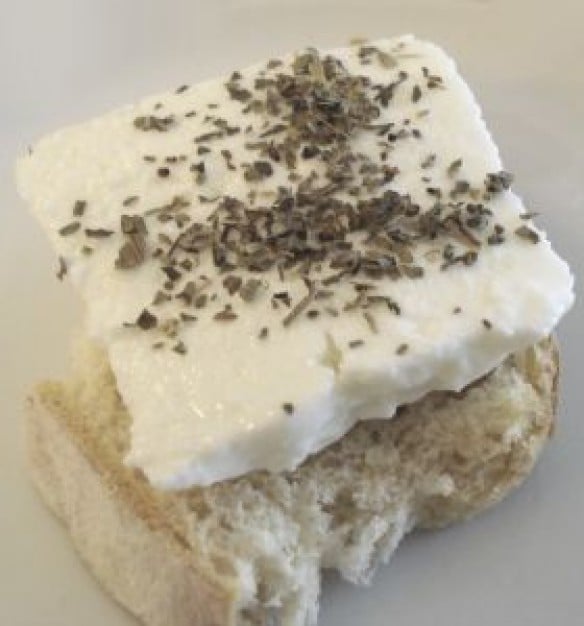
QueijoFresco.
QueijoFresco.

What Is Arachidonic Acid?

[dropcap]A[/dropcap]rachidonic acid is a major essential (must have/can’t make) omega-6 fatty acid.

Structurally, arachidonic acid is a key component of nerve membranes, together with docosahexaenoic acid (DHA), a major opposing omega-3 fatty acid, making up 15-20% of the brain’s dry mass and more than 30% of the retina.

In healthy human volunteers, positron emission tomography (PET) has shown that the normal human brain consumes 17.8 mg/day of arachidonic acid.1

Arachidonic acid is particularly concentrated in highly active membranes such as nerve synapses (junctions) and in photoreceptors in the eye retina.2

Arachidonic acid is an important building substance for the eicosanoids.

Q: What function do the eicosanoids have that are derived from arachidonic acid?

A: Eicosanoids are a large group of highly bioactive hormone-like substances including prostaglandins, leukotrienes, and thromboxanes that are involved in blood clotting, inflammation, and vasoconstriction. Eicosanoids derived from arachidonic acid are pro-inflammatory, pro-blood clotting, and constrict blood vessels in opposition to those derived from the omega-3 fatty acids which do the opposite in order to keep a balance in the body.

What Is Arachidonic Acid Deficiency In Celiac Disease and/or Gluten Sensitivity?

Sources:
  1. Rapoport SI. Brain arachidonic and docosahexaenoic acid cascades are selectively altered by drugs, diet and disease. Prostaglandins Leukot Essent Fatty Acids. 2008 Sep-Nov;79(3-5):153-6. Epub 2008 Oct 29. []
  2. Richardson AJ. The importance of omega-3 fatty acids for behavior, cognition, and mood. Scandinavian Journal of Nutrition. 2003;47(2):92-8. []

Vitamin B2 (Riboflavin) Deficiency

Chicken Kabobs on the Grill...umm Riboflavin.
Chicken Kabobs on the Grill…umm Riboflavin.

What Is Riboflavin?

[dropcap]R[/dropcap]iboflavin, also called vitamin B2, is fundamentally required for the breakdown of carbohydrates, amino acids, and fats in metabolism, enables oxygen to be used by cells, and acts as a component of more than a dozen enzymes involved in energy production and use.

Some enzymes needing riboflavin also require magnesium to properly function.

In other activities, riboflavin is required for healthy cardiovascular and nervous systems, eye health, and supports antioxidant protection.  Functions are more fully described below.

Urinary excretion of riboflavin cannot be detected when vitamin intake is below the required levels. On the other hand, when intake exceeds saturation in the body, the vitamin and/or its metabolites are actively excreted into urine to prevent excessive toxicity of the vitamins.1

There is no specific name for riboflavin deficiency disease.

What Is Riboflavin Deficiency In Celiac Disease and/or Gluten Sensitivity?

Sources:
  1. Shibata K, Hirose J, Fukuwatari T. Relationship Between Urinary Concentrations of Nine Water-soluble Vitamins and their Vitamin Intakes in Japanese Adult Males. Nutr Metab Insights. 2014 Aug 5;7:61-75. doi: 10.4137/NMI.S17245. []

Prolonged Prothrombin Time

bloodvialWhat Is Prolonged Prothrombin Time?

[dropcap]P[/dropcap]rolonged prothrombin time (PT) is a laboratory blood test result showing that blood clots too slowly which makes the patient subject to abnormal bleeding.

Q: What does the prothrombin time (PT) test measure?

A: The prothrombin test measures the clotting ability of blood protein factors I, II (prothrombin), V, VII, and X. If any of these factors are too low, it takes longer than normal for the blood to clot. Prothrombin is a vitamin K dependent factor meaning a deficiency of vitamin K will cause low prothrombin.

Blood is drawn into a blood collection tube with a light blue stopper which has a buffering additive. Tubes must be completely filled.

What Is Prolonged Prothrombin Time In Celiac Disease and/or Gluten Sensitivity?

Anti-Endomysium Antibodies (EMA) Present In Blood

Smooth muscle. Courtesy pathguy.com
Smooth Muscle. Courtesy pathguy.com

What Are Anti-Endomysium Antibodies?

[dropcap]A[/dropcap]nti-endomysium antibodies (EmA) are connective tissue autoantibodies produced in persons who have inherited the genes for celiac disease, an autoimmune disease, and are reacting to gluten in their diet.

Autoantibodies are abnormal in that they attack the body’s own tissue.

Q: What is endomysium?

A: Endomysium is the delicate connective tissue that surrounds individual muscle fibers. The autoantigen, or target, that stimulates the autoimmune response is the naturally occuring enzyme in endomysium called tissue transglutaminase (tTG), or more specifically tranglutaminase-2 (TG2). Anti-tissue transglutaminase antibodies recognize the same antigen as EmA, from which they differ in terms of detection method.

Anti-endomysium antibodies (EmA) are tested by the indirect immunofluorescence method and directed against “reticulin-like” fibres in connective tissue around smooth muscle fibres in the esophagus, liver, stomach, and bladder of monkeys, in the sections of the jejunum and kidneys of rats and in sections of the human umbilical cord. In comparison, for the determination of anti-tissue transglutaminase IgA and IgG antibodies, ELISA with human extractive or recombinant transglutaminase is recommended.1

EmA‐binding patterns in serum samples from patients with celiac disease have proved to be exclusively TG2‐targeted and the correlation between EmA and TG2 antibodies is therefore good. Evidence shows that celiac autoantibodies are produced in the small‐bowel mucosa.2

What Are Anti-Endomysium Antibodies In Celiac Disease and/or Gluten Sensitivity?

Sources:
  1. Trigoni E, Tsirogianni A, Pipi E, Mantzaris G, Papasteriades C. Celiac disease in adult patients: specific autoantibodies in the diagnosis, monitoring, and screening. Autoimmune Dis. 2014;2014:623514. doi: 10.1155/2014/623514. []
  2. Salmi TT, Collin P, Korponay-Szabó IR, Laurila K, Partanen J, Huhtala H, Király R, Lorand L, Reunala T, Mäki M, Kaukinen K. Endomysial antibody‐negative coeliac disease: clinical characteristics and intestinal autoantibody deposits.Gut. 2006 Dec;55(12):1746-53. []