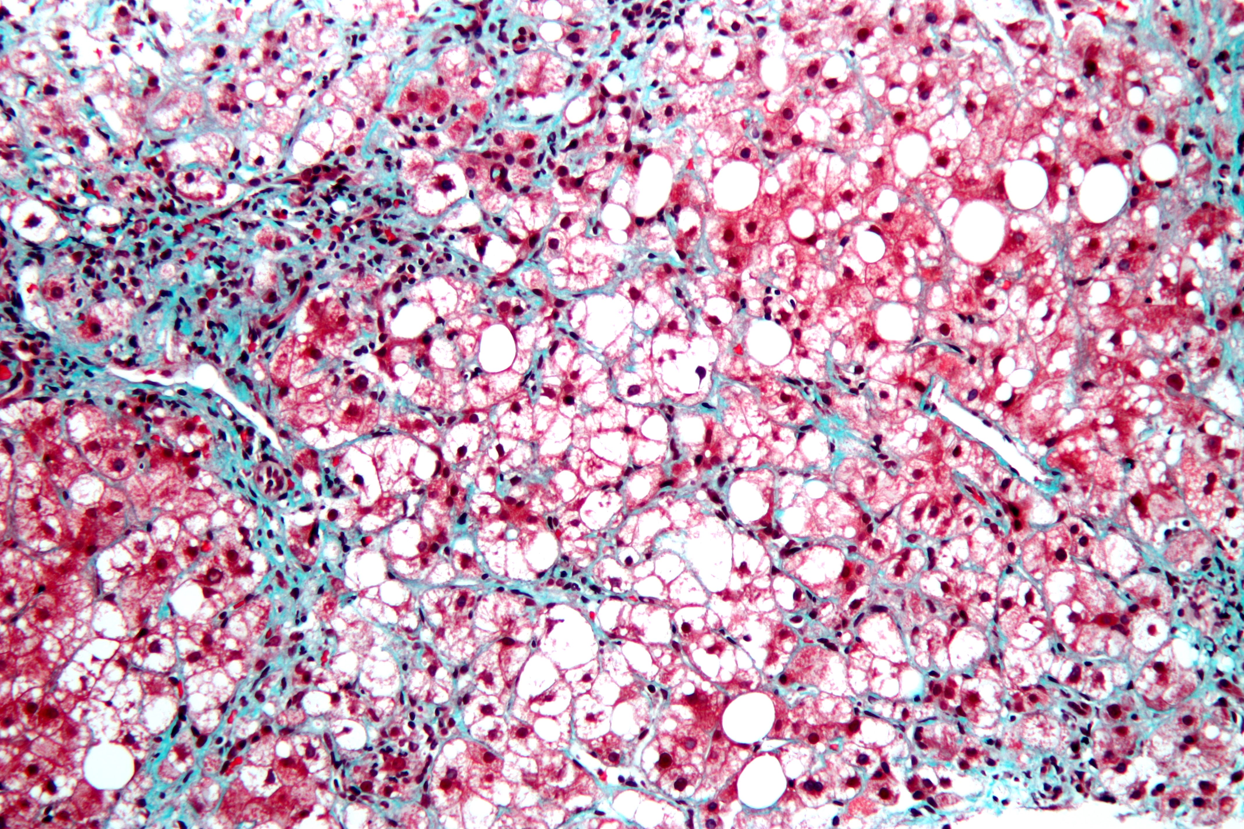
This is a stained liver biopsy sample showing advanced cellular changes in non-alcoholic fatty liver disease. Blue is fibrosis. White is fat accumulation in degenerated cells. Courtesy of Nephron’s work.
What Is Non-Alcoholic Fatty Liver Disease?
N on-alcoholic fatty liver is a non-inflammatory liver disorder characterized by degenerative changes in the liver caused by excessive accumulation of lipid (fat) in hepatocytes (liver cells) that is called free fatty acid-generated lipotoxicity.
Non-alcoholic fatty liver shows an increase in liver enzymes called transaminases.
Q: What are the enzymes that increase?
A: The transaminases that increase are ALT and AST. ALT is the abbreviation for alanine aminotransferase enzyme and AST is the abbreviation for aspartate aminotransferase enzyme. They are commonly measured in blood tests to determine liver function and when elevated indicate inflammation.
What Is Non-Alcoholic Fatty Liver In Celiac Disease and/or Gluten Sensitivity?
P rurigo nodularis is a chronic dermatitis characterized by hard, dry, deep seated, intensely itchy papules (small bumps like pimples) and/or nodules (large bumps) that erupt most commonly on the arms, legs, and back.
Papules and nodules vary in number and may become infected after picking or scratching.
Q: Does the itching go away?
A: New nodules develop from time to time, and existing nodules may remain itchy indefinitely, although some may regress spontaneously and leave scars. In most cases, the disease runs a very protracted course with exacerbations and remissions.1
Prurigo nodularis is an unusual disorder of unknown etiology, which is notoriously resistant to therapy. A variety of systemic conditions have been reported to be associated with prurigo nodularis. However, the mechanism by which these disorders may trigger prurigo nodularis is unknown.2
It has been shown to be associated with malnutriton and infection such as tonsillitis, which resolved after removal of tonsils.3
What Is Prurigo Nodularis In Celiac Disease and/or Gluten Sensitivity?
Sources:
Katotomichelakis M, Balatsouras DG, Bassioukas K, Kontogiannis N, Simopoulos K, Danielides V. Recurrent prurigo nodularis related to infected tonsils: a case report. J Med Case Rep. 2008 Jul 24;2:243. doi: 10.1186/1752-1947-2-243. [↩]
Lee MR, Shumack S. Prurigo nodularis: a review. Australas J Dermatol. 2005 Nov;46(4):211-18; quiz 219-20. [↩]
Katotomichelakis M, Balatsouras DG, Bassioukas K, Kontogiannis N, Simopoulos K, Danielides V. Recurrent prurigo nodularis related to infected tonsils: a case report. J Med Case Rep. 2008 Jul 24;2:243. doi: 10.1186/1752-1947-2-243. [↩]
The spleen is the darkish oval organ in the lower middle of this photo.
What Is Hyposplenism?
H yposplenism is the condition resulting from having lost spleen tissue, called atrophy of the spleen. Spleen atrophy impairs splenic functions or activities because there are insufficient tissues to do the work required.
Q: What splenic functions are impaired?
A: The spleen, apart from acting as a phagocytic filter, thus removing aging and damaged cells, is crucial in regulating immune homeostasis by linking innate and adaptive immunity, and in protecting against infections by encapsulated bacteria.1
Impaired function of the spleen therefore increases risk of infections with encapsulated bacteria because of inability to mount a proper defense and to filter and remove bacteria from the circulation.
The spleen is a highly vascular and solid organ about the size of a fist. It has a delicate structure inside that is enclosed by fibrous, elastic layers consisting of connective tissue.
The tissues within are made up of two different types of tissues, called white pulp and red pulp. White pulp carries out lymphoid functions. Red pulp filters and cleanses the blood. The spleen is situated above the stomach on the left side of the upper abdomen and firmly fixed in place by ligaments and ribs.
The spleen is an important organ of the lymph system, having the largest collection of lymph tissue in the body. It functions to produce antibodies (immunoglobulins) and white blood cells (T-cells and B-cells), help control the amount of blood in the body, keep body fluids in balance, destroy and filter out old and damaged cells2 and salvage the iron needed for producing new blood cells, and lastly, clear bacteria through production of substances that enable phagocytosis (engulfing bacteria and other unwanted particles, such as antigens, from blood).
Children and adults with hyposplenism are at risk for overwhelming infections. Management of hypospenism is directed towards preventing pneumonia by immununization against pneumonia and meningitis and treating bacterial infections as they arise, which may require hospitalization. For some patients, life-long treatment with antibiotics, such as erythromycin and penicillin, are recommended.
What Is Hyposplenism In Celiac Disease and/or Gluten Sensitivity?
Sources:
Di Sabatino A, Brunetti L, Carnevale Maffè G, Giuffrida P, Corazza GR. Is it worth investigating splenic function in patients with celiac disease? World J Gastroenterol. 2013 Apr 21;19(15):2313-8. doi: 10.3748/wjg.v19.i15.2313. [↩]
S teatorrhea is a condition of abnormal stool composition and consistency that is due to high fat content because fat from food is not digested and absorbed into the body but rather passes out with stool.1
Q: How does fatty stool look?
A: The stool in steatorrhea appears pale, bulky and may float in the toilet. If stool is loose, it varies from a pudding consistency to whipped cream. If it is solid, it can vary from thin sqiggles to big turds that smear the sides of the toilet bowl. All types have a foul smell.
Fat of any kind must be digested, or broken down, by enzymes into fatty acids and glycerol and then these parts only are absorbed through the small intestinal lining into the body. The failure to digest or absorb fats deprives the body of these foodstuffs needed for many functions such as blood clotting and vital cell parts such as eye and brain structure.
Fat malabsorption produces malabsorption of vitamins A, D, E, and K because these vitamins can only be absorbed along with fatty acids.
What Is Steatorrhea In Celiac Disease and/or Gluten Sensitivity?
Sources:
Murray JA, The widening spectrum of celiac disease. American Journal of Clinical Nutrition. Mar 1999;69 (3):354-365. [↩]
Image of Pancreas From Gray’s Anatomy. Courtesy Wikipedia.org
What Is Pancreatic Insufficiency?
P ancreatic insufficiency is a disorder characterized by insufficient exocrine production of pancreatic enzymes for normal digestion of fats, proteins, and carbohydrates that results in maldigestion of these foodstuffs.
Pancreatic insufficiency also causes malabsorption of the fat-soluble vitamins: vitamin A, vitamin D, vitamin E, and vitamin K.
Q: What are the exocrine enzymes produced by the pancreas?
A: Exocrine enzymes produced by the pancreas include amylase for the digestion of carbohydrates, lipase for the digestion of fats, and protease for the digestion of proteins.
While lipases and amylase are secreted in the active form, proteases are secreted as pro-enzymes (need to be activated). Trypsinogen is converted to its active form trypsin in the duodenum by enterokinase, a protease secreted by the enterocytes (surface cells that line the duodenum), and trypsin in turn activates the other pancreatic proteases.1
Pancreatic enzymes are released by the pancreas into the internal pancreatic duct that empties into the common bile duct from which they are pumped through the Sphincter of Odi directly into the duodenum as needed. Tthe Sphincter of Odi is a strong circular muscle that controls the entrance of pancreatic enzymes into the duodenum.
After a meal, enzyme secretion into the duodenum increases quickly reaching peak output within the first 20 to 60 minutes, then decreasing to a stable level before reaching an interdigestive level at the end of the digestive period, that is, about 4 hours after meal intake.2
Medical treatment is with medication that contains enzymes to break down carbohydrates, fats, and protein in food at mealtime. This treatment is safe, effective, and has few side effects.
What Is Pancreatic Insufficiency In Celiac Disease and/or Gluten Sensitivity?
Sources:
Fieker A, Philpott J, Armand M. Enzyme replacement therapy for pancreatic insufficiency: present and future. Clin Exp Gastroenterol. 2011;4:55-73. doi: 10.2147/CEG.S17634. [↩]
Fieker A, Philpott J, Armand M. Enzyme replacement therapy for pancreatic insufficiency: present and future. Clin Exp Gastroenterol. 2011;4:55-73. doi: 10.2147/CEG.S17634. [↩]
C achexia is a state of ill health involving deteriorating body composition that is characterized by general malnutrition and loss of lean tissue such as muscle.
Q: What are typical findings in cachexia?
A: Arm muscle triceps (the muscles at the back of the upper arm), skin folds, subscapular skin folds, fat area index, and bone mineral content are significantly lower than normal.
Cachexia may develop in protein-losing enteropathy such as celiac disease, chronic or severe infection such as pneumonia, tuberculosis, malaria, or many chronic diseases such as heart failure and cancer.
Cachexia can develop in persons of any age.
What Is Cachexia In Celiac Disease and/or Gluten Sensitivity?
A phthous ulcers, also called canker sores, are a chronic disorder of soft mouth tissue characterized by small, painful purpuric, papular, or erosive lesions that are often surrounded by erythematous (red) margins.1
Q: What soft mouth tissues develop canker sores?
A: Canker sores can erupt on the mucosal surface of the inside lip, sides of mouth, under the tongue and along the side of the tongue. When they are forming, the area swells into a papule, or small bump. Later, the surface erodes and a crater forms with firm pus.
What Are Aphthous Ulcers In Celiac Disease and/or Gluten Sensitivity?
Sources:
Lahteenoja H, Toivanen A, Viander M, Maki M, Irjala K, Raiha I, Syrjanen S. Oral mucosal changes in coeliac patients on a gluten-free diet. European Journal of Oral Sciences. Oct 1998;106(5):899,8p. [↩]
D ental enamel defects (DED) comprise a variety of abnormalities in the dental enamel of the second or permanent teeth and of primary or baby teeth.
Teeth may appear with pits, thin or missing enamel, non-white enamel (yellow, orange, or gray colored teeth), and demarcated opacities (very white spots) even if teeth are white.
Q: What is enamel?
A: Dental enamel is the shiny, hard, white, dense, inorganic substance covering the crowns of the teeth. The crown is the tooth portion above the gum. Under the enamel is hard dentin which surround the living pulp. In teeth with missing enamel, the areas with exposed dentin appear dull and tan colored.
What Are Dental Enamel Defects In Celiac Disease and/or Gluten Sensitivity?
B leeding and/or swollen gums is an inflammatory disorder characterized by changes in gum integrity that may result in tooth loss.
Sponginess of the gum (swelling) indicates inflammation, called gingivitis.
Q: What causes gingivitis?
A: Gingivitis that is caused by inadequate dental hygiene shows as pockets of swollen gum between or along the base of teeth where sticky plaque was allowed to build up. Plaque holds bacteria that inflames the gums where it occurs.
Gingivitis that is caused by vitamin C deficiency, pregnancy, certain drugs such as the anti-epileptic drug phenytoin, and leukemia affects the whole gum irrespective of mouth care.
What Are Bleeding / Swollen Gums In Celiac Disease and/or Gluten Sensitivity?
L aryngospasm is an acute disorder of the larynx that is characterized by life-threatening spasms of the muscles of the larynx located in the throat.
Laryngospasm is a rare feature of hypocalcemia (low blood calcium level) characterized by severe alteration in nerve conduction and muscle contraction.
What Is Laryngospasm In Celiac Disease and/or Gluten Sensitivity?
Cancel your subscription?
We're sorry to see you go.
Please select a reason for cancellation.
Please provide additional details.
Your subscription is cancelled
You won't be billed again. If you change your mind, you can subscribe again at any time.
Cancelled
Cancellation request submitted
We've received your request. Your subscription remains active while we process it. We'll email you once it's confirmed.
Cancellation pending
Pause subscription
Your subscription payments will be paused. Your existing access will remain active while your subscription is paused.
Please choose a valid future date.
Resume subscription
Your subscription payments will resume on the normal billing schedule.
Access Content
Affiliate Terms
By joining our Affiliate Program, you agree to the following terms:
1. Eligibility: You must be 18 years or older to participate. We reserve the right to refuse service to anyone for any reason at any time.
2. Promotional Guidelines: Affiliates must not engage in misleading advertising or spam. Promotions must accurately represent our brand and products.
3. Affiliate Links: You must use your unique tracking link to receive credit for referred sales.
4. Prohibited Use: You may not use our branding in a way that may confuse customers or misrepresent your relationship with us.
5. Termination: We reserve the right to terminate your account at any time for breach of these terms.
6. Changes to Agreement: We may update these terms at any time. Continued participation implies acceptance of any changes.
By continuing, you agree to the terms of this Affiliate Agreement.



![Aphthous_ulcer[1]](https://glutenfreeworks.com/health/wp-content/uploads/sites/10/Aphthous_ulcer1.jpg)
![Enamel_celiac[1]](https://glutenfreeworks.com/health/wp-content/uploads/sites/10/Enamel_celiac1.jpg)

