Skip to content

Mesenteric Lymph Node Cavitation And Hyposplenism

GERD Gluten Celiac Disease
Arrows Show Abnormal Movement of Gastric Acid in Gastroesophageal Reflux Disease.

What Is Gastroesophageal Reflux Disease (GERD)?

[dropcap]G ERD is an upper digestive disorder that is characterized by a decrease in lower esophageal sphincter pressure (LES,) which allows the abnormal reflux or backflow of stomach contents into the esophagus. It is also called erosive esophagitis or reflux esophagitis and is the most common disorder of the esophagus.

The esophagus is a muscular tube that transports swallowed substances to the stomach. It begins at the cricoid cartilage (Adam’s apple) as a continuation of the pharynx and ends at the lower esophageal sphincter (LES).

The lower esophageal sphincter is located at the junction of the esophagus and the stomach. It functions like a circular band to tighten after food is ingested in order to prevent its going back up the esophagus.

Q: How does reflux damage the esophagus?

A: Damage to the lining of the esophagus is induced by the caustic, chemical action of acid and pepsin in gastric juice and, in severe cases, also bile salts, that back upwards from the stomach through an impaired LES. Gastric acid combined with pepsin or bile salts seems to be more harmful to the esophageal epithelial layers than gastric acid alone.1

Pepsin is normally produced by the stomach to dissolve protein in swallowed food. Unfortunately, when the esophagus is inflamed, pepsin will act on it to break down the protein in its sore wall. These sores are called erosions.

Importantly, refluxate to the esophagus in patients with acid suppression therapy is different from those in patients without. Higher levels of secondary bile acids are detected in patients with acid suppression therapy. Even if acid suppression is successful, weakly acidic reflux with bile acids can damage the esophagus.1

Damage starts at the luminal surface (inside where food passes through) of the squamous epithelium (tough surface cells) and progresses through the underlying layers into the submucosa.

One of the primary functions of the esophageal epithelium is to protect the underlying tissue from mechanical and chemical damage by acting as a barrier. The epithelial layers of the distal esophagus need to withstand reflux from the stomach and its contents. When the epithelium fails to protect the underlying tissue from this damage, it leads to erosions, esophagitis, and may lead to Barrett’s esophagus.1

Barrett’s esophagus and esophageal small cell cancer are severe complications of GERD that can be fatal.

GERD can result from too much, or more commonly, too little stomach acid.

What Is Gastroesophageal Reflux Disease (GERD) In Celiac Disease and/or Gluten Sensitivity?

Sources:
  1. Chen X, Oshima T, Tomita T, Fukui H, Watari J, Matsumoto T, Miwa H. Acidic bile salts modulate the squamous epithelial barrier function by modulating tight junction proteins. Am J Physiol Gastrointest Liver Physiol. 2011 Aug;301(2):G203-9. doi: 10.1152/ajpgi.00096.2011. Epub 2011 May 26. [] [] []

Pulmonary Hemosiderosis, Idiopathic  (Lane-Hamilton Syndrome)

intest (2)What Is Obnoxious Gas?

[dropcap]O bnoxious gas, or flatus, is gas that is not only offensive when passed but also lingers in the air longer than ordinary gas does.

Gas is a natural digestive product within the colon that is composed mainly of hydrogen and carbon dioxide gases. These gases are given off in the necessary bacterial breakdown of undigested fermentable food entering the colon from the small intestine.

Q: What makes gas obnoxious?

A: The production of obnoxious gas depends on the type and quantity of undigested food residue that is passed into the colon from the small intestine, dysfunctional motility instead of normal peristalsis, and dysbiosis. Dysbiosis is the condition of  having unhealthy or insufficient populations of microbes responsible for digesting (fermenting) foodstuffs in the lower gut or colon. 

Gases produced by intestinal microbes may modulate intestinal motor function (muscle movement) in individuals with functional bowel disease. Methane, produced by enteric bacteria in the human gut, is associated with slowed intestinal transit and constipation.1

What Is Obnoxious Gas In Celiac Disease and/or Gluten Sensitivity?

Sources:
  1. Jahng J, Jung IS, Choi EJ, Conklin JL, Park H. The effects of methane and hydrogen gases produced by enteric bacteria on ileal motility and colonic transit time. Neurogastroenterol Motil. 2012 Feb;24(2):185-90, e92. doi: 10.1111/j.1365-2982.2011.01819.x. Epub 2011 Nov 20.

    Methane is produced in the colon by intestinal methanogens (microbes) that metabolize hydrogen, one of the end products of normal anaerobic (meaning without oxygen) bacterial fermentation.  Fermentation of the undigested starchy part of carbohydrates produces hydrogen in the intestine, which is the food for methane production by intestinal methanogens.

    Hydrogen and methane are excreted in the flatus and in breath giving the opportunity to indirectly measure their production using breath testing. ((Triantafyllou K, Chang C, Pimentel M. Methanogens, Methane and Gastrointestinal Motility. J Neurogastroenterol Motil. 2014 Jan;20(1):31-40. Epub 2013 Dec 30. []

Hashimoto’s Disease (Autoimmune Thyroiditis Causing Hypothyroidism)

hypoglycemia symptom of celiac disease and glutenWhat Is Hypoglycemia?

Hypoglycemia means the level of glucose within cells is too low to meet metabolic needs of the body for this essential sugar.

Q: What are the metabolic needs for glucose?

A: Glucose is the most important simple sugar in human metabolism mainly because it is the primary source of energy for most cells of the body.

Energy contained in the glucose molecule is obtained by the body from its reaction with oxygen (oxidation). This oxidation reaction occurs in power producing mitochondria structures that are located within cells.1

Hypoglycemia is characterized by alterations in neurologic, metabolic and muscular functions:

  1. Neurologic function because brain tissue is particularly dependent on glucose for energy,
  2. Metabolic function of glucose-dependent tissues which include red blood cells, white blood cells, bone marrow, eye, inner heart of the kidney, and peripheral nerves because these tissues cannot metabolize fatty acids as an alternate source of energy, and
  3. Muscle function because muscle cells continually require glucose for energy production.

Glucose is made available to cells through the regulating action of insulin, a hormone produced by specialized cells located on the surface of the pancreas.

What Is Hypoglycemia In Celiac Disease and/or Gluten Sensitivity?

Sources:
  1. http://hyperphysics.phy-astr.gsu.edu/hbase/organic/sugar.html accessed 11 14 12 []

Diarrhea, Chronic

chronic diarrhea gluten celiac disease symptomWhat Is Chronic Diarrhea?

[dropcap]C[/dropcap]hronic diarrhea is an intestinal motility disorder characterized by 1) alteration in stool formation causing loose to fluid movements and quantity of movements with or without abdominal pain and 2) interference with normal carbohydrate salvage by the intestinal microbe population.

The severity of diarrhea is determined by the frequency and quantity of fluid lost.

Q: What is carbohydrate salvage by microbes in the colon?

A: Carbohydrate salvage in the colon is the necessary process whereby billions of microbes normally present in the colon work to breakdown undigestible carbohydrate foodstuffs such as fiber that continually arrive from the small intestine.

The microbial action releases energy to the body that would otherwise be lost with defecation, generates short-chain fatty acids, and stimulates sodium and fluid absorption. In this important process, butyrate and proprionate are produced which nourish the colonocytes (cells that line the colon), and acetate is produced for the liver.

While diarrhea may be a common symptom of small bowel mucosal disease, the consequent malabsorption can lead to substantial malnutrition and nutrient deficiencies. The small intestine, unlike the colon, has been relatively inaccessible, and systematic evaluation is often necessary to identify and treat small intestinal mucosal diseases that lead to diarrhea. All patients with severe diarrhea or diarrhea associated with features suggestive of malabsorption may have a disease of the small intestinal mucosa that requires careful evaluation and targeted management.1

What Is Chronic Diarrhea In Celiac Disease and/or Gluten Sensitivity?

Sources:
  1. Murray JA1, Rubio-Tapia A. Diarrhoea due to small bowel diseases. Best Pract Res Clin Gastroenterol. 2012 Oct;26(5):581-600. doi: 10.1016/j.bpg.2012.11.013. []

Lymphoma, Enteropathy-Associated T-Cell (EATL) 

A real photomicrograph of carcinoma of the esophagus. Panorama of 6 photos of a slide at 40x through the microscope. Some areas may appear blurry due to shallow DOF.

What Is Cancer Of The Esophagus?

[dropcap]C ancer of the esophagus is a malignancy arising in the stratified squamous cell lining of any part of the esophagus and having a poor prognosis.

This tumor first invades the deeper layers of the esophagus which includes the layer beneath the mucosa (submucosa) and muscle before it may invade structures close to it including the aorta and trachea. Later, it may spread to organs further away such as the liver, lungs and bones.

Q: What is the stratified squamous cell lining of the esophagus?

A: Stratified squamous cells are flat epithelial cells that are composed of several layers, called the epithelium. This type of epithelium forms the surface mucosa that lines the inside of the esophagus.

The esophagus itself is a muscular tube that transports swallowed substances to the stomach. It begins at the cricoid cartilage (Adam’s apple) as a continuation of the pharynx in the throat and ends at the lower esophageal sphincter (LES).

The LES is a circular muscle surrounding the junction of the esophagus and stomach. The LES opens to allow swallowed food and liquids to enter the stomach and closes to prevent their travelling back into the esophagus.

Who is Affected in the General Population?

Esophageal cancer is the fifth leading cause of death in men from cancer worldwide.1

What Is Cancer Of The Esophagus In Celiac Disease and/or Gluten Sensitivity?

Sources:
  1. Feldman M, Friedman LS, Brandt LJ. Sleisenger & Fordtran’s Gastrointestinal and Liver Disease. ed 9. Philadelphia: Saunders; 2010. pp. 745–767. []

Weight Loss, Unexpected/ Inability To Gain Weight

unexpected weight loss celiac disease symptomWhat Is Unexpected Weight Loss?

[dropcap]U[/dropcap]nexpected weight loss is unintentional loss of body mass composition or inability to gain weight marked by decreased serum proteins and increased stool fat.1

What Is Unexpected Weight Loss In Celiac Disease and/or Gluten Sensitivity?

Sources:
  1. Krause’s Food, Nutrition, & Diet Therapy. 10th Edition. Kathleen Mahan, Sylvia Escott-Stump. 2000. W.B. Saunders Company. []

Alopecia Areata

neutrophilWhat Is Neutropenia?

[dropcap]N eutropenia  is a blood disorder characterized by presence of an abnormally low number of neutrophils.

Neutrophils are white blood cells (leukocytes) that serves as the primary defense against infections by destroying bacteria in the blood. 

Specfically, neutrophils are a type of granulocyte that contain granules filled with potent chemicals to break down the microbes they ingest. Some of these chemicals, such as histamine, also contribute to inflammation and allergy.

The process of eating and digesting microbes is called phagocytosis. Neutrophils are phagocytes.1

Q: How do neutrophils eat microbes?

A: Segmented neutrophils  are the mature phagocytes that migrate through tissues to destroy microbes and respond to inflammatory stimuli. Segmented neutrophils comprise 40-75 % of the peripheral leukocytes. They are usually 9 to 16 µm in diameter. The nuclear lobes, normally numbering from 2 to 5, may be spread out so that the connecting filaments are clearly visible, or the lobes may overlap or twist. The chromatin pattern is coarse and clumped. The cytoplasm is abundant with a few nonspecific granules and a full complement of rose-violet specific granules.1

What Is Neutropenia In Celiac Disease and/or Gluten Sensitivity?

Sources:
  1. http://www.wadsworth.org/chemheme/heme/microscope/seg.htm [] []

Alopecia, Diffuse (Balding Hair Loss)

Courtesy of slideshow.net

What Is Transient Erythroblastopenia?

[dropcap]T ransient erythroblastopenia is a rare disorder of red blood cell formation characterized by brief, reversible disappearance of erythroblasts (red blood cell precursors) in the bone marrow of children.

Q: What do the red blood cells look like?

A: Circulating red blood cells appear normal so that the anemia which develops is a normocytic (normal cellular) anemia in contrast to microcytic (small cell) anemia in iron deficiency or macrocytic (large cell) anemia in vitamin B12 or folic acid deficiency.

Because new red blood cells are failing to mature, packed red blood cell transfusions may be necessary in severe anemia.

In adults, this disorder is called pure red blood cell anemia.

What Is Transient Erythroblastopenia In Celiac Disease and/or Gluten Sensitivity?

Bone Fractures 

Proptosis and lid retraction are features of  Grave's disease, or hyperthyroidism. Courtesy of Wikimedia.
Proptosis and Lid Retraction are Features of Grave’s Disease, or Hyperthyroidism.

What Is Grave’s Disease (Hyperthyroidism)?

[dropcap]G rave’s disease, or hyperthyroidism, is an autoimmune thyroid disease characterized by diffuse nontender goiter, elevated thyroxine hormone levels (T4, T3), suppressed thyroid stimulating hormone (TSH), and presence of thyroid receptor antibodies in the blood.

The autoantibodies involved are anti-thyroid peroxidase and anti-thyroglobulin antibodies. They bind to the thyroid stimulating hormone receptors, causing thyroid stimulation. These antibodies are detected by blood tests.

Q: What happens to the thyroid gland in Grave’s disease?

A: The thyroid gland is located in the front of the neck. This butterfly shaped gland consists of a large number of closed vesicles that contain a homogenous substance called colloid, which contains the thyroglobulin. Thyroglobulin is an iodine-containing protein secreted by the thyroid gland and stored within its colloid, from which the thyroid hormones thyroxine (T4) and triiodothyroinine (T3) are derived.1

Thyroxine molecule, chemical structure. Thyroid gland hormone that plays a role in energy metabolism regulation. It is a iodine containing derivative of thyrosine. Atoms are represented as spheres with conventional color coding: hydrogen (white), carbon (grey), oxygen (red), nitrogen (blue), iodine (purple).
Thyroxine molecule. Atoms are represented as spheres with conventional color coding: hydrogen (white), carbon (grey), oxygen (red), nitrogen (blue), iodine (purple).

T3 is the active hormone and is made from T4. Thyroid hormones are released into the bloodstream as needed to control metabolism, brain development, breathing, heart and nervous system functions, body temperature, muscle strength, skin dryness, menstrual cycles, weight, and cholesterol levels.

Thyroid hormone production is regulated by thyroid-stimulating hormone (TSH), which is made by the pituitary gland in the brain. Normally, when thyroid hormone levels in the blood are low, the pituitary releases more TSH in response to stimulation by the nearby hypothalamus which is continually monitoring levels of thyroxin. When thyroid hormone levels are high, the pituitary decreases TSH production. So in Grave’s disease, release of TSH by the pituitary gland is suppressed by the hypothalamus because thyroid hormone is elevated. Goiter develops from growth stimulation by thyroid stimulating autoantibodies.

What Is Grave’s Disease In Celiac Disease and/or Gluten Sensitivity?

Sources:
  1. Taber’s Cyclopedic Medical Dictionary. 19th ed. F.A. Davis Company. Philadelphia, PA. []

Bronchiectasis

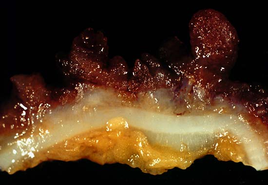
This photo was from a total colectomy done for clinically severe, intractable chronic ulcerative colitis. It shows a closer view of a longitudinal section through the colon wall. This demonstrates not only the angry red mucosa but also the tendency for the inflamed tissue to throw itself up into inflammatory pseudopolyps. Source: Ed Uthman, MD. Public domain.
This photo is from a total colectomy done for severe, intractable chronic ulcerative colitis. It shows a close view of a lengthwise section through the colon wall. This demonstrates not only the angry red mucosa, but also, the tendency for the inflamed tissue to throw itself up into inflammatory pseudopolyps.
Source: Ed Uthman, MD. Public domain.

What Is Ulcerative Colitis?

[dropcap]U lcerative colitis is an inflammatory disorder of the colon characterized by continuous inflammation of the mucosa and submucosa usually with small ulcers, extending from the rectum and typically involving the distal colon, rectum, and anus and producing bloody diarrhea.

While the severity of ulcerative colitis varies among patients, iron deficiency anemia often develops due to blood loss especially when there are many bloody bowel movements in a day.

The onset of ulcerative colitis is most commonly in young adulhood.

Q: Is this disease painful?

A: Yes with the passage of stool.

Psychological stress and subsequent dysbiosis exacerbate ulcerative colitis.

Psychological stress activates multiple physiological processes aimed at maintaining balance within the body. These physiological processes also have the capacity to influence the composition of microbial communities in the digestive tract, and research now indicates that exposure to stressful stimuli leads to gut microbiota dysbiosis.1

While the relative abundance of many different bacterial types can be altered during stressor exposure, findings in nonhuman primates and laboratory rodents, as well as humans, indicate that bacteria in the genus Lactobacillus are consistently reduced in the gut during stress.2

Presently, ulcerative colitis cannot be cured. This condition has a course of remissions, when symptoms subside, and flares, when symtpoms get worse. Treatment is aimed to reduce flares and promote remission. In all cases, correction of dysbiosis improves the condition.

Ulcerative colitis is associated with increased incidence of cancer of the colon.3

What Is Ulcerative Colitis In Celiac Disease and/or Gluten Sensitivity?

Sources:
  1. Galley JD, Bailey MT. Impact of stressor exposure on the interplay between commensal microbiota and host inflammation. Gut Microbes. 2014 May 1;5(3):390-396. Epub 2014 Apr 1. []
  2. Galley JD, Bailey MT. Impact of stressor exposure on the interplay between commensal microbiota and host inflammation. Gut Microbes. 2014 May 1;5(3):390-396. Epub 2014 Apr 1. []
  3. Taber’s Cyclopedic Medical Dictionary. F. A. Davis. Philadelphia, PA []