Skip to content

Anti-tissue Transglutaminase Antibodies (tTG) Present In Blood

Image depicts tissue transglutaminase (tTg).
Image Depicts Tissue Transglutaminase (tTG).

What Are Anti-tissue Transglutaminase Antibodies?

[dropcap]A[/dropcap]nti-tissue transglutaminase antibodies (anti-tTG) are connective tissue autoantibodies and can be detected in blood samples from affected persons who are reacting to gluten in the diet.

Autoantibodies are abnormal because they attack the body’s own tissue, which in the case of these antibodies is tissue transglutaminase 2 (TG2).

Q: What is tissue transglutaminase 2 (TG2)?

A: Tissue transglutaminase 2 (TG2) is an enzyme that appears in many cell locations and is particularly abundant in endothelial cells that line the small intestine. It has been implicated in a variety of cellular processes, such as differentiation, cell death, inflammation, cell migration and wound healing.

The cell appears to adapt the dynamics of this enzyme to meet specific sub-cellular needs or to respond to stress or other stimuli. Substantial evidence indicates that the location of TG2 within cells is critical for the regulation of its various biochemical activities, which subsequently trigger diverse downstream events,1

Although initially studied as an enzyme within cells, TG2 is now known to be secreted also into the extracellular space (between cells) or onto the cell surface.1

Abnormal activation of TG2 or deregulation of its function(s) is involved in a variety of human diseases, such as celiac disease, diabetes, neurodegenerative diseases, multiple sclerosis and rheumatoid arthritis. A role in inflammatory disorders and septic shock has also been shown. Moreover, multiple studies have revealed elevated TG2 expression in many types of cancer cells.1

What Are Anti-tissue Transglutaminase Antibodies In Celiac Disease and/or Gluten Sensitivity?

Sources:
  1. Piacentini M, D’Eletto M, Farrace MG, Rodolfo C, Del Nonno F, Ippolito G, Falasca L. Characterization of distinct sub-cellular location of transglutaminase type II: changes in intracellular distribution in physiological and pathological states. Cell Tissue Res. 2014 Dec;358(3):793-805. doi: 10.1007/s00441-014-1990-x. [] [] []

Bone Alkaline Phosphatase (BALP), Elevated

Vertebrae. Courtesy FreePik.com

What Is Elevated Bone Alkaline Phosphatase?

[dropcap]E[/dropcap]levated bone alkaline phosphatase (BALP) is a laboratory result that indicates an abnormal blood level of this bone enzyme.

A bone alkaline phosphatase blood level is one of the most frequently used biochemical markers of bone formation.

Q: Why is the purpose of  bone alkaline phosphatase?

A: Bone alkaline phosphatase is produced by bone cells called osteoclasts in normal bone maintenance for the purpose of breaking down old or damaged bone so that other bone cells called osteoblasts can fill in the excavated areas with new bone. This process keeps bone stong and healthy.

Elevated bone alkaline phosphatase shows that more bone is being broken down than is being replaced. It can be caused by hyperparathyroidism, bone tumors from cancer, and malnutrition.

What Is Elevated Bone Alkaline Phosphatase (BALP) In Celiac Disease and/or Gluten Sensitivity?

Vitamin B12 Deficiency Anemia

What Is Vitamin B12 Deficiency Anemia? [dropcap]V[/dropcap]itamin B12 deficiency anemia is a megaloblastic anemia that is characterized by defective DNA synthesis of red blood cells due to a lack of vitamin B12. Vitamin B12 is essential… 

Hypokalemia (Low Potassium Blood Level)

hypokalemia gluten celiac disease symptomWhat Is Hypokalemia?

[dropcap]H[/dropcap]ypokalemia means the level of potassium in the bloodstream is too low to meet metabolic needs of the body for this mineral and is characterized by metabolic acidosis, altered nerve conduction and muscle contraction.

Rapid potassium loss can result in life-threatening hypokalemic rhabdomyolysis which is destruction of muscle tissue that results in kidney damage.1

Q: What are metabolic needs of the body for potassium?

A: The metabolic needs of the body for potassium are great because this mineral is crucial for life and especially for normal nerve and muscle function.

Most potassium is intracellular, meaning it is found within cells while sodium, its opposing mineral (both electrolytes), is found in the fluid surrounding cells. In muscle contraction, exchange of potassium and sodium takes place so that potassium moves out of muscle cells and sodium moves into them. With muscle relaxation, potassium moves back into the cells and sodium moves out.

What Is Hypokalemia In Celiac Disease and/or Gluten Sensitivity?

Sources:
  1. Williams SG, Davison AG, Glynn MJ. Hypokalaemic rhabdomyolsis: an unusual presentation of celiac disease. European Journal of Gastroenterology and Hepatology. Feb 1995;7(2):183-4. []

Delayed Puberty in Boys

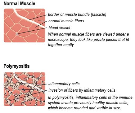
Drawing of Biopsy Showing Muscle Fibers Invaded by Immune Cells. Courtesy MDA.org

What Is Polymyositis?

[dropcap]P olymyositis is a body-wide connective tissue disease resulting from autoimmune attack of skeletal muscles that is characterized by inflammatory and degeneratory changes. The course is unpredictable being marked by spontaneous flare-ups and remissions.

Polymyositis can begin slowly or abruptly according to the factor that is triggering the onset such as infection, medications like phenytoin, and autoimmune disease.

Progressive muscle weakness starts in the proximal skeletal muscles (muscles closest to the trunk of the body).

Skeletal muscles, also called voluntary, are muscles that move the body as we want, such as walking and  lifting objects, as opposed to those we cannot voluntarily control, such as the muscles of digestion. 

Q: What are the degeneratory changes in skeletal muscles?

A: In polymyositis, degeneratory changes in skeletal muscles means that muscles are being destroyed (called necrosis), resulting in fibrosis, or scarring. When scar tissue takes the place of lost muscle tissue, it cannot act like muscle to contract and relax.   Muscle destruction is what causes muscle pain and weakness.

After the clinical work-up of exams and blood studies to determine muscle damage, the diagnosis of polymyositis is confirmed by muscle biopsy. See image at above left. The black dots are inflammatory cells. Edema (fluid) between cells caused by inflammation pushes muscle fibers apart.

There is no cure for polymyositis, but the symptoms can be treated.  Options include medication, physical therapy, exercise, heat therapy (including microwave and ultrasound), orthotics and assistive devices, and rest.  The standard treatment for polymyositis is a corticosteroid drug, given either in pill form or intravenously.  Immunosuppressant drugs, such as azathioprine and methotrexate, may reduce inflammation in people who do not respond well to prednisone. 

Periodic treatment using intravenous immunoglobulin can also improve recovery.  Other immunosuppressive agents used to treat the inflammation associated with polymyositis include cyclosporine A, cyclophosphamide, and tacrolimus.  Physical therapy is usually recommended to prevent muscle atrophy and to regain muscle strength and range of motion.1

Diagnosis is based on elevated muscles enzymes, increased urinary creatine level, and electromyograph abnormalities.

Polymyositis can affect people at any age. It is most common in adults between ages 50 and 70, and in children ages 5 to 15. It affects women twice as often as men and is more common in African Americans than Caucasians.2 The major causes of death from polymyositis are cancer and lung disease, including pneumonia.  The 5-year mortality rate can be as high as 1 in 5 patients.2

What Is Polymyositis In Celiac Disease and/or Gluten Sensitivity?

Sources:
  1. National Institute of Neurological Disorders and Stroke []
  2. http://www.nlm.nih.gov/medlineplus/ency/article/000428.htm [] []

Secondary Hyperparathyroidism

Hepatitis-C-Healthy-Liver-and-Inflammation-of-Liver

What Are Elevated Liver Enzymes?

[dropcap]H ypertransaminasemia is a chronic condition of elevated blood liver transaminase enzymes, commonly called “liver enzymes,” that signifies hepatocellular (liver) injury.

Q: What are serum transaminases?

A: Transaminases are the liver enzymes ALT and AST. ALT is the abbreviation for alanine aminotransferase enzyme and AST is the abbreviation for aspartate aminotransferase enzyme.

Transaminases are commonly measured in routine blood tests to determine liver function. Elevated ALT and AST transaminases  indicate inflammation of the liver. Other specific tests must follow to determine the cause of inflammation.

What Is Hypertransaminasemia In Celiac Disease and/or Gluten Sensitivity?

Recurrent Monoarthritis

Depiction of an antibody. Courtesy NIH

What Is Common Variable Immunodeficiency?

[dropcap]C ommon variable immunodeficiency (CVID) is a primary antibody deficiency disease characterized by the onset of recurrent bacterial infections resulting from markedly decreased immunoglobulin antibody production and antibody levels.

Q: What causes common variable immunodeficiency?

A: Common variable immunodeficiency is caused by a defect in any critical stage of B cell development and is characterized by impaired production of normal amounts of antigen-specific antibodies. This is a set up for infection, autoimmune disease, and cancer.

One important histologic feature is the absence or paucity of plasma cells in the biopsy that occurs in common variable immunodeficiency. The diagnosis is initially made by measuring quantitative immunoglobulins and then specific immune testing on these cell subsets in the circulation by flow cytometry.1

Early diagnosis and treatment with IgG therapy (immunoglobulin G) can decrease illness and mortality.2 

Pulmonary damage is the most frequent complication and may result from recurrent infections and/or immune dysregulation. Other complications due to the underlying immune dysregulation include lymphoproliferative disease (granulomatous disease, lymphadenopathy and hepatosplenomegaly), autoimmune disease, gastrointestinal disease such as chronic inflammation and an increased risk of cancer.3

It is important to recognize that common variable immunodeficiency can occur at any age, but an early onset may be associated with an increased risk of gastric cancers and lymphoma, particularly of the intestine.3

Patients with common variable immunodeficiency should be managed by an immunologist with experience in primary immunodeficiency states, given the complications of these patients.4

What Is Common Variable Immunodeficiency In Celiac Disease and/or Gluten Sensitivity?

Sources:
  1. Murray JA1, Rubio-Tapia A. Diarrhoea due to small bowel diseases. Best Pract Res Clin Gastroenterol. 2012 Oct;26(5):581-600. doi: 10.1016/j.bpg.2012.11.013. []
  2. Maarschalk-Ellerbroek LJ, Hoepelman AI, van Montfrans JM, Ellerbroek PM. The spectrum of disease manifestations in patients with common variable immunodeficiency disorders and partial antibody deficiency in a university hospital. J Clin Immunol. 2012 Oct;32(5):907-21. doi: 10.1007/s10875-012-9671-6. []
  3. Maarschalk-Ellerbroek LJ, Hoepelman AI, van Montfrans JM, Ellerbroek PM. The spectrum of disease manifestations in patients with common variable immunodeficiency disorders and partial antibody deficiency in a university hospital. J Clin Immunol. 2012 Oct;32(5):907-21. doi: 10.1007/s10875-012-9671-6. [] []
  4. Murray JA1, Rubio-Tapia A. Diarrhoea due to small bowel diseases. Best Pract Res Clin Gastroenterol. 2012 Oct;26(5):581-600. doi: 10.1016/j.bpg.2012.11.013. []

Pulmonary Permeability, Increased 

Erosions Of The Esophagus Can Be A Cause Of Gastrointestinal Bleeding.
Erosions Resulting From Esophagitis Can Be A Cause Of Gastrointestinal Bleeding.

What Is Occult Gastrointestinal Bleeding?

[dropcap]O ccult gastrointestinal bleeding is characterized by unseen or minute quantities of blood in stool. The origin of bleeding is from mucosa that lines the inside of the digestive tract at a site that must be found by medical testing and procedures to look at the lining.

Q: What tests and procedures are performed to locate the bleeding?

A: The simplest test to discover blood that cannot be seen is the stool test. This consists of a card with 3 spaces for 3 separate  stool collection over 3 separate days. A tiny smear of stool is placed on a test card space on each of three days. Then the card is examined for a reaction that indicates the existence of blood in the stool.

If the stool test is positive, the origin of bleeding must be located. This search requires certain procedures that look at the mucosal lining directly by a gastroenterologist, usually under light sedation.

  • Gastroscopy procedure can visualize the upper gastrointestinal tract, which includes the esophagus and stomach.
  • Endoscopy procedure can also visualize the upper small intestine. However, endoscopy procedure is limited in that it cannot visualize the middle small intestine (jejunum).
  • Colonoscopy can visualize the end of the small intestine and colon all the way to the rectum.
  • Capsule endoscopy procedure.  If  gastroendoscopy and colonoscopy fail to discover the source of bleeding, the physician may administer a capsule endoscopy which visualizes the entire gastrointestinal tract. In this procedure, the patient swallows a capsule size camera (encased) which takes constant pictures over the course of a day until expelled through the rectum.  Of course, the patient must be able to swallow the large size capsule.

What Is Occult Gastrointestinal Bleeding In Celiac Disease and/or Gluten Sensitivity?

Autoimmune Polyglandular Syndromes 

Zincemia low zinc level symptom of celiac disease and glutenWhat Is Zincemia?

[dropcap]Z incemia means the zinc level in blood plasma is too low to meet metabolic needs of the body for this mineral.

Q: How important is a normal blood level of zinc?

A: A low blood level of zinc is characterized by widespread alterations in energy metabolism, growth, hemoglobin, carbon dioxide transport, hormone activity, insulin storage, many enzyme activities, prostaglandin function, collagen production, male fertility, protein synthesis, and vitamin A metabolism.

What Is Zincemia In Celiac Disease and/or Gluten Sensitivity?

Antiphospholipid Syndrome

Testing Thigh Strength. Courtesy Charlie Goldberg, M.D., UCSD School of Medicine

What Is Hypophosphatemia?

[dropcap]H ypophosphatemia means the level of phosphates in the bloodstream is too low to meet metabolic needs of the body for this mineral.

Q: How important is phosphorus in metabolism?

A: Phosphorus is crucial to  life, being present in every cell of the body and constitutes 45% of skeletal bone weight along with 40% calcium needed to support the body as a framework.

A low blood phosphate level is characterized by alterations in blood acid-alkaline balance and serious neuromuscular, hematologic, renal, skeletal, and dental abnormalities.

Symptoms result primarily from decreased production of adenosine triphosphate (ATP), the main energy source in cells, and phosphocreatine, a secondary energy source for muscle contraction.

  • Acute phosphorus deficiency may precipitate rhabdomyolysis which is destruction of muscle.
  • Nervous system dysfunction is observed in severe hypophosphatemia.
  • Chronic phosphorus deficiency causes proximal myopathy (upper arms and thighs).1
  • Severe phosphorus deficiency has widespread and ultimately fatal consequences.

What Is Hypophosphatemia In Celiac Disease and/or Gluten Sensitivity?

Sources:
  1. Takeda E, Ikeda S, Nakahashi O. Lack of phosphorus intake and nutrition. Clin Calcium. 2012 Oct;22(10):1487-91. []