Skip to content

Delayed Puberty in Boys
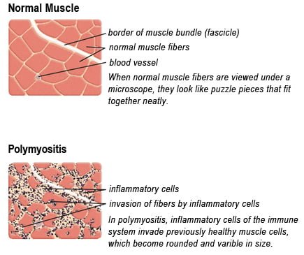
Drawing of Biopsy Showing Muscle Fibers Invaded by Immune Cells. Courtesy MDA.org

What Is Polymyositis?

[dropcap]P olymyositis is a body-wide connective tissue disease resulting from autoimmune attack of skeletal muscles that is characterized by inflammatory and degeneratory changes. The course is unpredictable being marked by spontaneous flare-ups and remissions.

Polymyositis can begin slowly or abruptly according to the factor that is triggering the onset such as infection, medications like phenytoin, and autoimmune disease.

Progressive muscle weakness starts in the proximal skeletal muscles (muscles closest to the trunk of the body).

Skeletal muscles, also called voluntary, are muscles that move the body as we want, such as walking and  lifting objects, as opposed to those we cannot voluntarily control, such as the muscles of digestion. 

Q: What are the degeneratory changes in skeletal muscles?

A: In polymyositis, degeneratory changes in skeletal muscles means that muscles are being destroyed (called necrosis), resulting in fibrosis, or scarring. When scar tissue takes the place of lost muscle tissue, it cannot act like muscle to contract and relax.   Muscle destruction is what causes muscle pain and weakness.

After the clinical work-up of exams and blood studies to determine muscle damage, the diagnosis of polymyositis is confirmed by muscle biopsy. See image at above left. The black dots are inflammatory cells. Edema (fluid) between cells caused by inflammation pushes muscle fibers apart.

There is no cure for polymyositis, but the symptoms can be treated.  Options include medication, physical therapy, exercise, heat therapy (including microwave and ultrasound), orthotics and assistive devices, and rest.  The standard treatment for polymyositis is a corticosteroid drug, given either in pill form or intravenously.  Immunosuppressant drugs, such as azathioprine and methotrexate, may reduce inflammation in people who do not respond well to prednisone. 

Periodic treatment using intravenous immunoglobulin can also improve recovery.  Other immunosuppressive agents used to treat the inflammation associated with polymyositis include cyclosporine A, cyclophosphamide, and tacrolimus.  Physical therapy is usually recommended to prevent muscle atrophy and to regain muscle strength and range of motion.1

Diagnosis is based on elevated muscles enzymes, increased urinary creatine level, and electromyograph abnormalities.

Polymyositis can affect people at any age. It is most common in adults between ages 50 and 70, and in children ages 5 to 15. It affects women twice as often as men and is more common in African Americans than Caucasians.2 The major causes of death from polymyositis are cancer and lung disease, including pneumonia.  The 5-year mortality rate can be as high as 1 in 5 patients.2

What Is Polymyositis In Celiac Disease and/or Gluten Sensitivity?

Sources:
  1. National Institute of Neurological Disorders and Stroke []
  2. http://www.nlm.nih.gov/medlineplus/ency/article/000428.htm [] []

Angina Pectoris

What Is Angina Pectoris?Coronary Artery Lesion

[dropcap]A[/dropcap]ngina pectoris, or simply angina, is a coronary syndrome characterized by an oppressive substernal pain (pain under breastbone) or pressure brought on by exertion and relieved by rest that results from failure of coronary arteries to deliver adequate oxygen to heart tissue due to ischemic heart disease.

Q: Why do coronary arteries fail to deliver adequate oxygen to heart tissue?

A: Coronary arteries are the blood vessels that serve the heart. In angina, these vessels fail to deliver adequate oxygen to heart tissue because they are narrowed or blocked by fatty buildups, called atherosclerotic plaques or by a blood clot which impair their ability to carry adequate blood that carries the oxygen. Diseased coronary arteries cannot deliver adequate oxygenated blood pumped by the heart to its own muscle cells.

The heart is a muscular organ that is working all the time without rest, so it needs a constant supply of oxygen. When heart muscle has to work harder, it needs more oxygen. Lack of oxygen causes pain which makes the affected person stop activity and rest.

Angina can be stable or unstable. Unstable angina is much more serious and can be life-threatening.

  • Stable angina produces predictable pain and responds to rest and/or medication. It is less serious than unstable angina but can be very painful or uncomfortable. Anything that makes the heart muscle need more oxygen can cause an angina attack in someone with heart disease, including: smoking, cold weather, exercise, emotional stress, obesity, and large meals. Other causes of angina include: abnormal heart rhythms (usually ones that cause the heart to beat quickly), anemia, coronary artery spasm, heart failure, heart valve disease, and hyperthyroidism (overactive thyroid).1
  • Unstable angina produces unpredictable pain that may occur at rest, lasting more than 20 minutes. It is more severe than stable angina and less responsive to medication. Atherosclerosis is by far the most common cause of unstable angina. Oxidized low-density lipoprotein, so-called bad cholesterol, and oxysterols play an important role in atherogenesis, the development of atherosclerosis. Coronary arteries that are narrowed by atherosclerotic plaques can rupture causing injury to the coronary blood vessel resulting in blood clotting which blocks the flow of blood to the heart muscle. Blood clots may form, partially dissolve, and later form again and angina can occur each time a clot blocks blood flow in an artery. People with unstable angina are at increased risk of having a heart attack.2

What Is Angina In Celiac Disease and/or Gluten Sensitivity?

Sources:
  1. http://www.ncbi.nlm.nih.gov/pubmedhealth/PMH0001247/ []
  2. http://www.heart.org/HEARTORG/Conditions/HeartAttack/SymptomsDiagnosisofHeartAttack/Unstable-Angina_UCM_437513_Article.jsp# []

Coronary Artery Disease

Image on left shows how atherosclerosis impedes blood flow through coronary arteries while blood clots block blood flow. Courtesy Google.
Figure on right shows how atherosclerosis impedes blood flow through coronary arteries while blood clots block blood flow. Courtesy Google.

What Is Coronary Artery Disease (CAD)?

[dropcap]C[/dropcap]oronary artery disease (CAD), also called ischemic heart disease, is a gradual narrowing of medium and large arteries of the heart by fatty buildups, called atherosclerotic plaques.

It is characterized by slowly developing interference with blood flow to heart tissue itself, resulting in oppressive chest pain called angina and, ultimately, thrombosis (clot) causing heart attack.  

The heart is a muscular organ that is working all the time, so it needs a constant supply of oxygen. Oxygen is brought to the working heart tissue by the coronary arteries with each beat of the heart. When heart muscle has to work harder, it needs more oxygen delivered to itself. Lack of oxygen causes pain.

In fact, failure of diseased coronary arteries to deliver adequate oxygen to heart tissue is the most common cause of angina pectoris – substernal pain (under breastbone) or pressure brought on by exertion and relieved by rest. 

Thrombosis, or clot formation, occurs when blood cells within a narrowed artery can no longer get through. Trapped, blood cells pile up and block the artery thus triggering a cascade of events called heart attack. Coronary arteries that are narrowed by atherosclerotic plaques can rupture causing injury to the coronary blood vessel resulting in blood clotting which blocks the flow of blood to the heart muscle. Blood clots may form, partially dissolve, and later form again and angina can occur each time a clot blocks blood flow in an artery.1

Q: How does coronary artery disease develop?

A: Coronary artery disease slowly develops from this combination of events:

  • Dysfunction of epithelial cells that line the inside of arteries cause the vessels to stiffen, and subsequently

  • Accumulation of lipid (fat) in smooth muscle cells beneath the inside lining of arteries and in foam cells cause buildup of fatty deposits on the inside walls progressing to fibrous plaque formation.

Oxidized low-density lipoprotein (oxLDL), so-called bad cholesterol, and oxysterols play important roles in the development of  atherosclerosis. OxLDL triggers the immune system to produce autoantibodies against oxLDL that are detectable in serum. These antibodies are called anti-oxLDL. Anti-oxLDL antibody and oxysterol concentrations are associated with coronary artery stenosis. Oxidative stress may be greatly increased in unstable angina.2 and Chronic inflammation in the general population is a major risk factor for ischemic heart disease.

The pathophysiology of atherosclerosis is, clearly, different in women when compared to the men. The women have a higher risk of blood coagulability making them at high risk for the blood clot formation. In a large number of women endothelial dysfunction, small vessel size and diffuse atherosclerosis have been identified as causes of ischemia without evidence of blockade in the coronary arteries.3

Also, atherosclerotic plaque in women is less fibrotic and contains more lipid filled foam cells, implying greater potential for reversibility but also potentially greater vulnerability for plaque rupture and thrombosis.4

Who is Affected in the General Population?

  • Coronary artery disease remains the leading cause of death in developed countries despite significant progress in primary prevention and treatment strategies.

  • It is the leading cause of death in women, as well as an important cause of disability.

  • Older patients are at particularly high risk of poor outcomes following acute coronary syndrome.5

What Is Coronary Artery Disease In Celiac Disease and/or Gluten Sensitivity?

Ischemic heart disease is the leading cause of death in the United States, making cardiovascular risk assessments and potential interventions or treatments imperative for patients with celiac disease.6

Sources:
  1. http://www.heart.org/HEARTORG/Conditions/HeartAttack/SymptomsDiagnosisofHeartAttack/Unstable-Angina_UCM_437513_Article.jsp# []
  2. Yasunobu Y, Hayashi K, Shingu T, Yamagata T, Kajiyama G, Kambe M. Coronary atherosclerosis and oxidative stress as reflected by autoantibodies against oxidized low-density lipoprotein and oxysterosis. Atherosclerosis. Apr 2001;155(2):445-53. []
  3. Kunadian V, Ford GA, Bawamia B, Qiu W, Manson JE. Vitamin D deficiency and coronary artery disease: A review of the evidence. Am Heart J. 2014 Mar;167(3):283-291. doi: 10.1016/j.ahj.2013.11.012. Epub 2013 Dec 19. []
  4. Kunadian V, Ford GA, Bawamia B, Qiu W, Manson JE. Vitamin D deficiency and coronary artery disease: A review of the evidence. Am Heart J. 2014 Mar;167(3):283-291. doi: 10.1016/j.ahj.2013.11.012. Epub 2013 Dec 19. []
  5. Kunadian V, Ford GA, Bawamia B, Qiu W, Manson JE. Vitamin D deficiency and coronary artery disease: A review of the evidence. Am Heart J. 2014 Mar;167(3):283-291. doi: 10.1016/j.ahj.2013.11.012. []
  6. Robinson BL, Davis SC, Vess J, Lebel, J. Primary care management of celiac disease. Nurse Practitioner. February 2015: Vol 40 – Issue 2; 28–34. []

Delayed Puberty In Girls 

sick woman on bed, symptom of cold, flu, insomnia, stress, headache, hangover, dizzinessWhat Is Hypokalemic Rhabdomyolysis?

[dropcap]H ypokalemic rhabdomyolysis is an acute and sometimes fatal disease due to its rapid progression of muscle destruction when untreated.

It is characterized by the accumulation of by-products of skeletal muscle destruction in the renal (kidney) tubules and producing acute kidney failure caused by rapid potassium loss.

This condition puts you in bed because the legs muscles cannot support the body and arms are too weak to move.

What Is Hypokalemic Rhabdomyolysis In Celiac Disease and/or Gluten Sensitivity and Dermatitis Herpetiformis?

Spina Bifida 

pityriasis rubraWhat Is Pityriasis Rubra Pilaris?

[dropcap]P ityriasis rubra pilaris (PRP) is a chronic generalized exfoliative dermatitis (sloughing skin) characterized by erythema (redness), scaling, dilated plugged hair follicles, and keratoderma (thickened skin) of the hands and feet that is often associated with anemia and low serum albumin.

It may manifest either as Type I classical adult onset PRP, Type II atypical adult (onset) PRP, or Type VI PRP (HIV-associated PRP pityriasis rubra pilaris) in contrast to classical juvenile (Type III) and circumscribed juvenile (Type IV) encountered among children.1

Q: Who is affected in the general population?

A: All ages are affected. Pityriasis rubra pilaris occurs all over the world but with racial variations – it is 1 in 5,000 in Great Britain and 1 in 50,000 in India.2[/box]

What Is Pityriasis Rubra Pilaris In Celiac Disease and/or Gluten Sensitivity?

Sources:
  1. Sehgal VN, Srivastava G, Dogra S. Adult onset pityriasis rubra pilaris. Indian J Dermatol Venereol Leprol. 2008 Jul-Aug;74(4):311-21. []
  2. Sehgal VN, Srivastava G, Dogra S. Adult onset pityriasis rubra pilaris. Indian J Dermatol Venereol Leprol. 2008 Jul-Aug;74(4):311-21. []

Hangnail

Aphthous_ulcer[1]What Are Aphthous Ulcers?

[dropcap]A phthous ulcers, also called canker sores, are a chronic disorder of soft mouth tissue characterized by small, painful purpuric, papular, or erosive lesions that are often surrounded by erythematous (red) margins.1

Q: What soft mouth tissues develop canker sores?

A: Canker sores can erupt on the mucosal surface of the inside lip, sides of mouth, under the tongue and along the side of the tongue. When they are forming, the area swells into a papule, or small bump. Later, the surface erodes and a crater forms with firm pus.

What Are Aphthous Ulcers In Celiac Disease and/or Gluten Sensitivity?

Sources:
  1. Lahteenoja H, Toivanen A, Viander M, Maki M, Irjala K, Raiha I, Syrjanen S. Oral mucosal changes in coeliac patients on a gluten-free diet. European Journal of Oral Sciences. Oct 1998;106(5):899,8p. []

Ichthyosis, Acquired  

cheilosisWhat Is Cheilosis?

[dropcap]C heilosis is a painful feature of nutritional deficiencies, especially riboflavin and/or pyridoxine deficiency, and less frequently, from vitamin B12, folic acid, and iron. It is characterized by redness of the lips with cracking and weeping in the corner of the mouth.

Q: What is the consequence of weeping at the corners of the mouth?

A: The open sores that develop at the corners of the mouth invite infection by candida albicans, a yeast organism, and less commonly by bacteria normally present on the skin such as staphylococcus aureus.

These sores make opening the mouth very painful and so restrict eating, talking, socializing, and dental care.

What Is Cheilosis In Celiac Disease and/or Gluten Sensitivity?

Cortical Calcifying Angiomatosis

What Is Carbohydrate Malabsorption? [dropcap]C arbohydrate malabsorption is a digestive disorder characterized by the inability to properly digest and absorb carbohydrates within the small intestine to supply needed energy to the body. Q: What carbohydrates should be… 

Epilepsy (Convulsions)

chronic diarrhea gluten celiac disease symptomWhat Is Chronic Diarrhea?

[dropcap]C hronic diarrhea is an intestinal motility disorder characterized by 1) alteration in stool formation causing loose to fluid movements and quantity of movements with or without abdominal pain and 2) interference with normal carbohydrate salvage by the intestinal microbe population.

The severity of diarrhea is determined by the frequency and quantity of fluid lost.

Q: What is carbohydrate salvage by microbes in the colon?

A: Carbohydrate salvage in the colon is the necessary process whereby billions of microbes normally present in the colon work to breakdown undigestible carbohydrate foodstuffs such as fiber that continually arrive from the small intestine.

The microbial action releases energy to the body that would otherwise be lost with defecation, generates short-chain fatty acids, and stimulates sodium and fluid absorption. In this important process, butyrate and proprionate are produced which nourish the colonocytes (cells that line the colon), and acetate is produced for the liver.

While diarrhea may be a common symptom of small bowel mucosal disease, the consequent malabsorption can lead to substantial malnutrition and nutrient deficiencies. The small intestine, unlike the colon, has been relatively inaccessible, and systematic evaluation is often necessary to identify and treat small intestinal mucosal diseases that lead to diarrhea. All patients with severe diarrhea or diarrhea associated with features suggestive of malabsorption may have a disease of the small intestinal mucosa that requires careful evaluation and targeted management.1

What Is Chronic Diarrhea In Celiac Disease and/or Gluten Sensitivity?

Sources:
  1. Murray JA1, Rubio-Tapia A. Diarrhoea due to small bowel diseases. Best Pract Res Clin Gastroenterol. 2012 Oct;26(5):581-600. doi: 10.1016/j.bpg.2012.11.013. []

Aphthous Ulcers (Canker Sores) 

Aphthous_ulcer[1]What Are Aphthous Ulcers?

[dropcap]A[/dropcap]phthous ulcers, also called canker sores, are a chronic disorder of soft mouth tissue characterized by small, painful purpuric, papular, or erosive lesions that are often surrounded by erythematous (red) margins.1

Q: What soft mouth tissues develop canker sores?

A: Canker sores can erupt on the mucosal surface of the inside lip, sides of mouth, under the tongue and along the side of the tongue. When they are forming, the area swells into a papule, or small bump. Later, the surface erodes and a crater forms with firm pus.

What Are Aphthous Ulcers In Celiac Disease and/or Gluten Sensitivity?

Sources:
  1. Lahteenoja H, Toivanen A, Viander M, Maki M, Irjala K, Raiha I, Syrjanen S. Oral mucosal changes in coeliac patients on a gluten-free diet. European Journal of Oral Sciences. Oct 1998;106(5):899,8p. []