Or maybe you have trouble falling asleep at night and wake up completely drained as if you were running a marathon in your sleep. Sure, you could chalk it up to a busy, stress-filled life, but the answer is more complex and hits closer to home that you even realize.
Dermatitis Herpetiformis On Forearm. Skin Is Darkened Where Old Blisters Healed.
What Is Dermatitis Herpetiformis?
D ermatitis herpetiformis (DH) is an autoimmune extremely itchy, painful bullous skin rash (blistering eruptions) arising from the underlying dermis layer of skin as a consequence of gluten sensitivity.
Dermatitis herpetiformis is characterized by multiple intensely itchy, red blisters appearing on the elbows which can extend down the forearm to the wrist and the knees. Less usual areas involve the back, buttocks, scalp, and abdomen.
Q: Do the blisters leave a mark when healed?
A: Crops of skin eruptions begin with itching or a burning sensation in reddened papules. There are grouped vesicles and tense blisters. The blister contents may be serous or bloody, with symmetrical distribution (eg, both knees or both elbows). Fluid filled elements rupture leaving denuded areas of sore skin and crust. Subsequently, there is residual hypopigmentation (a white area) or hyperpigmentation (dark area).1
Rupture of blisters begins relief from intense burning and itching.
Dermatitis Herpetiformis Eruptions On Knees. Notice White Areas Showing Loss of Pigmentation From Healed Blisters.
What Is Dermatitis Herpetiformis In Celiac Disease and/or Gluten Sensitivity?
Primary care providers should be aware of this skin condition, as they are more likely than a gastroenterologist to be confronted with this type of presentation of celiac disease.2
Sources:
Mendes FB, Hissa-Elian A, de Abreu MA, Gonçalves VS. Review: dermatitis herpetiformis. An Bras Dermatol. 2013 Jul-Aug;88(4):594-9. [↩]
Robinson BL, Davis SC, Vess J, Lebel, J. Primary care management of celiac disease. Autoimmune Disorders. Nurse Practitioner. February 2015: Vol 40 – Issue 2; 28–34. [↩]
P soriasis is a chronic, autommune relapsing skin disorder characterized by scaling, erythema (redness), and less commonly, pustulation.1
The body surface area affected and the degree to which psoriasis is a problem varies considerably among patients and over time.2 Often there are additional manifestations in the nails and in joints.3
Q: Are there different forms of psoriasis?
A: There are five forms of psoriasis. The lesions in all forms are itchy and red but vary in appearance and severity. Plaque psoriasis is the most common form observed in more than 80% of patients. Atypical forms include guttate, inverse, pustular, and erythrodermic psoriasis.4
Plaque psoriasis features thickened or raised red areas that have a distinct edge and are covered with silvery white buildup of flaky skin typically on elbows, knees, scalp and buttocks.
Gutate psoriasis appears as small, flat red patches with shiny buildup that are not usually painful, just itchy. There may be a few or many patches and they can group together.
Inverse psoriasis affects folds of skin, armpits and the groin area. Lesions are deep red with shiny buildup. It can be a thin red area along a crease line or involve, for example, the whole armpit.
Pustular psoriasis features an itchy, red base followed by blisters of white, non-infectious pus that appears glossy after a day or two and then sloughs in cycles. These areas may be limited to certain areas such as the hands and feet or be more widespread.
Erythrodermic psoriasis involves large areas of the body’s surface, inflaming normal skin and changing it into very red, raw looking flesh that is painful, swollen and itchy. This form requires extensive treatment, and complications can be life-threatening. Fortunately, this form of psoriasis is the least common.
Psoriasis in children has been reported to differ from that among adults being more frequently itchy and plaque lesions are relatively thinner, softer, and less scaly, face and flexural involvement is common and guttate type is the characteristic presentation.5
In children, psoriasis is a common skin disorder with about one third of all patients having onset of disease in the first or second decade of life. A chronic disfiguring skin disease, such as psoriasis, in childhood is likely to have profound emotional and psychological effects, and hence requires special attention.6
What Is Psoriasis In Celiac Disease and/or Gluten Sensitivity?
Sources:

Addolorato G, Parente A, de Lorenzi G, et al. Rapid regression of psoriasis in a coeliac patient after gluten-free diet. A case report and review of the literature. Digestion. 2003;68(1):9-12. [↩]
Stern, R. S., Nijsten, T., Feldman, S. R., Margolis, D. J. and Rolstad, T.
Psoriasis is common, carries a substantial burden even when not extensive, and is associated with widespread treatment dissatisfaction. J. Invest. Dermatol. Symp.. 2004 Mar;9(2):136-9.. [↩]
Weigle N, McBane S. Psoriasis. Am Fam Physician. 2013 May 1;87(9):626-33. [↩]
Dogra S, Kaur I. Childhood psoriasis. Indian J Dermatol Venereol Leprol. 2010 Jul-Aug;76(4):357-65. doi: 10.4103/0378-6323.66580. [↩]
T etany is a painful feature of low blood calcium or low blood magnesium causing hyperexcitability of all nerves which in turn stimulates involuntary sudden, intermittent and repetitious tonic spasms of muscles. Tonic spasms are steady rather than jerky.
Causes include low function of the parathyroid glands which regulate calcium blood levels, vitamin D deficiency which inhibits calcium absorption from the gut that leads to calcium deficiency, and alkalosis.
In alkalosis, the chemical pH (acid/alkaline measure) of blood is too high, or alkaline, which inhibits adequate free calcium ions from being available for cell use.
Q: Why do low blood levels of calcium or magnesium cause muscles to spasm?
A: Calcium controls the readiness of muscle to respond to nerve stimulation. In muscle action, calcium and magnesium are minerals that oppose each other. Calcium allows a muscle to contract while magnesium allows it to relax. Inadequate levels of these opposing minerals affect the ability of muscles to properly function.
A simple test for tetany is called Trouseau’s named after the doctor who developed it. A blood pressure cuff is placed on an upper arm as usual and pumped up to a pressure sufficient to stop blood flow. The pressure must be held for 3 minutes. A positive result is shown in the above photo of carpopedal spasm: the wrist and hand flexes inward while the fingers extend (opposite of wrist and hand) and the thumb goes against the palm.
What Is Tetany In Celiac Disease and/or Gluten Sensitivity?
C hronic constipation is an intestinal motility disorder characterized by abnormal stool formation, consistency, and evacuation.
Motility disorder means the normal rhythmic movement of intestinal muscles, called peristalsis, that moves food matter through the gut is hampered or dysfunctional.
Studies show that methane gas present in the colon induces constipation by delaying transit time, which is the time it takes for stool to pass through the colon.
Researchers investigating the relationship between methane and constipation found that methane positivity was detected in 75% of patients with slow transit, 44% of patients with normal transit and and 28% of the patients who were controls. However, methane positivity was not related with stool consistency.1
Other researchers investigating the total amount of methane produced found that there was significantly more methane production in patients with constipation (21.1 ppm vs. 6.1 ppm, respectively) than in controls without constipation.2
Q. How does methane get into the colon?
A. Methane is produced in the colon by intestinal methanogens (microbes) that metabolize hydrogen, one of the end products of normal anaerobic (meaning without oxygen) bacterial fermentation. Fermentation of the undigested starchy part of carbohydrates produces hydrogen in the intestine which is the substrate (food) for methane production by intestinal methanogens.
Hydrogen and methane are excreted in the flatus and in breath giving the opportunity to indirectly measure their production using breath testing. Methane is detected in 30%-50% of the healthy adult population worldwide.3
Other common causes of constipation include not getting enough exercise, not drinking enough fluids, not eating enough fiber in the diet, not eating foods that supply microbes needed by the colon (probiotics), not eating foods that nourish the good microbe population (prebiotics) and supply minerals needed for healthy movement of stool, and food sensitivities. Too much cows milk is a common cause of stool that forms into balls.
Who is Affected in the General Population? Chronic constipation is a remarkably common and costly condition that can negatively impact the quality of life and result in a major social and economic burden. Based on the definition, either self-reported or using Rome criteria, chronic constipation can affect up to 27% of the population. There is strong evidence that constipation occurs more frequently in women.4
What Is Chronic Constipation In Celiac Disease and/or Gluten Sensitivity?
Sources:
Triantafyllou K, Chang C, Pimentel M. Methanogens, Methane and Gastrointestinal Motility. J Neurogastroenterol Motil. 2014 Jan;20(1):31-40. Epub 2013 Dec 30. [↩]
Sanchez MI, Bercik P. Epidemiology and burden of chronic constipation. Can J Gastroenterol. 2011 Oct;25 Suppl B:11B-15B. [↩]
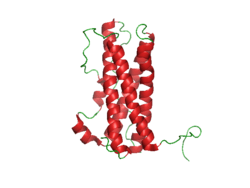
Prolactin Structure. Courtesy Boris TM at Wikipedia.
What Is Hyperprolactinemia?
H yperprolactinemia is an excess secretion of prolactin hormone causing an elevated blood level that is characterized by its effects on reproduction: in females causing amenorrhea and in males causing reduced sexual potency.
Q: What is prolactin?
A: Prolactin is a hormone produced by the the anterior pituitary gland that is transported by the bloodstream to stimulate androgen hormone production in male and estrogen hormone production in female sex glands. However, too much prolactin inhibits hormone production of estrogen in women and testosterone in men.
In pregnant females upon delivery of a baby, prolactin stimulates breast development and milk production for the purpose of feeding the infant. Sucking by the infant stimulates the release of prolactin which keeps the milk supply adequate.
Prolactin also promotes autoimmune mechanisms. Prolactin interferes specifically with B cell tolerance induction, enhances proliferative response to antigens and mitogens and increases the production of immune globulins, cytokines and autoantibodies.1
Other conditions that can stimulate prolactin release include nutritional deficiencies, thyroid stimulating hormone (TSH) produced by the pituitary gland, stress, exercise, and anterior pituitary tumors.
What Is An Elevated Prolactin Level In Celiac Disease and/or Gluten Sensitivity?
Sources:

Shelly S, Boaz M, Orbach H. Prolactin and autoimmunity. Autoimmun Rev. 2012 May;11(6-7):A465-70. doi: 10.1016/j.autrev.2011.11.009. [↩]
H ypocalcemia, or low plasma calcium, means the level of calcium in blood is too low to meet metabolic needs of the body for calcium.
Low blood calcium is characterized by bone and tooth demineralization (loss of calcium causing weak teeth and fragile bones), and these impaired functions: nerve conduction, muscle contraction, blood clotting, blood pressure regulation, glycogen to glucose conversion, many hormone actions, many enzyme activities, and acetylcholine production.
Q: Where is calcium found in the body?
A: Calcium is the most abundant mineral in the body, with 99% residing in bones and teeth where it constitutes 40% of skeletal bone weight along with 45% phosphorus. As a component of bone (hard tissue), calcium fulfills a structural role to maintain body size and act as attachments for musculoskeletal tissues. The remaining 1% of calcium is present in blood and soft tissues.
Calcium levels in the blood are maintained within very strict limits by dietary intake, hormonal regulation by the parathyroid gland and a rapidly exchangeable pool in bone tissue.
What Is Hypocalcemia In Celiac Disease and/or Gluten Sensitivity?
Enlarged heart showing dilated cardiomyopathy at autopsy.
What Is Idiopathic Dilated Cardiomyopathy?
I diopathic dilated cardiomyopathy (IDCM) is a disorder of myocardial (heart muscle) function characterized by dilation or enlargement of the cardiac chambers and reduction in the ability of the ventricles (lower chambers) to contract with the required force needed to pump blood out of the heart to the body.
Idiopathic means the cause is not known.
Q: What is the prognosis for idiopathic dilated cardiomyopathy?
A: This disorder often results in symptomatic heart failure due to the inability of the heart to pump blood as required to supply the body with oxygen and meet metabolic needs.
What Is Idiopathic Dilated Cardiomyopathy In Celiac Disease and/or Gluten Sensitivity?
What Is Dysphagia? D ysphagia is a functional upper digestive disorder characterized by difficulty in swallowing or food getting stuck and swallowing that takes longer than 10 seconds to complete. Q: What determines the symptoms?…
A 3D Image From Magnetic Resonace Cholangiography showing biliary tree. NIHMS
What Is Primary Sclerosing Cholangitis?
Primary sclerosing cholangitis (PSC) is an uncommon, slowly progressive bile duct disease that results in stagnation or build-up of bile in the liver, called cholestasis.
Primary sclerosing cholangitis is characterized by sclerosis, or scarring inflammation in bile ducts both within the liver (intra-hepatic ducts), and outside the liver (extra-hepatic ducts), causing progressive narrowing and, eventually, obliteration of the bile ducts.
Primary sclerosing cholangitis comes under the umbrella term autoimune liver disease in which the end result is immune-mediated hepatocellular (liver cell) or hepatobiliary (bile duct) injury.1
Q:What happens when scarred bile ducts can no longer transport bile out of the liver?
A:Bile that cannot be removed from the liver by the biliary duct system backs up and damages the liver, causing cirrhosis.
Bile is continually made by the liver from phospholipids salt, cholesterol, and aging blood cells that it removes from circulation to be carried out of the liver. Bile also carries away waste products produced by normal metabolism and toxic substances that are removed by the liver for eventual elimination in stool. As such, bile must continually flow out of the liver to prevent build-up in the liver.
Bile is a greenish brown liquid made by the liver. Bile ducts carry it out of the liver to the gall bladder for storage until needed to aid in the digestion and absorption of fat from the small intestine. Bile emulsifies fat eaten in the diet so that the pancreatic enzyme called lypase can break it down into its fatty acid and glycerol components.
The liver is the largest organ within the body. It lies mostly in the upper part of the abdomen on the right side just under the diaphragm. About 70% of liver tissue is made up of cube shaped cells called hepatocytes that do the main work of the liver. Other cells (epithelial) form structure and are arranged in single layers around blood vessels, sinusoids, and bile ducts.
Build-up of bile in the liver is the end result of the inflammatory process in primary sclerosing cholangitis, that by swelling and scarring of bile ducts impedes and eventually prevents bile flow out of the liver, leading to liver failure. There is no curative treatment available for primary sclerosing cholangitis, besides liver transplantation.2
The appearance of the intrahepatic and extrahepatic biliary ducts can be assessed by use of cholangiography, and magnetic resonance (MR) imaging is the best way to identify patients. See image above.3
MR cholangiography offers a noninvasive method of obtaining images of the biliary system without the use of a contrast agent. There is no radiation exposure. Pulse sequences can be chosen to obtain bright bile or black bile cholangiograms. Image processing algorithms can be selected to obtain a three-dimensional representation of biliary anatomy and pathology, and those images can be rotated in any plane so that ductal anatomy and pathology can be seen to best advantage.4
There is no cure for primary sclerosing cholangitis but there are symptom treatments one of which is supplementation for low levels of vitamins A,D,E, and K. Liver transplant is the only effective option.
What Is Primary Sclerosing Cholangitis In Celiac Disease and/or Gluten Sensitivity?
Sources:
Trivedi PJ, Adams DH. Mucosal immunity in liver autoimmunity: a comprehensive review. J Autoimmun. 2013 Oct;46:97-111. doi: 10.1016/j.jaut.2013.06.013. [↩]
Kummen M, Schrumpf E, Boberg KM. Liver abnormalities in bowel diseases. Best Pract Res Clin Gastroenterol. 2013 Aug;27(4):531-42. doi: 10.1016/j.bpg.2013.06.013. [↩]
Eaton JE, Talwalkar JA, Lazaridis KN, Gores GJ, Lindor KD. Pathogenesis of primary sclerosing cholangitis and advances in diagnosis and management. Gastroenterology. 2013 Sep;145(3):521-36. doi: 10.1053/j.gastro.2013.06.052. Epub 2013 Jul 1. [↩]
Meakem TJ 3rd, Schnall MD. Magnetic resonance cholangiography. Gastroenterol Clin North Am. 1995 Jun;24(2):221-38. [↩]