Skip to content

Osteitis Fibrosa Cystica 

Deep Vein Thrombosis in the Right Leg with Swelling and Redness. Coutesy wikipedia.
Deep Vein Thrombosis in the Right Leg with Swelling and Redness. Courtesy wikipedia.

What Is Antiphospholipid Syndrome?

Antiphospholipid syndrome (APS) is an autoimmune disease and a blood clotting disorder characterized by these clinical and laboratory criteria:

Clinical criteria – recurrent vascular thrombosis (clots in veins/arteries) from hypercoagulability (abnormal excessive clotting) and/or recurrent complications of pregnancy that include loss of the fetus  (miscarriage) and pre-eclampsia or eclampsia.

Laboratory criteria – persistently elevated anticardiolipin, anti–beta-2 glycoprotein I, and/or lupus anti-coagulant antibodies in blood.

In antiphospholipid syndrome autoantibodies are produced by the body and directed against negatively charged phospholipids that are found in the outer layer of cell membranes and platelets. B2-glycoprotein-I (a protein in blood plasma) has been found as a major target antigen for antiphospholipid antibodies.

Q: Are phospholipids important in the body?

A: Yes.  Phospholipid molecules are an essential part of cell membranes. They form a barrier around cells that protect the cell, allow movement of oxygen in and carbon dioxide out of the cell, and regulate other small molecules through the cell wall. Because phospholipids are widespread in the body, this disorder can produce a large variety of symptoms and affect many organs.

One severe effect of APS is the development of a blood clot in a vein deep in the arm or leg, called deep vein thrombosis (DVT). DVT can cause pain, swelling, redness, or increased warmth in the affected limb. Deep vein clots can break off, travel to the lungs, and cause pulmonary embolism.1 Pulmonary embolism is a medical emergency.

Treatment is with anticoagulant medications and blood monitoring.

What Is Antiphospholipid Syndrome In Celiac Disease and/or Gluten Sensitivity?

Sources:
  1. http://www.nhlbi.nih.gov/health/health-topics/topics/ebc/signs.html []

Hypocalciuria (Low Urine Calcium)

 IgA Molecule.
Depiction of the IgA Molecule

What Is IgA Deficiency?

I gA deficiency (IgAD) is an immunodeficiency disease characterized by lack of immunoglobulin A type antibody production, called IgA antibody, with no detectable levels in blood or secretions.

Q: What is an IgA antibody?

A: IgA is an antibody of the immune system that is secreted by plasma cells (specialized white blood cells) through epithelial cell linings of mucosal surfaces into mucosa secretions to protect the lining from microbe invasion.

In fact, immunoglobulin class A is the main protein of the mucosal immune system. This includes mucosa of the eye surface, digestive tract, respiratory tract, urinary tract, and genital tract.

Both major histocompatibility complex (MHC) and non-MHC genes contribute to susceptibility to the disease. The former genes appear to be located in different parts of the MHC region depending on the HLA haplotype. The latter show a marked overlap with genes associated with a variety of autoimmune disorders including Graves’ disease, systemic lupus erythematosus, type 1 diabetes and celiac disease, suggesting common pathophysiological mechanisms. The involvement of genes associated with autoimmunity may suggest that IgAD in itself is an autoimmune disease.1

IgA deficiency may progress into a common variable immunodeficiency (CVID).2

What Is IgA Deficiency (IgAD) In Celiac Disease and/or Gluten Sensitivity?

Sources:
  1. Wang N, Hammarström L. IgA deficiency: what is new? Curr Opin Allergy Clin Immunol. 2012 Dec;12(6):602-8. doi: 10.1097/ACI.0b013e3283594219. []
  2. Binek A, Jarosz-Chobot P.Selective immunoglobulin A deficiency. Pediatr Endocrinol Diabetes Metab. 2012;18(2):76-8. []

Infertility In Males

Cutaneous Vasculitis. Courtesy quizlet.com

What Is Cutaneous Vasculitis?

Cutaneous vasculitis is a vascular disease of small blood vessels serving the skin that is characterized by segmental (spotty) inflammation of affected vessel walls resulting in their necrosis (destruction). Scarring is seen in the affected skin that lies over the destroyed blood vessels.

Skin appearance varies from small flat, purplish spots to raised purplish areas over damaged vessels.

In most cases, cutaneous vasculitis results from deposits of toxic immune complexes in the affected vessel walls that is caused by autoimmune activity targeting small blood vessels.1

Cutaneous vasculitis can also result from infection, drug reactions, or malignancies.

Who is Affected in the general Population? Cutaneous vasculitis affects all ages.

The overall annual incidence of cutaneous vasculitis was 38.6 persons per million in 1998.2

What Is Cutaneous Vasculitis In Celiac Disease and/or Gluten Sensitivity?

Sources:
  1. Abenavoli L, Proietti I, Leggio L, Ferrulli A, Vonghia L, Capizzi R, Rotoli M, Amerio PL, Gasbarrini G, Addolorato G. Cutaneous manifestations in celiac disease. World J Gastroenterol. 2006 Feb 14;12(6):843-52. []
  2. Watts RA, Jolliffe VA, Grattan CE, Elliott J, Lockwood M, Scott DG. Cutaneous vasculitis in a defined population–clinical and epidemiological associations. J Rheumatol. 1998 May;25(5):920-4. []

Hypogonadism In Adult Males That Is Unexplained  

Nail Anatomy. A. Nail plate; B. lunula; C. root; D. sinus; E. matrix; F. nail bed; G. hyponychium; H. free margin. Courtesy Wikipedia.org
Nail Anatomy. A. Nail plate; B. lunula; C. root; D. sinus; E. matrix; F. nail bed; G. hyponychium; H. free margin. Courtesy Wikimedia.org

What Are Rounded Nails With Curved Ends?

R ounded, curved, dark and dry nails are a well known feature of vitamin B12 deficiency.

Q: How does vitamin B12 deficiency cause the nails to grow this way?

A: Vitamin B12 deficiency affects the nail plate. The nail plate is the hard keratin cover of the finger tip and toe tip, simply called “the nail.” The nail plate is produced by the living nail matrix at its base.

What Are Rounded Nails With Curved Ends In Celiac Disease and/or Gluten Sensitivity?

Inflammatory Vaginitis

The tiny brown streak is a splinter hemorrhage.

What Are Splinter Hemorrhages In Nails?

S ubungual (under the nail) splinter hemorrhage is characterized by fragility of the underlying capillaries resulting in bleeding under the nail . Bleeding that results looks like a brown streak or splinter.1

Because the streak of blood is not in the nail but under the nail, it does not grow out with the nail. It can be a feature of vitamin C deficiency which resolves with adequate nutrition.

Nail Anatomy. Nail Anatomy. A. Nail plate; B. lunula; C. root; D. sinus; E. matrix; F. nail bed; G. hyponychium; H. free margin. Courtesy Wikipedia.org.

A. Nail plate; B. lunula; C. root; D. sinus; E. matrix; F. nail bed; G. hyponychium; H. free margin. Courtesy Wikipedia.

Splinter hemorrhage can be seen in a diet poor in vitamin C, disorders of food absorption, systemic disease such as psoriasis, nail lichen planus, an aggressive disease that may destroy the nail plate, and endocarditis. Some drugs, such as oral contraceptives, cause vitamin C deficiency.

Who is Affected in the General Population? Both sexes are commonly affected, though it occurs significantly more frequently in males. Where present in females, they were confined to a single digit.2

What Are Splinter Hemorrhages In Nails In Celiac Disease and/or Gluten Sensitivity?

Sources:
  1. Krause’s Food, Nutrition, & Diet Therapy. 10th Edition. Kathleen Mahan, Sylvia Escott-Stump. 2000. W.B. Saunders Company. []
  2. Monk BE. The prevalence of splinter haemorrhages. Br J Dermatol. 1980 Aug;103(2):183-5. []

Dysmenorrhea (Painful Periods)

Beaus lines in thin nails (The tiny brown streak is a splinter hemorrhage.)
Beau”s lines in a thin nail. (The tiny brown streaks are splinter hemorrhages due to vitamin C deficiency.)

What Are Horizontal Ridges In Fragile Nails?

Horizontal ridges, also called “beau’s lines,” are abnormalities of the nail plate that appear as rumpling from the base to the tips of nails and are characterized by poor nail structure of both fingernails and toenails.

The nail plate is the hard keratin cover of the finger tip and toe tip which we ordinarily call “nails.” The nail plate is produced by the nail matrix. 

Q: Why do Beau’s lines develop in nails?

A: Beau’s lines occur due to temporary cessation of proliferation (growth) of proximal nail matrix at the nail base. As the finger nail grows at the rate of 0.1 mm/day, the time course of the illness can be estimated from the position of the Beau’s line from proximal nail fold.1

Nail Anatomy. Nail Anatomy. A. Nail plate; B. lunula; C. root; D. sinus; E. matrix; F. nail bed; G. hyponychium; H. free margin. Courtesy Wikipedia.org.

A. Nail plate; B. lunula; C. root; D. sinus; E. matrix; F. nail bed; G. hyponychium; H. free margin. Courtesy Wikipedia.

Beau’s lines are frequently seen in nutritional deficiency states, bacterial illness, acute stress, and systemic disease. The conditions where Beau’s lines have been described include severe systemic illness, chemotherapy, malnutrition, zinc deficiency, trauma, paronychia, pemphigus, and Kawasaki disease.2 Beau’s lines are commonly seen in patients undergoing chemotherapy.3

 This condition of nails was named after Joseph Honoré Simon Beau (1806-1865).

What Are Horizontal Nail Ridges In Celiac Disease and/or Gluten Sensitivity?

Sources:
  1. Naik GS1, Harikrishna J. Beau’s lines. Indian J Med Res. 2013 Jan;137(1):220. []
  2. Naik GS1, Harikrishna J. Beau’s lines. Indian J Med Res. 2013 Jan;137(1):220. []
  3. Patel LM, Lambert PJ, Gagna CE, Maghari A, Lambert WC. Cutaneous signs of systemic disease. Clin Dermatol. 2011 Sep-Oct;29(5):511-22. doi: 10.1016/j.clindermatol.2011.01.019. []

PMS (Premenstrual Syndrome) 

DSCN4758aWhat Are Brittle Nails?

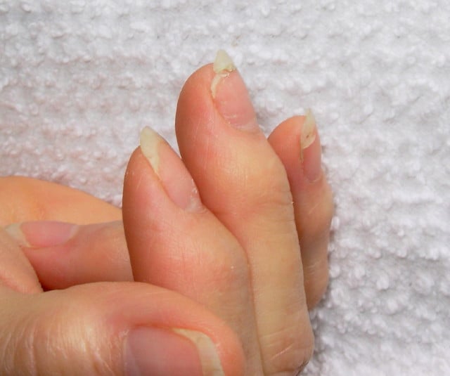
B rittle nails are abnormalities of the nail plate that are characterized by poor nail structure affecting all fingernails and toenails seen as thin, dry nails that easily chip, split, and are difficult to maintain a clean edge. Usually longitudinal ridging occurs from the nail base to the tips.

Q: What is the nail plate?

A: The nail plate is the hard keratin cover protecting the finger tip and toe tip. The nail plate (non-living tissue) is produced by the nail matrix (living tissue) at the base of the nail plate under the lunula (moon), which is the site of brittle nail development.

Nail Anatomy. A. Nail plate; B. lunula; C. root; D. sinus; E. matrix; F. nail bed; G. hyponychium; H. free margin. Courtesy Wikipedia.org

Poor nail structure affecting all nails may be a feature of nutritional deficiency in poor diet or malabsorption such as occurs in celiac disease.

Some other causes are: idiopathic (unknown cause), the result of aging, the effects of certain drugs, or an association with systemic autoimmune disorders such as vitiligo, alopecia areata (with pitting), psoriasis (with pitting), and lichen planus. 

External (non-nutritional or disease) causes of dry, brittle nails, such as detergents and cleaners, would only affect fingernails but not toenails.

Note: It has been shown that working with your hands in water or soaking them through activities like swimming does not cause dry, brittle nails but will worsen them.

What Are Brittle Nails In Celiac Disease and/or Gluten Sensitivity?

Late Menarche (Start of Periods)

Courtesy quizlet.com

What Is Diffuse Alopecia?

D iffuse alopecia is characterized by abnormal hair loss or baldness.

Hair loss usually develops gradually and may be patchy or all over (diffuse). The average scalp contains about 100,000 hairs. Roughly 100 hairs are shed from the head every day.

Diffuse baldness not related to male pattern or heredity can be related to aging, nutritional deficiencies, some froms of dermatitis, radiation, endocrine disorders, especially thyroid hormone imbalance and diabetes, and undue stress.

Q: Can sudden stress cause hair loss?

A: A sudden physical or emotional stress may cause one-half to three-quarters of the hair throughout the scalp to shed. Other causes that need to be evaluated include use of common medications such as birth control pills, blood thinners, and anti-inflammatory pain drugs, and continued exposure to environmental chemicals such cleaning products.

What Is Diffuse Alopecia In Celiac Disease and/or Gluten Sensitivity?

Bladder Infection (Cystitis)

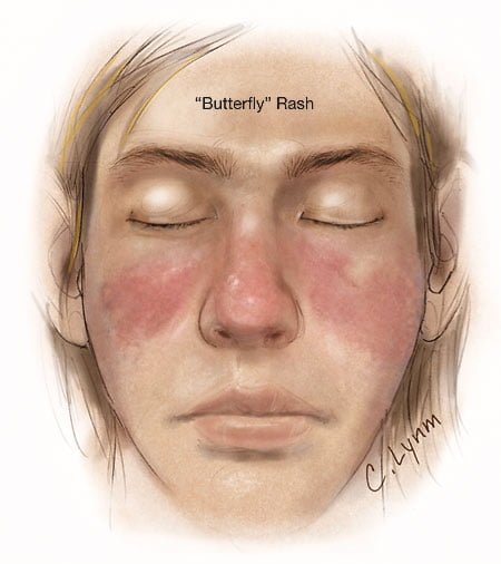
Image showing butterfly rash of SLE. Courtesy JAMA.
Image showing butterfly rash typical of SLE. Courtesy JAMA.

What Is Systemic Lupus Erythematosus?

S ystemic lupus erythematosus (SLE) is a chronic autoimmune inflammatory disease that is characterized by involvement of multiple organs due to the production of antibodies to components of the cell nucleus.1 SLE has an unpredictable course of acute flare-ups and remissions.

Severity depends on the extent of organs affected with skin and nail involvement, called discoid lupus, being the least serious and inflammmation of the kidney, called lupus nephritis, being the worst.

Nevetheless, a classic presentation is development of a rash over the cheeks and nose that resembles a butterfy with wings spread hence the name “butterfly rash.”

Symptoms are many and varied depending on the tissues affected and are often not specific, for example hair loss has a variety of causes. Symptoms can be confused by co-existence with other autoimmune disease such as Sjogren’s syndrome.

Systemic lupus erythematosus should be managed by a specialist. Symptoms can be controlled with steroid therapy, but this disease can be a cause of premature death  mainly from active disease, organ failure (e.g., kidneys), infection, or cardiovascular disease from accelerated atherosclerosis.

Certain common medicines known to cause drug-induced lupus are:

  • Isoniazid
  • Hydralazine
  • Procainamide

Other less common drugs may also cause the condition. These may include:

  • Anti-seizure medications
  • Capoten
  • Chlorpromazine
  • Etanercept
  • Infliximab
  • Methyldopa
  • Minocycline
  • Penicillamine
  • Quinidine
  • Sulfasalazine

Symptoms tend to occur after taking the drug for at least 3 to 6 months.2

Although there is a strong familial aggregation, the disease is relatively uncommon and most cases are sporadic.1 According to the Center for Diseases (CDC), lupus most commonly affects women of childbearing age but also occurs in infants, children, adolescents, and men with peak occurrence between ages 15 and 40. Blacks (and possibly Hispanics, Asians, and Native Americans) are affected more than Whites.

What Is Systemic Lupus Erythematosus In Celiac Disease and/or Gluten Sensitivity?

Sources:
  1. http://www.cdc.gov/arthritis/basics/lupus.htm [] []
  2. www.nlm.nih.gov/medlineplus/ency/article/000446.htm []

IgA Nephropathy

X-ray of lungs in sacroidosis showing honeycoming. Courtesy Wikimedia.
X-ray of Lungs in Sacroidosis Showing Honeycoming. Courtesy Wikimedia.

What Is Sarcoidosis?

S arcoidosis is a multisystem granulomatous disease characterized by hard granulomas and inflammation of the alveoli in lungs that occurrs in 80% of patients.

Alveoli are the tiny one cell thick round structures that are clustered at the ends of air passages in the lungs where oxygen from air inhaled is exchanged for carbon dioxide from blood which is breathed out.

Q: What are granulomas?

A: Granulomas are collections of macrophages (white blood cells) that targeted and enclosed but failed to destroy a foreign substance that entered the body.  In turn, the macrophages become encased by other immune cells and fibroblasts and collagen. They can occur in any organ.

Presently, the etiology, or cause, of sarcoidosis is unknown, although it is thought the predisposition is genetic with a local immune mechanism. This disease has flares when active and remissions when symptoms subside.

Referral to a specialist is important for proper treatment and monitoring. The main treatment for severe active sarcoidosis is prednisone, a type of steroid that reduces inflammation in the body. In most people, prednisone relieves symptoms within a couple of months, although most people need to take prednisone for 12 months or longer. Long-term use of prednisone, especially at high doses, can cause serious side effects. Side effects include bone loss, elevated blood sugar, and high blood pressure.

When wheezing and coughing are a problem, inhaler devices are used to deliver medicine into the lungs in order to help open the airways.1

Ibuprofen is prescribed if sarcoidosis is causing joint pain. Steroid creams are prescribed for skin problems.

Sarcoidosis affects 10 to 20 person per 100,000.

What Is Sarcoidosis In Celiac Disease and/or Gluten Sensitivity?

Sources:
  1. National Heart, Lung, and Blood Institute []