Skip to content

Uveitis, Non-Infectious  

Courtesy Wikipedia.com
Parathyroid Glands in the Neck. Courtesy Wikipedia.com

What Is Secondary Hyperparathyroidism?

S econdary hyperparathyroidism is a parathyroid disorder resulting from hypocalcemia (low blood calcium level) that is characterized by excessive production of parathyroid hormone in the attempt to normalize the low blood calcium by releasing calcium from bone.

Parathyroid hormone is produced by the four pea sized parathyroid glands that are located on the thyroid gland in the front of the neck. In part, because the thyroid and parathyroid glands share the same anatomic place in the body and because they have similar names, they are often confused although they have completely different actions.

Parathyroid hormone regulates calcium and the opposing mineral phosphorus in the blood. In secondary hyperparathyroidism, calcium blood levels are low to normal while phosphorus levels are increased which stimulates the outpouring of parathyroid hormone.

Q: How does secondary hyperparathyroidism differ from primary hyperparathyroidism?

A: In primary hyperparathyroidism blood calcium is high and phosphorus is low, which is the opposite of secondary hyperparathyroidism.

The most common cause of secondary hyperparathyroidism is kidney disease causing failure to reabsorb calcium followed by vitamin D deficiency and malabsorption.

What Is Secondary Hyperparathyroidism In Celiac Disease and/or Gluten Sensitivity?

Endocrine Glands: definition

Glands which empty their hormone substances into the bloodsteam and from there to a targeted part of the body such as insulin produced by the pancreas and thyroxine produced by the thyroid gland. They are… 

Tremors

chronic abdominal distention celiac disease gluten symptomWhat Is Chronic Abdominal Distention?

A bdominal distention, or bloating, is characterized by enlargement in normal size of abdomen not due to fatty tissue.

Q: Why does the abdomen enlarge?

A: The abdomen enlarges because the bowel is dysfunctional. Loops of bowel usually lack normal peristalsis (rhythmic wave-like muscle movement) trapping gas in stagnant loops of bowel, inflammation swells loops of bowel also trapping gas, and dysbiosis overproduces gas. All these factors distend the abdomen.

What Is Chronic Abdominal Distention In Celiac Disease and/or Gluten Sensitivity?

Psoriasis

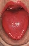
niacin deficiencyWhat Is A Fiery Red, Smooth, Burning Tongue?

A fiery red, smooth, burning tongue is an alteration in tongue tissue that is characteristic of advanced niacin deficiency.1

Q: How does niacin deficiency cause the tongue to be red and sore?

A: Niacin is an essential B vitamin that is required for a healthy tongue and by all body cells as well. Deficiency first shows in tissues with rapid cellular turnover, such as mucosal cells of the tongue.

When absorbed from the small intestines, niacin (the form in food) becomes the active form niacinamide. Niacinamide is converted by the body into co-enzymes which are present in all cells. These enzymes function in oxidation-reduction reactions essential for release of energy from carbohydrates, fats, and proteins and are needed as components for more than 200 enzymes involved in metabolism.

In addition to producing energy, niacinamide is essential for healthy skin and the mucosal lining of the digestive tract, normal functioning of the brain and nervous system, and production of steroid hormones from adrenal glands and hormones from sex glands.

What Is A Fiery Red, Smooth, Burning Tongue In Celiac Disease and/or Gluten Sensitivity?

Sources:
  1. Krause’s Food, Nutrition, & Diet Therapy. 10th Edition. Kathleen Mahan, Sylvia Escott-Stump. 2000. W.B. Saunders Company. []

Smell, Loss of  

CT Scan of Parathyroid Carcinoma Displacing the Trachea. Courtesy Orphanet Journal of Rare Diseases
CT Scan of Parathyroid Carcinoma Displacing the Trachea by Doina Piciu. Courtesy Orphanet Journal of Rare Diseases

What Is Parathyroid Carcinoma?

P arathyroid carcinoma is a slow growing rare malignancy involving overactive parathyroid glands and is characterized by profound hypercalcemia (elevated blood calcium level), parathyroid hormone levels of more than 3 times upper normal limits, and palpable neck mass.1

Parathyroid carcinoma is a cause of severe hyperparathyroidism. Hyperparathyroidism results from overproduction of parathyroid hormone by the parathyroid glands.

Research comparing patients with carcinoma to patients with primary hyperparathyroidism showed that patients with parathyroid carcinoma had significantly higher serum parathyroid hormone and calcium levels compared with patients with primary hyperparathyroidism.2

Q: Why is the blood calcium level elevated?

A: Blood calcium level is elevated due to overproduction of parathyroid hormone by the diseased gland(s).

The function of parathyroid hormone (PTH) is to keep calcium blood levels normal, which it does by drawing calcium out of bones as needed. The problem of too much PTH is that bone is constantly robbed of calcium and this depletion causes bone demineralization, resulting in osteopenia progressing to osteoporosis. Of course, weakened bones are subject to breakage with light trauma.

On the other hand, too much calcium in the blood can cause kidney stone formation because urine production is the way the body excretes excess calcium. That is, highly concentrated calcium precipitates out of the urine solution to form stones.

With an estimated incidence of 0.015 per 100,000 population and an estimated prevalence of .005% in the United States, parathyroid cancer is one of the rarest of all human cancers according to the National Cancer Institute.

What Is Parathyroid Carcinoma In Celiac Disease and/or Gluten Sensitivity?

Sources:
  1. Schaapveld M, Jorna FH, Aben KK, Haak HR, Plukker JT, Links TP. Incidence and prognosis of parathyroid gland carcinoma: a population-based study in The Netherlandsestimating the preoperative diagnosis. Am J Surg. 2011 Nov;202(5):590-7. doi: 10.1016/j.amjsurg.2010.09.025. Epub 2011 Aug 20. []
  2. Schaapveld M, Jorna FH, Aben KK, Haak HR, Plukker JT, Links TP. Incidence and prognosis of parathyroid gland carcinoma: a population-based study in The Netherlandsestimating the preoperative diagnosis. Am J Surg. 2011 Nov;202(5):590-7. doi: 10.1016/j.amjsurg.2010.09.025. Epub 2011 Aug 20. []

Xerophthalmia

Parathyroid gland anatomyWhat Is Idiopathic Hypoparathyroidism?

I diopathic hypoparathyroidism is a metabolic condition that results from reduced secretion or impaired action of parathyroid hormone (PTH) which results in a combination of low calcium and elevated phosphorus levels in the body.

Calcium and phosphorus are minerals that act in opposition to each other in the body. Idiopathic means the cause is not known.

Parathyroid hormone is produced by the four pea sized parathyroid glands that are located on the thyroid gland in the front of the neck. Partly because the thyroid and parathyroid glands share the same anatomic place in the body and partly because they have similar names, they are often confused although they have completely different actions.

Q: What does parathyroid hormone do in the body?

A: This vital hormone maintains a correct balance of calcium and phosphorous in the bloodstream and ultimately has an effect on all organs because of the complexity of intracellular calcium physiology.1 

Parathyroid hormone is required to sustain life, therefore undetected or misdiagnosed hypoparathyroidism may pose a significant threat to health outcomes, as its presence may increase disease and mortality in affected individuals.

The clinical consequences of parathyroid hormone deficiency or impaired receptor action involve many body functions. In some patients, however, its manifestation may be non-specific, and in these cases the correct diagnosis may be easily missed.

Digestive manifestations of hypoparathyroidism are few and consist mainly of steatorrhea due to insufficient meal-stimulated cholecystokinin secretion by the duodenal mucosa.2

Laboratory measurements show hypocalcemia (low blood calcium level), hyperphosphatemia (high blood phosphate level), and inappropriately low or undetectable parathyroid hormone levels. Treatment consists of oral calcium supplementation and vitamin D derivatives.3

Idiopathic hypoparathyroidism is rare, although there is a growing incidence of the autoimmune form of hypoparathyroidism, which may occur in combination with other autoimmune diseases such as celiac disease.

What Is Idiopathic Hypoparathyroidism In Celiac Disease and/or Gluten Sensitivity?

Sources:
  1. Abboud B, Daher R, Boujaoude J. Digestive manifestations of parathyroid disorders. World J Gastroenterol. 2011 Sep 28;17(36):4063-6. doi: 10.3748/wjg.v17.i36.4063. []
  2. Abboud B, Daher R, Boujaoude J. Digestive manifestations of parathyroid disorders. World J Gastroenterol. 2011 Sep 28;17(36):4063-6. doi: 10.3748/wjg.v17.i36.4063. []
  3. Krysiak R, Handzlik-Orlik G, Kedzia A, Machnik G, Okopień B. Hypoparathyroidism: the present state of art. Wiad Lek. 2013;66(1):18-29. []

Tetany

VomitingWhat Is Vomiting?

V omiting is the regurgitation of swallowed food back out of the mouth.

What Is Vomiting In Celiac Disease and/or Gluten Sensitivity?

Tuberculosis – Non-Response to Treatment 

Underarm showing skin darkening, which is a feature of Addison's Disease
Underarm showing skin darkening, which is a feature of Addison’s Disease

What Is Primary Addison’s Disease?

A ddison’s disease is an autoimmune destruction of the adrenal glands by autoantibodies that target the adrenal cortex, or outer part of these glands, and is characterized by a slow progressive failure of the adrenal glands to adequately produce its steroid hormones.

Symptoms of adrenal fatigue or failure may not develop until the majority of adrenal tissue is destroyed. When untreated, progression leads to coma, called Addisonian crisis, which is a medical emergency.

There are two adrenal glands each located on top of a kidney and enclosed in a connective tissue capsule. Each is a small, triangular shape that is made of two parts: the outer region and the inner region.

The inner region, called the adrenal medulla, produces epinephrine and norepinephrine chemicals that are needed to deal with stress.

The outer region, called the adrenal cortex, produces adrenocortical (steroid) hormones and releases them into the bloodstream in response to pituitary stimulating hormone from the brain.

Q: What is the function of steroid hormones produced by the adrenal glands?

A: Functions of the three steroid hormones produced by the adrenal glands are:

  1. Glucocorticoids restrain inflammation and metabolism of carbohydrates, fats and proteins to maintain a normal glucose blood level. The major glucocorticoid is hydrocortisone.
  2. Mineralocorticoids regulate the retention and excretion of fluids and electrolytes by the kidneys. The most important mineralocorticoid is aldosterone.
  3. Androgen (testosterone) is a male sex hormone.

Secondary adrenal insufficiency may develop from other causes that are not immune related such as chronic infections, tumor, and medications.

What Is Addison’s Disease In Celiac Disease and/or Gluten Sensitivity?

Vasculitis, Cerebral (Cause of Stroke, TIA, and Seizure)

Lactose IntoleranceWhat Is Lactose Intolerance?

L actose intolerance is a well known symptom of carbohydrate malabsorption characterized by inability to properly digest lactose, the sugar in milk, due to low lactase digesting activity in the small intestine.

Lactase is an enzyme produced by specialized cells in the tips of villi within the brush border of the small intestinal lining that digests lactose.

Lactose is a disaccharide, or double sugar, made up of a molecule of glucose and a molecule of galactose (simple sugars). Lactose cannot be absorbed as is into the body unless it is broken down by lactase into glucose and galactose.

Q: How does lactose cause diarrhea?

A: If lactose is not broken down, undigested lactose moves into the colon where it acts osmotically to draw water from the walls of the colon into the lumen, which causes diarrhea by the unnatural amount of water. How much water? Can be 2 liters! Meanwhile, the normal population of colonic bacteria ferment the undigested lactose, generating short-chain fatty acids and hydrogen gas, resulting in bloating pain and flatulence.

Testing for intolerance is based on the action of lactose to increase intestinal permeability.1 Positive response to a breath hydrogen test (BHT), involving 1 – 3 hours of time post ingestion of lactose test dose, signifies malabsorption in the small intestine and fermentation in the colon. If BHT is positive before 60 minutes, the result implies bacteria is abnormally present in the small intestine, causing fermentation there.

Endoscopy is used to measure activity of lactase in a tissue sample.

Treatment is avoidance of milk and dairy products that contain lactose. Nevertheless, milk treated with lactase enzyme can be safely consumed. Fermented milk products like yogurt or kefir are safe because the lactose has been digested by organisms. If there is also a problem with milk protein, then no milk is acceptable that comes from the cow or animal that is causing the allergy.

What Is Lactose Intolerance In Celiac Disease and/or Gluten Sensitivity?

Sources:
  1. Farhadi A, Banan A, Fields J, Keshavarzian A. Intestinal barrier: an interface between health and disease. Journal of Gastroenterology and Hepatology. 2003;18:479-91. []

Attention Deficit Hyperactive Disorder (ADHD)

A small intestinal mucosa from a case of refractory coeliac disease immunostained sequentially for CD3 (alkaline phosphatase-blue) and CD8 (peroxidase-brown). Most intraepithelial lymphocytes are CD3+, CD8-.Courtesy pubcan.org
Mucosa  in refractory celiac  disease immunostained sequentially for CD3 (alkaline phosphatase-blue) and CD8 (peroxidase-brown). Most intraepithelial lymphocytes are CD3+, CD8-. Courtesy pubcan.org

What Is Refractory Celiac Disease?

R efractory celiac disease, formerly called refractory sprue, is a severe complication characterized by persistence of symptoms and intestinal inflammation despite gluten free diet after 12 months.1

Refractory celiac disease appears in two forms, ulcerative jejunitis (RCD I) and cryptic intestinal T-cell lymphoma (RCD II).

Patients with RCD I seem to profit from immunosuppressive treatment, but positive response to corticosteroid treatment does not exclude underlying enteropathy–associated T-cell lymphoma (EATL).

Patients with RCD II have a high percentage of abberant T-cells and is usually resistant to medical therapies. The presence of an aberrant clonal intraepithelial T-cell population has led to the designation of refractory celiac disease with this population as a cryptic intestinal T-cell lymphoma, characterized by frequent dissemination to the blood and the entire gastrointestinal lining.2

  • Refractory sprue may occur after an initial response to gluten free diet or without any evidence of preexisting celiac disease. All other causes of malabsorption must be excluded, such as collagenous colitis.
  • In a subgroup of patients with enteropathy-associated T-cell lymphoma (EATL) there is progressive deterioration of a refractory form of celiac disease. The prognosis is poor, although some patients respond to corticosteroids and immunosuppressive agents.3
  • A nationwide Finnish study showed that patients of male gender, older age, severe symptoms or seronegativity (negative antibody result) at the diagnosis of celiac disease are at risk of future refractory coeliac disease and should be followed up carefully.4
  • Chorea has been described as a paraneoplastic phenomenon in patients with non-Hodgkin’s lymphoma and has been described as associated with lymphoma arising from a background of refractory celiac disease. The finding of chorea in association with celiac disease should prompt a search for possible underlying intestinal T-cell lymphoma.5

How Prevalent Is Refractory Celiac Disease?

Sources:
  1. Murray JA, The widening spectrum of celiac disease. American Journal of Clinical Nutrition. Mar 1999;69 (3):354-365. []
  2. Culliford AN, Green PH. Refractory sprue. Current Gastroenterology Reports. Oct 2003;5(5):373-8. []
  3. Culliford AN, Green PH. Refractory sprue. Current Gastroenterology Reports. Oct 2003;5(5):373-8. []
  4. Ilus T, Kaukinen K, Virta LJ, Huhtala H, Mäki M, Kurppa K, Heikkinen M, Heikura M, Hirsi E, Jantunen K, Moilanen V, Nielsen C, Puhto M, Pölkki H, Vihriälä I, Collin P. Refractory coeliac disease in a country with a high prevalence of clinically-diagnosed coeliac disease. Aliment Pharmacol Ther. 2014 Feb;39(4):418-25. doi: 10.1111/apt.12606. []
  5. Kitiyakara T, Jackson M, Gorard DA. Refractory coeliac disease, small-bowel lymphoma and chorea. J R Soc Med. 2002 Mar;95(3):133-4. []