Skip to content

Adenocarcinoma Of Small Intestine (Cancer)

Section of small bowel surgically removed for adenocarcinoma that grew through the wall. By: CDC/ Dr. Edwin P. Ewing, Jr.

What Is Adenocarcinoma Of Small Intestine?

Adenocarcinomas are malignant tumors, or cancer, of the small bowel arising out of glandular tissue. They fall in the category of rare neoplasm, comprising only 3% of all gastrointestinal malignancies.

Primary adenocarcinoma is the most common histological (cell) subtype constituting 35–50% of cases.1 

Q: What does adenocarcinoma look like?

A: Adenocarcinoma may manifest as strictures, nodules, excavating masses, or annular lesions.2

What Is Adenocarcinoma Of Small Intestine In Celiac Disease and/or Gluten Sensitivity?

Sources:
  1. Benhammane H, El M’rabet FZ, Serhouchni KI, El yousfi M, Charif I, Toughray I, et al. Small Bowel Adenocarcinoma Complicating Coeliac Disease: A Report of Three Cases and the Literature Review. Case Rep Oncol Med. 2012; 2012: 935183. Published online 2012 December 1. doi: 10.1155/2012/935183 []
  2. Ramachandran I, Sinha R, Rajesh A, Verma R. Multidetector row CT of small bowel tumors.  Clinical Radiology. 2007; 62:607-614. []

Tremors

chronic abdominal distention celiac disease gluten symptomWhat Is Chronic Abdominal Distention?

A bdominal distention, or bloating, is characterized by enlargement in normal size of abdomen not due to fatty tissue.

Q: Why does the abdomen enlarge?

A: The abdomen enlarges because the bowel is dysfunctional. Loops of bowel usually lack normal peristalsis (rhythmic wave-like muscle movement) trapping gas in stagnant loops of bowel, inflammation swells loops of bowel also trapping gas, and dysbiosis overproduces gas. All these factors distend the abdomen.

What Is Chronic Abdominal Distention In Celiac Disease and/or Gluten Sensitivity?

Turner’s Syndrome 

Hyperkeratosis Back of Arm. GFW Photo.
Hyperkeratosis Back of Arm. GFW Photo

What Is Follicular Hyperkeratosis?

F ollicular hyperkeratosis is an abnormal skin condition characterized by disordered overgrowth of the horny layer of the epidermis with horny plugs filling the openings of hair follicles. The plugs look like bumps in hair follicles.

Follicular hyperkeratosis with corkscrew hairs (coiled hairs in the follicles) appears in scurvy, which is advanced vitamin C deficiency.1

Typical  hyperkeratotic papules first appear on the extensor surfaces of the extremities (e.g. the back of upper arms), shoulders, and buttocks.

Phrynoderma is a distinctive form of follicular hyperkeratosis with papule formation in dilated hair follicles believed to be a manifestation of severe malnutrition.

Q: Why does hyperkeratosis develop?

A: Follicular hyperkeratosis develops from lack of necessary nutrients to support healthy skin. Although originally thought to represent only vitamin A deficiency or vitamin C in the case of scurvy, several studies have demonstrated deficiencies of B vitamins and vitamin E. General malnutrition seems to be the strongest association. The clinical picture typically improves with enhanced nutritional status.2

What Is Follicular Hyperkeratosis In Celiac Disease and/or Gluten Sensitivity?

Sources:
  1. Krause’s Food, Nutrition, & Diet Therapy. 10th Edition. Kathleen Mahan, Sylvia Escott-Stump. 2000. W.B. Saunders Company. []
  2. Maronn M, Allen DM, Esterly NB. Phrynoderma: a manifestation of vitamin A deficiency?… The rest of the story. Pediatr Dermatol. 2005 Jan-Feb;22(1):60-3. []

Short Stature

What Is Vitiligo? V itiligo is a pigmentation disorder of the skin characterized by permanent loss of melanocytes in defined areas and, in some patients, antibodies to melanin. Vitiligo has significant psychological impact if occurring before… 

Xerophthalmia

Parathyroid gland anatomyWhat Is Idiopathic Hypoparathyroidism?

I diopathic hypoparathyroidism is a metabolic condition that results from reduced secretion or impaired action of parathyroid hormone (PTH) which results in a combination of low calcium and elevated phosphorus levels in the body.

Calcium and phosphorus are minerals that act in opposition to each other in the body. Idiopathic means the cause is not known.

Parathyroid hormone is produced by the four pea sized parathyroid glands that are located on the thyroid gland in the front of the neck. Partly because the thyroid and parathyroid glands share the same anatomic place in the body and partly because they have similar names, they are often confused although they have completely different actions.

Q: What does parathyroid hormone do in the body?

A: This vital hormone maintains a correct balance of calcium and phosphorous in the bloodstream and ultimately has an effect on all organs because of the complexity of intracellular calcium physiology.1 

Parathyroid hormone is required to sustain life, therefore undetected or misdiagnosed hypoparathyroidism may pose a significant threat to health outcomes, as its presence may increase disease and mortality in affected individuals.

The clinical consequences of parathyroid hormone deficiency or impaired receptor action involve many body functions. In some patients, however, its manifestation may be non-specific, and in these cases the correct diagnosis may be easily missed.

Digestive manifestations of hypoparathyroidism are few and consist mainly of steatorrhea due to insufficient meal-stimulated cholecystokinin secretion by the duodenal mucosa.2

Laboratory measurements show hypocalcemia (low blood calcium level), hyperphosphatemia (high blood phosphate level), and inappropriately low or undetectable parathyroid hormone levels. Treatment consists of oral calcium supplementation and vitamin D derivatives.3

Idiopathic hypoparathyroidism is rare, although there is a growing incidence of the autoimmune form of hypoparathyroidism, which may occur in combination with other autoimmune diseases such as celiac disease.

What Is Idiopathic Hypoparathyroidism In Celiac Disease and/or Gluten Sensitivity?

Sources:
  1. Abboud B, Daher R, Boujaoude J. Digestive manifestations of parathyroid disorders. World J Gastroenterol. 2011 Sep 28;17(36):4063-6. doi: 10.3748/wjg.v17.i36.4063. []
  2. Abboud B, Daher R, Boujaoude J. Digestive manifestations of parathyroid disorders. World J Gastroenterol. 2011 Sep 28;17(36):4063-6. doi: 10.3748/wjg.v17.i36.4063. []
  3. Krysiak R, Handzlik-Orlik G, Kedzia A, Machnik G, Okopień B. Hypoparathyroidism: the present state of art. Wiad Lek. 2013;66(1):18-29. []

Uveitis, Non-Infectious  

Courtesy Wikipedia.com
Parathyroid Glands in the Neck. Courtesy Wikipedia.com

What Is Secondary Hyperparathyroidism?

S econdary hyperparathyroidism is a parathyroid disorder resulting from hypocalcemia (low blood calcium level) that is characterized by excessive production of parathyroid hormone in the attempt to normalize the low blood calcium by releasing calcium from bone.

Parathyroid hormone is produced by the four pea sized parathyroid glands that are located on the thyroid gland in the front of the neck. In part, because the thyroid and parathyroid glands share the same anatomic place in the body and because they have similar names, they are often confused although they have completely different actions.

Parathyroid hormone regulates calcium and the opposing mineral phosphorus in the blood. In secondary hyperparathyroidism, calcium blood levels are low to normal while phosphorus levels are increased which stimulates the outpouring of parathyroid hormone.

Q: How does secondary hyperparathyroidism differ from primary hyperparathyroidism?

A: In primary hyperparathyroidism blood calcium is high and phosphorus is low, which is the opposite of secondary hyperparathyroidism.

The most common cause of secondary hyperparathyroidism is kidney disease causing failure to reabsorb calcium followed by vitamin D deficiency and malabsorption.

What Is Secondary Hyperparathyroidism In Celiac Disease and/or Gluten Sensitivity?

Short Duration of Breast Feeding 

dermatomyositisWhat Is Dermatomyositis?

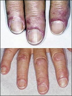
D ermatomyositis is a rare autoimmune systemic disease of the connective tissue that is characterized by inflammatory and debilitating degenerative changes in the muscles and in the skin. 

Dermatomyositis results in symmetric, proximal muscle weakness of limbs (upper arms and legs), and skin manifestations. 50-70% of patients have circulating myositis-specific auto-antibodies.

The course of dermatomyositis is unpredictable being marked by spontaneous flare-ups and remissions. It can begin slowly or abruptly according to the factor that is triggering the onset such as infection, medications like phenytoin, and autoimmune disease.

Q: What are the skin manifestations of dermatomyositis?

A: Classic skin manifestations of dermatomyositis include these features:

  • The heliotrope rash (lilac color) on upper eyelids.
  • Rash on face, neck, shoulders, upper chest, elbows, knees, knuckles, and back.
  • Gottron’s papules (scaly, red eruptions or  patches over the knuckles, elbows, and knees).
  • The V-sign (rash front of neck and chest).
  • The shawl sign (rash distribution on shoulders and back).1

Additional cutaneous manifestations are described below under symptoms.

Dermatomyositis is associated with an increased risk of  cancer, other autoimmune diseases, such as lupus and psoriasis, and it can be a complication of interferon-α therapy. About 1 person in 100,000 are affected according to various studies. While it affects all ages, women have twice the occurence of men.

There is no cure for dermatomyositis, but the symptoms can be treated.  Options include medication, physical therapy, exercise, heat therapy (including microwave and ultrasound), orthotics and assistive devices, and rest.  The standard treatment for dermatomyositis is a corticosteroid drug, given either in pill form or intravenously.  Immunosuppressant drugs, such as azathioprine and methotrexate, may reduce inflammation in people who do not respond well to prednisone.2

What Is Dermatomyositis In Celiac Disease and/or Gluten Sensitivity?

Sources:
  1. Marvi U, Chung L, Fiorentino DF. Clinical presentation and evaluation of dermatomyositis. Indian J Dermatol. 2012 Sep;57(5):375-81. doi: 10.4103/0019-5154.100486. []
  2. National Institute of Neurological Disorders and Stroke. []

Sperm Abnormalities 

This 21 year-old woman (left) appears as old as her 70 year-old granny (right). Courtesy Prof Dr Chua Chung Nen
This 21 year-old woman (right) appears as old as her 70 year-old grandmother (left). Courtesy Prof Dr Chua Chung Nen

What Is Acquired Cutis Laxa?

A cquired cutis laxa is an uncommon skin disorder characterized by abnormal reduction and degeneration of elastic fibers of the skin that can appear simply as thick, saggy skin with loose folds to severe involvement showing a premature aged appearance.

Q: What are elastic fibers of the skin?

A: Elastic fibers of the skin are connective tissue found in the dermis, which is the layer of skin under the epidermis, or surface layer. They hold the shape of skin and are important for wound healing in the development of scars.

What Is Cutis Laxa In Celiac Disease and/or Gluten Sensitivity?

Rickets

Seborrhea patches at the inner eyebrows. GFW

What Is Seborrhea Dermatitis?

S eborrhea dermatitis is a recurring inflammatory disorder of sebaceous glands characterized by scaly patches of skin, often with bumps.

Seborrhea dermatitis results from the body’s inflammatory reaction to invasion by pityrosporum yeast that naturally inhabits the scalp and skin.1 Inflammation is the normal response to tissue injury and germ invasion.

Pityrosporum is a yeast that is commonly present worldwide. Its development depends on various factors that predispose to pityriasis versicolor, a chronic and mild superficial yeast infection. These infections usually are asymptomatic without itching or pain and without cellular and/or antibody responses.2

Q: Why are the sebaceous glands particularly affected by this yeast?

A: Pityrosporum yeast is an organism that needs oil produced by sebaceous glands to grow. If conditions permit, this yeast invades the superficial layer of skin and hair shafts to reproduce, causing infection. Such conditions include weakened skin due to nutritional deficiencies, excessive build-up of oil on skin, and altered immunity due to systemic disease such as psoriasis.

In adults, areas of skin that are the most affected have the greatest number of sebaceous glands especially the scalp, back, underarms, and the face including the eyelids, eyebrows and side folds of the nose.

All ages are subject to seborrhea dermatitis, and males have a higher occurence than females.

What Is Seborrhea In Celiac Disease and/or Gluten Sensitivity?

Sources:
  1. Krause’s Food, Nutrition, & Diet Therapy. 10th Edition. Kathleen Mahan, Sylvia Escott-Stump. 2000. W.B. Saunders Company. []
  2. Zarei-Mahmoudabadi A, Zarrin.  M, Mehdinezhad F. Seborrheic dermatitis due to Malassezia species in Ahvaz, Iran. Iran J Microbiol. 2013 Sep;5(3):268-71. []

Endocrine Glands: definition

Glands which empty their hormone substances into the bloodsteam and from there to a targeted part of the body such as insulin produced by the pancreas and thyroxine produced by the thyroid gland. They are…