
 PART 1: Our Digestive System

Our digestive system consists of many organs and parts. Each performs its own function while interacting in harmony with others every time we eat. These interesting and complex organs and parts turn the food we eat into specific components we need to fuel, build, maintain, repair, regulate, and protect our bodies.
Muscles, nerves, reflexes, enzymes, and hormones of the digestive tract all work together to change our food into nutrients that can be absorbed into our main transport systems: our bloodstream and our lymph system. These two transport systems then carry fresh supplies of nutrients throughout our body to nourish every individual cell.
Our brain monitors our nutritional needs and employs a hunger mechanism to alert us when fresh food is needed. Hunger causes appetite which is the desire for food. The tempting sight, smell, taste and expectation of food stimulate involuntary sensory nerves. By reflex action, these nerves cause muscle and sensory activity in various digestive organs. Salivary glands begin to secrete saliva as stomach glands and muscles become active.
How Digestion Works – A Walk Through…
First we chew our food. After we chew it, our throat swallows it, and our esophagus transports it to our stomach. Our stomach dissolves and sends it on to our small intestine, where it is digested and absorbed into our blood and lymph systems. Our small intestine then sends the undigestible substances to our large intestine where water is absorbed. Normal bacteria living there go to work on the residue to extract more nutrients, while producing nourishment for our colon and substances for our body. The waste is then sent on to our rectum, which forms it. Finally, our anus expels it.
Now, that we have the basics of digestion, let’s take a closer look. The following is based in part on an excerpt from the The National Digestive Diseases Information Clearinghouse.
Movement of Food Through the System
 The first major muscle movement occurs when food or liquid is swallowed. Although we are able to start swallowing by choice, once the swallow begins, it becomes involuntary and proceeds under the control of the nerves.
The large, hollow organs of the digestive system contain muscle that enables their walls to move. The movement of organ walls can propel food and liquid and also can mix the contents within each organ. Typical movement of the esophagus, stomach, and intestine is called peristalsis.
The action of peristalsis looks like an ocean wave moving through the muscle. The muscle of the organ produces a narrowing and then propels the narrowed portion slowly down the length of the organ. These waves of narrowing push the food and fluid in front of them through each hollow organ.
The esophagus is the organ into which the swallowed food is pushed. It connects the throat above with the stomach below. At the junction of the esophagus and stomach, there is a ringlike valve closing the passage between the two organs. However, as the food approaches the closed ring, the surrounding muscles relax and allow the food to pass.
The food then enters the stomach, which has three mechanical tasks to do. First, the stomach must store the swallowed food and liquid. This requires the muscle of the upper part of the stomach to relax and accept large volumes of swallowed material. The second job is to mix up the food, liquid, and digestive juice produced by the stomach. The lower part of the stomach mixes these materials by its muscle action. The third task of the stomach is to empty its contents slowly into the small intestine.
Several factors affect emptying of the stomach, including the nature of the food (mainly its fat and protein content) and the degree of muscle action of the emptying stomach and the next organ to receive the contents (the small intestine). As the food is digested in the small intestine and dissolved into the juices from the pancreas, liver, and intestine, the contents of the intestine are mixed and pushed forward to allow further digestion.
Finally, all of the digested nutrients are absorbed through the intestinal walls. The waste products of this process include undigested parts of the food, known as fiber, and older cells that have been shed from the mucosa. These materials are propelled into the colon, where they remain to be further broken down by the millions of normal microbes that live there. This usually takes a day or two, until the feces are expelled by a bowel movement.
Production of Digestive Juices
 Mouth. The digestive glands that act first are in the mouth – the salivary glands. Saliva produced by these glands contains an enzyme called ptyalin (or salivary amylase) that begins to digest into smaller molecules any starch that is contained in food.
Use those teeth! It is very important to thoroughly chew food in order to mix it with ptyalin. Resist the urge to wash food down with a beverage! Gulping food with liquids not only diminishes the action of ptyalin but also washes away the ptyalin…such a waste of enzyme production.
An important action to improve overall digestion is to drink clear liquids before eating in order to provide the digestive tissue and glands with water for optimum function.
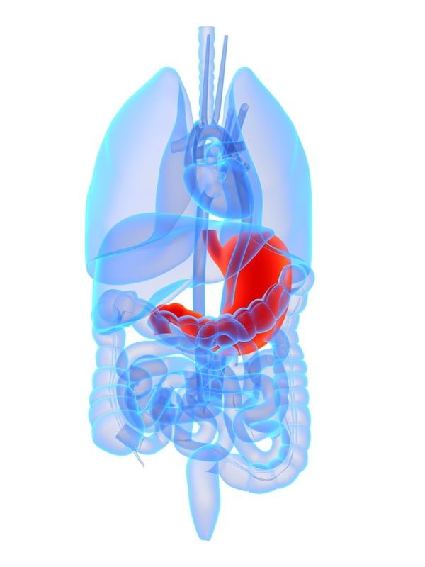 Stomach. The next set of digestive glands is in the stomach lining. They produce the enzymes pepsin that begins the digestion of protein and lipase that begins the digestion of fat.
Just as importantly, as soon as food enters the mouth or even at the sight of food, the stomach produces acid to dissolve food that is eaten. Stomach acid is the powerful hydrochloric acid! One of the unsolved puzzles of the digestive system is why the acid juice of the stomach does not dissolve the tissue of the stomach itself. In healthy people, the stomach mucosa is able to resist the juice, although food and other tissues of the body cannot.
Partially digested food can take 1½ to 6 hours before passing on to the small intestine, depending on the type and amount of food eaten. Rich food, raw food, and/or large quantities substantially delay passage.
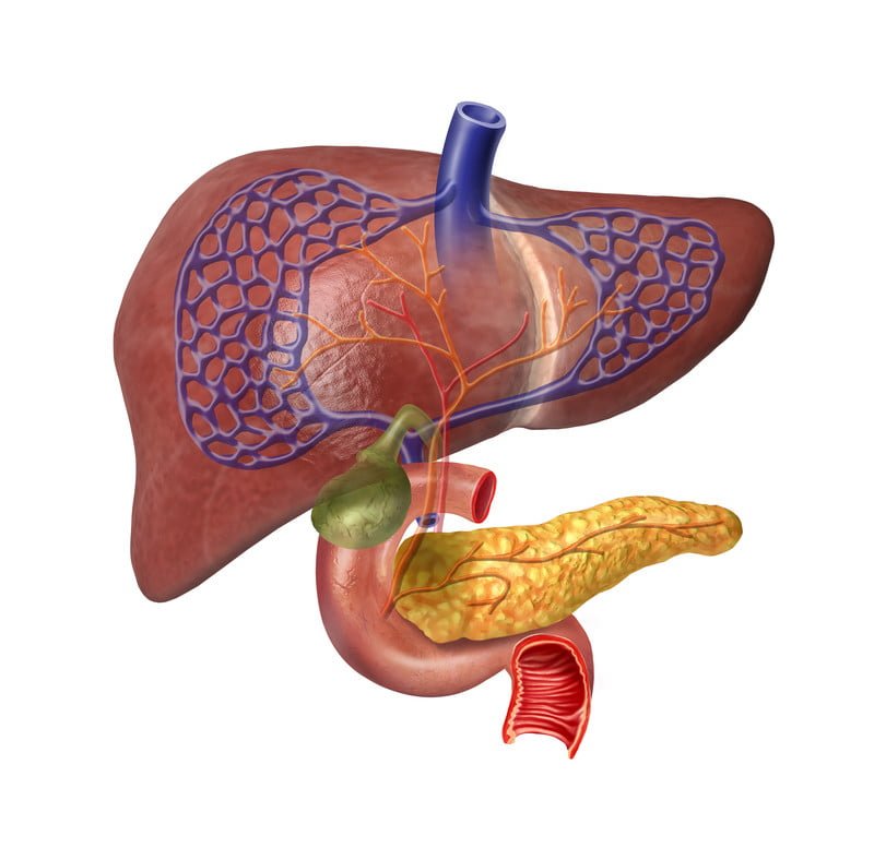
Pancreas and duodenum. After the stomach empties the food and juice mixture into the small intestine, the juices of two other digestive organs mix with the food to continue the process of digestion. One of these organs is the pancreas. It produces a juice that contains a wide array of enzymes to break down the carbohydrate, fat, and protein in food. These are lipase to digest fats, protease to digest proteins, and amylase to digest complex carbohydrates.
Other enzymes that are active in the process come from glands in the wall of the intestine or even a part of that wall. Peptidases finish the digestion of protein and disaccharidases finish the digestion of sugars.
Liver and gallbladder. The liver produces a digestive juice called bile. The bile is stored between meals in the gallbladder. At mealtime, it is squeezed out of the gallbladder into the bile ducts to reach the intestine (duodenum) and mix with the fat in our food.
Bile acids dissolve the fat into the watery contents of the intestine, much like detergents that dissolve grease from a frying pan. After the fat is dissolved, it is digested by enzymes from the pancreas and also the lining of the intestine.
Absorption and Transport of Nutrients
Digested molecules of food, as well as water and minerals from the diet, are absorbed from the cavity of the upper small intestine. Most absorbed materials cross the mucosa into the blood and are carried off in the bloodstream to other parts of the body for storage or further chemical change. As already noted, this part of the process varies with different types of nutrients.
- Carbohydrates. It is recommended that about 55 to 60 percent of total daily calories be from carbohydrates. Some of our most common foods contain mostly carbohydrates. Examples are GF bread, potatoes, legumes, rice, fruits, and vegetables. Many of these foods contain both starch and fiber.
The digestible carbohydrates are broken into simpler molecules by enzymes in the saliva, in juice produced by the pancreas, and in the lining of the small intestine. Starch is digested in two steps. First, an enzyme in the saliva and pancreatic juice breaks the starch into molecules called maltose. Then an enzyme in the lining of the small intestine (maltase) splits the maltose into glucose molecules that can be absorbed into the blood. Glucose is carried through the bloodstream to the liver, where it is stored or used to provide energy for the work of the body.
Table sugar is another carbohydrate that must be digested to be useful. An enzyme in the lining of the small intestine digests table sugar into glucose and fructose, each of which can be absorbed from the intestinal cavity into the blood. Milk contains yet another type of sugar, lactose, which is changed into absorbable molecules by an enzyme called lactase, also found in the intestinal lining.
- Protein. Foods such as meat, eggs, and beans consist of giant molecules of protein that must be digested by enzymes before they can be used to build and repair body tissues. An enzyme in the juice of the stomach starts the digestion of swallowed protein. Further digestion of the protein is completed in the small intestine. Four pancreatic enzymes, as part of pancreatic juice, finish the bulk of protein digestion that was begun by pepsin in the stomach. Trypsin and chymotrypsin break large chains of proteins into smaller amino acids, called peptides. Carboxypeptidase and aminopeptidase split off one amino acid at a time from peptides. Enzymes in the villi will finish digestion to simple amino acids.
- Fats. Fat molecules are a rich source of energy for the body. The first step in digestion of a fat such as butter is to dissolve it into the watery content of the intestinal cavity. The bile acids produced by the liver act as natural detergents to dissolve fat in water and allow the enzymes to break the large fat molecules into smaller molecules, some of which are fatty acids and cholesterol. The bile acids combine with the fatty acids and cholesterol and help these molecules to move into the cells of the mucosa. In these cells the small molecules are formed back into large molecules, most of which pass into vessels (called lymphatics) near the intestine. These small vessels carry the reformed fat to the veins of the chest, and the blood carries the fat to storage depots in different parts of the body.
- Vitamins. Another vital part of our food that is absorbed from the small intestine is the class of chemicals we call vitamins. The two different types of vitamins are classified by the fluid in which they can be dissolved: water-soluble vitamins (all the B vitamins and vitamin C) and fat-soluble vitamins (vitamins A, D, E and K). Most vitamins, excepting vitamin A, which is changed into retinol, and niacin, which is changed into nicotinimide and nicotinic acid, are absorbed here unchanged into the bloodstream.
- Minerals. Acid in the stomach gives an ionic charge (+ or -) to minerals so they can be absorbed into the small intestinal lining. These mainly include potassium, sodium, calcium, chloride, sulfur, iron, copper, chromium, magnesium, phosphorus, and zinc.
- Water and Salt. Most of the material absorbed from the cavity of the small intestine is water in which salt is dissolved. The salt and water come from the food and liquid we swallow and the juices secreted by the many digestive glands.
How Is the Digestive Process Controlled?
1. Hormone Regulators
A fascinating feature of the digestive system is that it contains its own regulators. The major hormones that control the functions of the digestive system are produced and released by cells in the mucosa of the stomach and small intestine. These hormones are released into the blood of the digestive tract, travel back to the heart and through the arteries, and return to the digestive system, where they stimulate digestive juices and cause organ movement.
The hormones that control digestion are gastrin, secretin, and cholecystokinin (CCK):
- Gastrin causes the stomach to produce an acid for dissolving and digesting some foods. It is also necessary for the normal growth of the lining of the stomach, small intestine, and colon.
- Secretin causes the pancreas to send out a digestive juice that is rich in bicarbonate. It stimulates the stomach to produce pepsin, an enzyme that digests protein, and it also stimulates the liver to produce bile.
- CCK causes the pancreas to grow and to produce the enzymes of pancreatic juice, and it causes the gallbladder to empty.
Additional hormones in the digestive system regulate appetite:
- Ghrelin is produced in the stomach and upper intestine in the absence of food in the digestive system and stimulates appetite.
- Peptide YY is produced in the GI tract in response to a meal in the system and inhibits appetite.
Both these hormones work on the brain to help regulate the intake of food for energy.
2. Nerve Regulators
Two types of nerves help to control the action of the digestive system. Extrinsic (outside) nerves come to the digestive organs from the unconscious part of the brain or from the spinal cord. They release a chemical called acetylcholine and another called adrenaline. Acetylcholine causes the muscle of the digestive organs to squeeze with more force and increase the “push” of food and juice through the digestive tract. Acetylcholine also causes the stomach and pancreas to produce more digestive juice. Adrenaline relaxes the muscle of the stomach and intestine and decreases the flow of blood to these organs.
Even more important, though, are the intrinsic (inside) nerves, which make up a very dense network embedded in the walls of the esophagus, stomach, small intestine, and colon. The intrinsic nerves are triggered to act when the walls of the hollow organs are stretched by food. They release many different substances that speed up or delay the movement of food and the production of juices by the digestive organs.
_____________________________________________________________________________________
PART 2: Impact Of Gluten and Celiac Disease On Digestion
_____________________________________________________________________________________
Celiac disease interferes with digestion by way of inflammation and damage to tissues and structures of the small intestinal lining, the mucosa. Alteration in form and function of the mucosa interferes with the proper absorption of nutrients, causing malabsorption.
Celiac disease causes a variety of gastrointestinal health problems. It affects the harmony of organs by disrupting the action of muscles, nerves, reflexes, enzymes, and hormones. Structural and functional changes lead to disorders such as difficulty swallowing, reflux, dyspepsia, nausea, vomiting, abdominal bloating, constipation and diarrhea.
-
Impact on Hunger and Appetite
Our brain monitors nutritional needs and employs a hunger mechanism to alert us when fresh food is needed. Hunger causes appetite which is the desire for food. The tempting sight, smell, taste and expectation of food stimulate involuntary sensory nerves. By reflex action, these nerves cause muscle and sensory activity in various digestive organs. Salivary glands begin to secrete saliva as stomach glands and muscles become active.
While hunger prompts us to get food, it is up to us to choose nutritious foods and avoid toxic foods containing gluten.
Conditions Affecting Hunger and Appetite in Celiac Disease.
Anorexia can result from:
- Just one or a combination of nutrient deficiencies. These include zinc, iron, magnesium, phosphorus, potassium, thiamin, vitamin B12, and protein.*
- Anxiety.*
- Depression.*
- Primary hyperparathyroidism causes anorexia.*
- Cachexia.*
- Associated autoimmune disease such as sarcoidosis.
Hunger results primarily from:
- Protein and glucose deficiencies.*
Increased thirst results from:
- Deficiencies of omega fatty acids and potassium.*
*Search Health Conditions for Details.
-
Impact On The Mouth
Chewing food begins digestion. Food is made ready to swallow by the tearing and grinding action of the teeth and the chemical breakdown action of saliva. Saliva adapts to the type of food being chewed, dissolving substances and diluting material that would be too concentrated for the stomach.
Saliva consists of mucin and salivary amylase. Mucin is a slippery carbohydrate-protein complex which protects the lining of the mouth. It lubricates food for swallowing, neutralizes acids present in food and kills many bacteria that enter the mouth with food. Salivary amylase works best at breaking down starch when the mouth is neutral or has a slightly alkaline Ph (not acidic).
Conditions and Disease of the Mouth in Celiac Disease.
- Beefy, red smooth tongue with burning from vitamin B12 deficiency.*
- Burning of the lips, mouth and tongue which appears magenta with enlarged and/or atrophied papillae from riboflavin deficiency (geographic tongue).*
- Candida yeast infection.*
- Cheilosis or cracking at the corners of the mouth.
- Defective dental enamel resulting from phosphorus and vitamin D deficiencies.*
- Gingival (gum) inflammation, bleeding, infection and eventual tooth loss from vitamin C deficiency.
- Impaired taste from zinc, vitamin B12, and niacin deficiencies.*
- Increased susceptibility to infection from iron and vitamin A deficiencies.*
- Lowered saliva Ph (acidic), predisposing to dental cavities and poor starch digestion.*
- Mouth ulcers, both aphthous ulcers (canker sores) and non-aphthous ulcers.*
- Oral inflammation from folate and vitamin B6 deficiencies.*
- Pale, sore and swollen tongue from iron deficiency.*
- Scarlet, swollen tongue with burning of the mouth from niacin deficiency.*
*Search Health Conditions for Details.
-
Impact On The Pharynx And Larynx
Swallowing that is started by voluntary action of the mouth is completed by reflex through the larynx and pharynx, or throat. The larynx separates the mouth from the pharynx and controls the passage of food into the esophagus while closing access to the trachea (windpipe). The opening of the trachea (glottis) is closed by the automatic action of the attached epiglottis leaning backward over it which keeps food from entering the lungs. Breathing is held by reflex action to prevent choking.
Conditions and Disease of the Pharynx and Larynx in Celiac Disease.
- Burning of the throat from niacin deficiency.*
- Dysphagia (difficulty or inability to swallow.*
- Cancer of the pharynx.*
- Post-cricoid carcinoma.*
- Laryngospasm from calcium, magnesium or vitamin D deficiencies.*
*Search Health Conditions for Details.
-
Impact On The Esophagus
Located in the middle of the chest, the esophagus is a hollow muscular tube positioned behind the heart and lungs that generally follows the curvature of the spine. The average person’s esophagus is approximately 10 inches long (24 cm). In its resting state, the walls of the esophagus flatten front to back measuring almost 1 inch wide (about 2 cm).
The esophagus performs swallowing through highly coordinated voluntary and involuntary muscle actions (peristalsis) that quickly move the food into the stomach at a rate of 2-3 cm per second. Liquids are able to rapidly pass into the stomach without peristalsis.
The thickened end of the esophagus works in concert with the lower esophageal sphincter (LES) to reliably pass food into the stomach. The LES is a strong muscle band surrounding the stomach opening. It relaxes to allow food into the stomach and closes to keep food from going back up the esophagus, preventing reflux.
Conditions and Disease of the Esophagus in Celiac Disease.
- Acanthosis, Glycogenic (esophageal plaques). *
- Burning of the esophagus from niacin deficiency.*
- Cancer of the esophagus.*
- Candida albicans infection.*
- Dysphagia (difficulty or inability to swallow)*
- Esophageal motor abnormalities.*
- Esophageal small cell carcinoma.*
- Gastroesophageal reflux disease (GERD).*
- Heartburn.*
- Occult bleeding.*
- Plummer-Vinson syndrome from iron deficiency.*
*Search Health Conditions for Details.
-
Impact On The Stomach
The stomach is a complex organ that is chemically monitored and controlled by the brain and nervous system. This form-changing sack acts as a holding tank, mixing food until it is dissolved and broken down into a liquid state called chyme.
The shape and size of the stomach can vary considerably, depending on the position of the body and the amount of filling. When empty, the front and back practically touch. A rough organ, the stomach can hold nearly 2 liters of food and fluid because its wall is elastic and made of stretchable folds called rugae.
Located in the upper abdomen, the stomach is composed of four specialized areas: cardiac, fundus, body and pylorus. Each is distinctly different in muscle and gland structure. When food enters the cardia, it first falls to the pylorus initiating strong muscle contractions that push the food up to the fundus. Muscle contractions of the main body follow, one after another, alternating with pylorus contractions. These powerful muscles churn food and mix it with gastric juice, dissolving and breaking it down into liquid. The pylorus regulates the mixing of food and gastric juice for proper consistency before allowing it to pass into the small intestine.
Gastric juice is made of high concentration of hydrochloric acid and enzymes, pepsin and gastric lipase. Hydrochloric acid dissolves food tissues and kills most bacteria that are swallowed with food. Pepsin begins the digestion of proteins, working best in an acid environment. Gastric lipase is the enzyme that begins fat digestion.
Cells lining the stomach secrete about 3 liters of gastric juice from numerous, microscopic glands every day. The secretion of gastric juice is controlled by a hormone called gastrin. Stomach cells are replaced every three days due to the erosive effect of gastric juice.
Conditions and Disease of the Stomach in Celiac Disease.
- Delayed gastric emptying.*
- Gastric ulcer and ulcerations.*
- Gastritis, collagenous.*
- Gastritis lymphocytic.*
- Increased permeability of gastric mucosa.*
- Increased susceptibility to H. Pylori Bacter infection, leading to ulcers.*
- Indigestion from deficiency of thiamin.
- Low hydrochloric acid levels causing low pepsin output, resulting in poor protein digestion.*
- Nausea and vomiting from deficiencies of magnesium, potassium, and niacin.*
- Occult bleeding.*
*Search Health Conditions for Details.
-
Impact On The Small Intestine
The liquid mass, chyme, enters the small intestine where the work of digestion and absorption of proteins, carbohydrates, fats, vitamins, minerals, and bioflavanoids takes place. The small intestine is a complex, hollow muscular tube comprised of three segments: the duodenum, jejunum, and ileum. It has an overall length of 22 feet (6.8 meters) and is the longest section of the digestive tract.
The inside lining of the small intestine is formed into tightly packed folds that overlay the muscle layer. This folding greatly increases the lining’s surface area contacting the chyme. The folds are covered with multitudinous, tiny, hollow projections called villi. The barely visible villi (0.5 to 1.5 mm long) jut out into the passing chyme. By this formation, more than 4,000,000 villi provide significantly more surface area for absorbing nutrients than the folds alone can provide.
The walls of villi are made up of microscopic absorptive cells that are continually exposed to digested nutrients moving along their surface. These cells do the actual work of absorbing nutrients. In addition, each absorptive cell has numerous microscopic projections called microvilli. The microvilli are collectively called the “brush border.”
The prodigious numbers of microvilli dramatically increase absorption. Altogether, the folds, villi and microvilli in a healthy small intestine make up a huge surface area for absorbing nutrients calculated to equal the size of a tennis court.
Enzymes that finish digestion of the disaccharides, lactose, sucrose and maltose, into simple sugars and small peptide chains into their amino acids are produced in the villi.
Conditions and Disease of the Small Intestine in Celiac Disease.
- Abdominal distention resulting from gas, intestinal edema, and deficiency of niacin.*
- Abdominal pain resulting from inflammation, spasm, gas and deficiencies of thiamin and vitaminB12.*
- Acute onset of diarrhea.*
- Adenocarcinoma of small intestine.*
- Bleeding.*
- Bovine beta casein enteropathy.*
- Candida albicans mucosal infection.*
- Chronic diarrhea resulting from poorly absorbed nutrients and deficiencies of niacin and folic acid, and is made worse by deficiency of zinc and potassium depletion.*
- Colonic volvulus and jejunoileitis.*
- Cryptic intestinal T-cell lymphoma (refractory sprue).*
- Enteropathy-associated T-cell lymphoma (EATL).*
- Food allergies.*
- Increased permeability of the small intestinal mucosa.*
- Intermittent diarrhea and constipation resulting from deficiency of vitamin B12.*
- Intestinal edema resulting from inflammation.*
- Loss of membrane integrity of the gastrointestinal lining, thickening of epithelial cells and increased susceptibility to microbe invasion resulting from deficiency of vitamin A.*
- Lymphocytosis.*
- Malabsorption of nutrients including fat, protein, carbohydrate, minerals and vitamins; worsened by folic acid deficiency.*
- Small bowel intussusception (telescoping bowel).*
- Steatorrhea (foul-smelling stool that floats) as a consequence of fat malabsorption.*
- Sugar intolerance – lactose, sucrose, and maltose.*
*Search Health Conditions for Details.
-
Impact On The Duodenum
The duodenum is the first part of the small intestine, measuring 8 to 11 inches (20 to 28 cm). Muscular, vigorous, freely moveable, and capable of adapting its course according to the stomach contents, it is centrally located in the upper abdomen, touching parts of the pancreas, liver, gall bladder, right kidney, large intestine and stomach.
The duodenum continuously receives chyme from the stomach. Because the duodenum is naturally alkaline, the strong acid contents entering from the stomach stimulate the release of secretin, a hormone, from the duodenal wall. Secretin signals the release of bicarbonate, a natural antacid, from the adjacent pancreas. The strongly alkaline bicarbonate quickly neutralizes the acidic chyme.
Three juices act on chyme. Duodenal juice splits proteins. Bile, produced by the liver and delivered by way of the gallbladder, emulsifies fat to render it into smaller particles. Pancreatic juice digests starches, proteins and fats.
Other nutrients are also absorbed through the wall of the duodenum. Most vitamins, excepting vitamin A, which is changed into retinol, and niacin, which is changed into nicotinimide and nicotinic acid, are absorbed here unchanged into the bloodstream. Minerals absorbed here include calcium, chloride, flouride, sulfur, iron, copper, magnesium, and zinc.
Conditions and Disease of the Duodenum in Celiac Disease.
- Abdominal pain resulting from inflammation, spasm, gas and deficiencies of thiamin and vitaminB12.*
- Diminished active transport of minerals resulting from vitamin D deficiency.*
- Duodenal erosions and ulceration.*
- Malabsorption of water soluble vitamins thiamin, riboflavin, niacin, folates, and vitamin C.
- Malabsorption of fat soluble vitamins A ,D,E and K.
- Malabsorption of minerals including calcium, chloride, fluoride, sulfur, iron, copper, magnesium, and zinc.*
- Occult bleeding.*
- Pancreatic insufficiency resulting in fat malabsorption.*
- Postbulbar duodenal ulceration and stenosis.*
- Scalloping of the duodenal folds.*
- Unabsorbed fat binds minerals, causing loss in the feces and loose bowels or diarrhea.*
*Search Health Conditions for Details.
-
Impact On The Jejunum
Liquid chyme enters the jejunum, the second part of the small intestine. The jejunum continues the work of dismantling and preparing nutrients for entry into the body.
The jejunum is about 8 feet (2.5 meters) in length with an average diameter measuring 1 ¼ to 1 1/2 inches (3 to 3.5 cm). The lining forms tightly packed, circular folds on the inner muscle wall varying in height from 3 to 10 millimeters. Allowing for huge numbers of villi that cover them, the tiny folds maximize absorption area in a limited space and physically slow the passage of chyme for longer contact with villi.
Three carbohydrate-splitting enzymes, (disaccharidases), are embedded in microvillus cells. These enzymes finish carbohydrate digestion by splitting complex sugars, or disaccharides, into simple sugars:
- Lactase, located at the tips of villi, splits lactose, the sugar in milk, into galactose and glucose.
- Sucrase, located further down the villi, splits sucrose into fructose and glucose.
- Maltase, located at the base of villi, splits maltose, the sugar in starch, into glucose.
Protein-splitting enzymes (dipeptidases) are also located in the microvillus. Dipeptidases finish the digestion of protein by breaking down protein fragments (dipeptides) into amino acids. Without these enzymes, proteins cannot be properly absorbed.
Other important nutrients that are absorbed in the jejunum include vitamin C, thiamin, riboflavin, pyridoxine, magnesium, and folic acid.
Conditions and Disease of the Jejunum in Celiac Disease.
- Adenocarcinoma.*
- Edema.*
- Jejunal ulceration.*
- Maldigestion and malabsorption of protein, carbohydrate and fat.*
- Malabsorption of vitamins C, thiamin, riboflavin, pyridoxine, and folic acid.*
- Malabsorption of magnesium.*
- Non-Hodgkin’s lymphoma.*
*Search Health Conditions for Details.
-
Impact On The Ileum
The last section of the small intestine is the ileum where final absorption of digested nutrients occurs. The ileum is about 13 feet long (4 meters), making it the longest segment of the small intestine. Its diameter is narrower than the jejunum, measuring an inch or less (2.5). The end of the ileum is closed by the ileocecal valve, which separates the small intestine from the colon.
The mucosa is composed of less tightly packed, circular folds covered by villi. The villi are more slender here and fewer in number than in the jejunum. They produce the same enzymes and do the same work as the jejunal villi.
The work of the ileum ensures that all the nutrients that can be absorbed are fully absorbed before leaving the small intestine. Nutrients that are digested and/or absorbed in the ileum include disaccharides, peptides, vitamin A, vitamin D, vitamin E, vitamin K and vitamin B12.
Waste products leaving the small intestine pass through the ileocecal valve. This smooth muscle, circling the end of the ileum, controls liquid mass as it enters the colon and prevents stool from reentering the ileum. The ileocecal sphincter responds to the gastrocecal reflex. This reflex acts to allow the ileum to empty its contents into the colon, making room for incoming new food. In this way chyme in the upper regions of the small intestine moves down into the emptying ileum.
Conditions and Disease of the Ileum in Celiac Disease.
- Adenocarcinoma.*
- Maldigestion and malabsorption of peptides.*
- Maldigestion and malabsorption of complex sugars.*
- Maldigestion and malabsorption of vitamins A, D, E, K and vitamin B12.*
*Search Health Conditions for Details.
-
Impact On The Large Intestine
The large intestine is the hollow, tube shaped organ that provides for the breakdown of indigestible nutrients coming from the small intestine. In addition, the large quantity of fluid that has entered along the digestive tract is reabsorbed with sodium and potassium minerals. In the large intestine the food mass waste is called feces or stool, changing from liquid to solid as it travels along by peristalsis. Stool takes 12 to 24 hours to reach the rectum.
The large intestine averages 4 to 6 feet in length, mainly consisting of the colon and rectum. The colon is segmented into four parts: ascending, transverse, descending, and sigmoid colon. The colon has the same mucus lining as the small intestine, but without villous structures. Cells that make up the lining are called colonocytes. There are no digestive enzymes secreted from these cells. Rather, digestion that takes place in the colon is largely for nourishment of the colon itself and is due to the important action of billions of beneficial microbiota, called flora. Intestinal flora thrives on arriving undigested nutrients coming from the small intestine. They break down complex molecules in food that otherwise would be wasted, through a chemical reaction called fermentation. For example, fermentation is the only way nutrients can be obtained from cellulose. Smelly gases like hydrogen sulfide can result from their action.
When flora ferment undigested carbohydrates, short-chain fatty acids are made. These new nutrients become food for colonocytes. Excess fatty acids can also be absorbed into the bloodstreams a source of fuel for the body and can easily be stored as fat. Some flora produces vitamin K, necessary for blood clotting, which is the absorbed into the bloodstream. This important bacterial mechanism is the main source of vitamin K and assures a ready supply. Bacteria in the colon eventually die. When they do, they become part of stool. Dead bacteria make up a third of the bulk of stool. If the normal bacteria are not thriving, there will be fewer dead ones in turn, so that the total amount of stool will be reduced. This is an indication of poor colon health. Stool that has reached the sigmoid colon is moved into the rectum where it is formed for exit through the anus.
Conditions and Disease of the Large Intestine in Celiac Disease.
- Collagenous colitis.*
- Constipation.*
- Constipation alternating with diarrhea.*
- Crohn’s disease.*
- Gas.*
- Irritable bowel syndrome.*
- Lymphocytic colitis.*
- Ulcerative colitis.*
*Search Health Conditions for Details.
Resources:
Libonati, Cleo. “Recognizing Celiac Disease.” GFWorks Publishing. Fort Washington, PA. 2007.