Skip to content

Lymphoma, Enteropathy-Associated T-Cell (EATL) 

A real photomicrograph of carcinoma of the esophagus. Panorama of 6 photos of a slide at 40x through the microscope. Some areas may appear blurry due to shallow DOF.

What Is Cancer Of The Esophagus?

C ancer of the esophagus is a malignancy arising in the stratified squamous cell lining of any part of the esophagus and having a poor prognosis.

This tumor first invades the deeper layers of the esophagus which includes the layer beneath the mucosa (submucosa) and muscle before it may invade structures close to it including the aorta and trachea. Later, it may spread to organs further away such as the liver, lungs and bones.

Q: What is the stratified squamous cell lining of the esophagus?

A: Stratified squamous cells are flat epithelial cells that are composed of several layers, called the epithelium. This type of epithelium forms the surface mucosa that lines the inside of the esophagus.

The esophagus itself is a muscular tube that transports swallowed substances to the stomach. It begins at the cricoid cartilage (Adam’s apple) as a continuation of the pharynx in the throat and ends at the lower esophageal sphincter (LES).

The LES is a circular muscle surrounding the junction of the esophagus and stomach. The LES opens to allow swallowed food and liquids to enter the stomach and closes to prevent their travelling back into the esophagus.

Who is Affected in the General Population?

Esophageal cancer is the fifth leading cause of death in men from cancer worldwide.1

What Is Cancer Of The Esophagus In Celiac Disease and/or Gluten Sensitivity?

Sources:
  1. Feldman M, Friedman LS, Brandt LJ. Sleisenger & Fordtran’s Gastrointestinal and Liver Disease. ed 9. Philadelphia: Saunders; 2010. pp. 745–767. []

Alopecia, Diffuse (Balding Hair Loss)

Courtesy of slideshow.net

What Is Transient Erythroblastopenia?

T ransient erythroblastopenia is a rare disorder of red blood cell formation characterized by brief, reversible disappearance of erythroblasts (red blood cell precursors) in the bone marrow of children.

Q: What do the red blood cells look like?

A: Circulating red blood cells appear normal so that the anemia which develops is a normocytic (normal cellular) anemia in contrast to microcytic (small cell) anemia in iron deficiency or macrocytic (large cell) anemia in vitamin B12 or folic acid deficiency.

Because new red blood cells are failing to mature, packed red blood cell transfusions may be necessary in severe anemia.

In adults, this disorder is called pure red blood cell anemia.

What Is Transient Erythroblastopenia In Celiac Disease and/or Gluten Sensitivity?

Short Stature

What Is Vitiligo? V itiligo is a pigmentation disorder of the skin characterized by permanent loss of melanocytes in defined areas and, in some patients, antibodies to melanin. Vitiligo has significant psychological impact if occurring before… 

Attention Deficit Hyperactive Disorder (ADHD)

A small intestinal mucosa from a case of refractory coeliac disease immunostained sequentially for CD3 (alkaline phosphatase-blue) and CD8 (peroxidase-brown). Most intraepithelial lymphocytes are CD3+, CD8-.Courtesy pubcan.org
Mucosa  in refractory celiac  disease immunostained sequentially for CD3 (alkaline phosphatase-blue) and CD8 (peroxidase-brown). Most intraepithelial lymphocytes are CD3+, CD8-. Courtesy pubcan.org

What Is Refractory Celiac Disease?

R efractory celiac disease, formerly called refractory sprue, is a severe complication characterized by persistence of symptoms and intestinal inflammation despite gluten free diet after 12 months.1

Refractory celiac disease appears in two forms, ulcerative jejunitis (RCD I) and cryptic intestinal T-cell lymphoma (RCD II).

Patients with RCD I seem to profit from immunosuppressive treatment, but positive response to corticosteroid treatment does not exclude underlying enteropathy–associated T-cell lymphoma (EATL).

Patients with RCD II have a high percentage of abberant T-cells and is usually resistant to medical therapies. The presence of an aberrant clonal intraepithelial T-cell population has led to the designation of refractory celiac disease with this population as a cryptic intestinal T-cell lymphoma, characterized by frequent dissemination to the blood and the entire gastrointestinal lining.2

  • Refractory sprue may occur after an initial response to gluten free diet or without any evidence of preexisting celiac disease. All other causes of malabsorption must be excluded, such as collagenous colitis.
  • In a subgroup of patients with enteropathy-associated T-cell lymphoma (EATL) there is progressive deterioration of a refractory form of celiac disease. The prognosis is poor, although some patients respond to corticosteroids and immunosuppressive agents.3
  • A nationwide Finnish study showed that patients of male gender, older age, severe symptoms or seronegativity (negative antibody result) at the diagnosis of celiac disease are at risk of future refractory coeliac disease and should be followed up carefully.4
  • Chorea has been described as a paraneoplastic phenomenon in patients with non-Hodgkin’s lymphoma and has been described as associated with lymphoma arising from a background of refractory celiac disease. The finding of chorea in association with celiac disease should prompt a search for possible underlying intestinal T-cell lymphoma.5

How Prevalent Is Refractory Celiac Disease?

Sources:
  1. Murray JA, The widening spectrum of celiac disease. American Journal of Clinical Nutrition. Mar 1999;69 (3):354-365. []
  2. Culliford AN, Green PH. Refractory sprue. Current Gastroenterology Reports. Oct 2003;5(5):373-8. []
  3. Culliford AN, Green PH. Refractory sprue. Current Gastroenterology Reports. Oct 2003;5(5):373-8. []
  4. Ilus T, Kaukinen K, Virta LJ, Huhtala H, Mäki M, Kurppa K, Heikkinen M, Heikura M, Hirsi E, Jantunen K, Moilanen V, Nielsen C, Puhto M, Pölkki H, Vihriälä I, Collin P. Refractory coeliac disease in a country with a high prevalence of clinically-diagnosed coeliac disease. Aliment Pharmacol Ther. 2014 Feb;39(4):418-25. doi: 10.1111/apt.12606. []
  5. Kitiyakara T, Jackson M, Gorard DA. Refractory coeliac disease, small-bowel lymphoma and chorea. J R Soc Med. 2002 Mar;95(3):133-4. []

Abscess Formation Causing Lung Cavities

Bristol Stool Chart Showing Normal and Abnormal Stool.

What Is Chronic Constipation Alternating With Diarrhea?

C hronic constipation alternating with diarrhea is an intestinal motility disorder, or irregularity, characterized by alteration in stool formation, consistency, and evacuation which results in a bowel movement that consists of some hard or balled stool along with some loose stool that can cause leakage.

Q: How do irregular movement patterns develop in the colon?

A: The colon produces irregular movements as a result of problems that originate in  the colon (large intestine) itself and/or the small intestine which then affects function of the colon.

Here are listed the many types of problems or diseases that cause these abnormal bowel movements:

  • Disorders that adversely affect the colon, an organ which must propel stool, remove excess water, absorb electrolytes, ferment undigested food material that passes into it, and produce nutrients from the fermentation process:
  • Poor diet that does not contain adequate nutrition, fiber, probiotics, prebiotics, and water to form normal stool.
  • Diet that contains irritating, toxic or allergenic food that cause spasms.
  • Diseases that inflame the mucosa lining such as collagenous colitis, altering the proper absorption of water and electrolytes.
  • Diseases that damage and swell the colon walls, such as Crohn’s disease, ulcerative colitis, and diverticulitis.
  • Diseases that obstruct the lumen or passageway so that stool passes with difficulty.
  • Diseases that hamper normal peristalsis (muscle action), such as irritable bowel syndrome (IBS), diabetes and thyroid disease.
  • Disorders that adversely affect the small intestine, an organ which must digest and absorb nutrients needed by the body while passing unabsorbed food material to the colon:  
  • Diet that conatins too much fat, sugar or artifical sweeteners, causing diarrhea.
  • Disorders that result in malabsorption, such as gluten enteropathy, milk enteropathy, steatorrhea (fat malabsorption), lactose intolerance, sucrose intolerance, maltose intolerance, and bacterial overgrowth, passing abnormal amounts of undigested food material to the colon where it is fermented producing excessive gas, diarrhea and spasm.
  • Disorders that impair peristalsis, such as active celiac disease, diabetes, scleroderma, and thyroid disease.
  • Tumors like cancer and lymphoma impair regular passage of material to colon.
  • Drugs that impair peristalsis, such as iron supplements, aluminum containing antacids, narcotics, some anti-depressants, some anti-seizure, and some diuretics.

What Is Chronic Constipation Alternating With Diarrhea In Celiac Disease and/or Gluten Sensitivity?

Blepharitis, Non-Infectious (Swollen Eyelids)

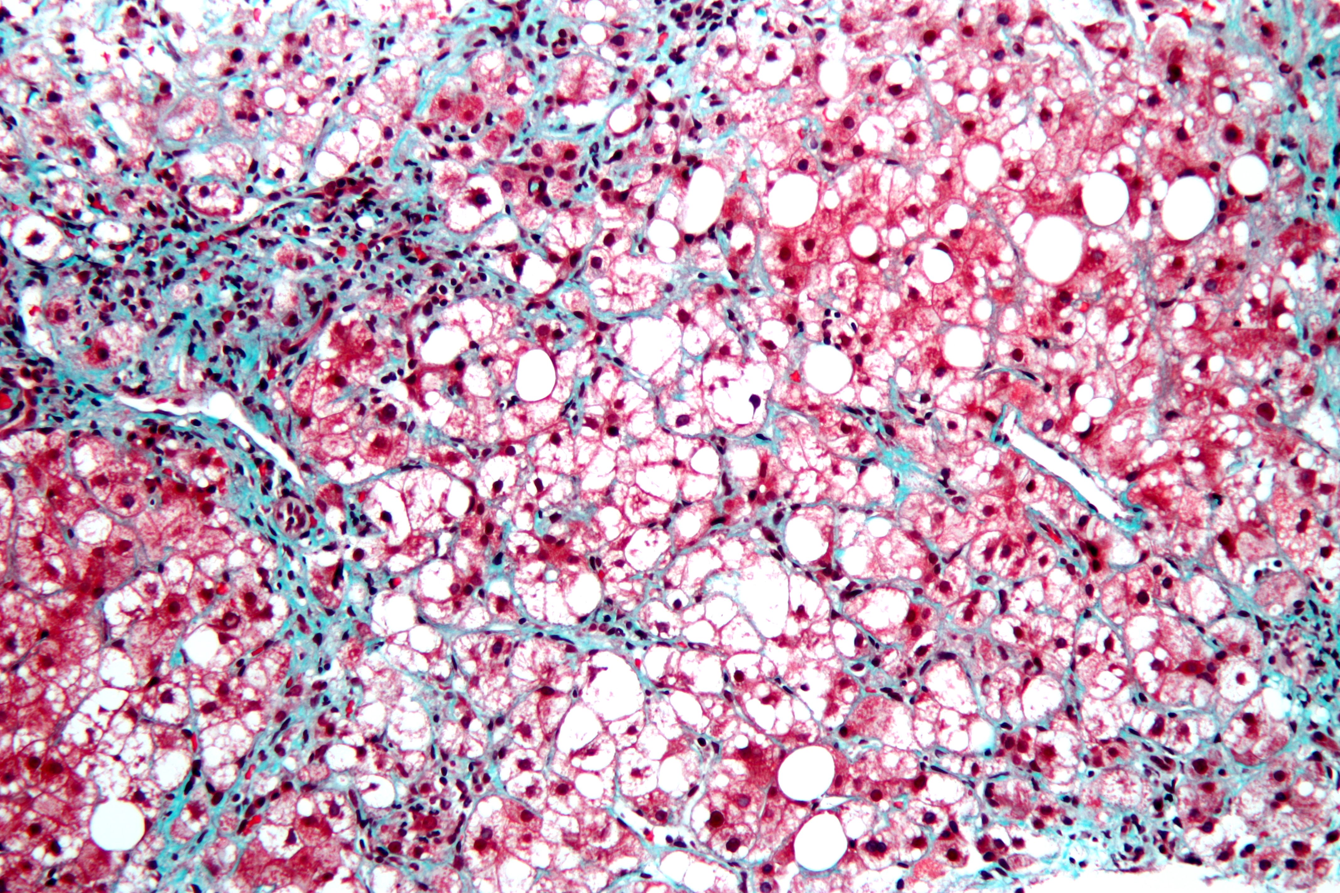
This is a stained liver biopsy sample showing advanced cellular changes in non-alcoholic fatty liver disease. Blue is fibrosis. White is fat accumulation in degenerated cells. Courtesy of Nephron's work.
This is a stained liver biopsy sample showing advanced cellular changes in non-alcoholic fatty liver disease. Blue is fibrosis. White is fat accumulation in degenerated cells. Courtesy of Nephron’s work.

What Is Non-Alcoholic Fatty Liver Disease?

N on-alcoholic fatty liver is a non-inflammatory liver disorder characterized by degenerative changes in the liver caused by excessive accumulation of lipid (fat) in hepatocytes (liver cells) that is called free fatty acid-generated lipotoxicity.

Non-alcoholic fatty liver shows an increase in liver enzymes called transaminases.

Q: What are the enzymes that increase?

A: The transaminases that increase are ALT and AST. ALT is the abbreviation for alanine aminotransferase enzyme and AST is the abbreviation for aspartate aminotransferase enzyme. They are commonly measured in blood tests to determine liver function and when elevated indicate inflammation.

What Is Non-Alcoholic Fatty Liver In Celiac Disease and/or Gluten Sensitivity?

Failure To Thrive And Growth Retardation

Hyde's Prurigo. Courtesy quizlet.com
Hyde’s Prurigo. Courtesy quizlet.com

What Is Prurigo Nodularis (Hyde’s Prurigo)?

P rurigo nodularis is a chronic dermatitis characterized by hard, dry, deep seated, intensely itchy papules (small bumps like pimples) and/or nodules (large bumps) that erupt most commonly on the arms, legs, and back.

Papules and nodules vary in number and may become infected after picking or scratching.

Q: Does the itching go away?

A: New nodules develop from time to time, and existing nodules may remain itchy indefinitely, although some may regress spontaneously and leave scars. In most cases, the disease runs a very protracted course with exacerbations and remissions.1

Prurigo nodularis is an unusual disorder of unknown etiology, which is notoriously resistant to therapy. A variety of systemic conditions have been reported to be associated with prurigo nodularis. However, the mechanism by which these disorders may trigger prurigo nodularis is unknown.2

It has been shown to be associated with malnutriton and infection such as tonsillitis, which resolved after removal of tonsils.3

What Is Prurigo Nodularis In Celiac Disease and/or Gluten Sensitivity?

Sources:
  1. Katotomichelakis M, Balatsouras DG, Bassioukas K, Kontogiannis N, Simopoulos K, Danielides V. Recurrent prurigo nodularis related to infected tonsils: a case report. J Med Case Rep. 2008 Jul 24;2:243. doi: 10.1186/1752-1947-2-243. []
  2. Lee MR, Shumack S. Prurigo nodularis: a review. Australas J Dermatol. 2005 Nov;46(4):211-18; quiz 219-20. []
  3. Katotomichelakis M, Balatsouras DG, Bassioukas K, Kontogiannis N, Simopoulos K, Danielides V. Recurrent prurigo nodularis related to infected tonsils: a case report. J Med Case Rep. 2008 Jul 24;2:243. doi: 10.1186/1752-1947-2-243. []

Hypotonia

In this photo, the spleen is the darkish oval organ in the lower middle of this photo. In your body, it is located in your upper abdoman to the left of your stomach under your ribs.
The spleen is the darkish oval organ in the lower middle of this photo.

What Is Hyposplenism?

H yposplenism is the condition resulting from having lost spleen tissue, called atrophy of the spleen. Spleen atrophy impairs splenic functions or activities because there are insufficient tissues to do the work required.

Q: What splenic functions are impaired?

A: The spleen, apart from acting as a phagocytic filter, thus removing aging and damaged cells, is crucial in regulating immune homeostasis by linking innate and adaptive immunity, and in protecting against infections by encapsulated bacteria.1

Impaired function of the spleen therefore increases risk of infections with encapsulated bacteria because of inability to mount a proper defense and to filter and remove bacteria from the circulation.

The spleen is a highly vascular and solid organ about the size of a fist. It has a delicate structure inside that is enclosed by fibrous, elastic layers consisting of connective tissue.

The tissues within are made up of two different types of tissues, called white pulp and red pulp. White pulp carries out lymphoid functions. Red pulp filters and cleanses the blood. The spleen is situated above the stomach on the left side of the upper abdomen and firmly fixed in place by ligaments and ribs.

The spleen is an important organ of the lymph system, having the largest collection of lymph tissue in the body. It functions to produce antibodies (immunoglobulins) and white blood cells (T-cells and B-cells), help control the amount of blood in the body, keep body fluids in balance, destroy and filter out old and damaged cells2 and salvage the iron needed for producing new blood cells, and lastly, clear bacteria through production of substances that enable phagocytosis (engulfing bacteria and other unwanted particles, such as antigens, from blood).

Children and adults with hyposplenism are at risk for overwhelming infections. Management of hypospenism is directed towards preventing pneumonia by immununization against pneumonia and meningitis and treating bacterial infections as they arise, which may require hospitalization. For some patients, life-long treatment with antibiotics, such as erythromycin and penicillin, are recommended.

What Is Hyposplenism In Celiac Disease and/or Gluten Sensitivity?

Sources:
  1. Di Sabatino A, Brunetti L, Carnevale Maffè G, Giuffrida P, Corazza GR. Is it worth investigating splenic function in patients with celiac disease? World J Gastroenterol. 2013 Apr 21;19(15):2313-8. doi: 10.3748/wjg.v19.i15.2313. []
  2. http://www.nlm.nih.gov/medlineplus/spleendiseases.html []

Irritability

Small Bowel Volvulus, Courtesy  African Journal of Pediatric Surgery.
Small Bowel Volvulus Caused the  Enlarged Dark Loops. Courtesy African Journal of Pediatric Surgery.

What Is Volvulus?

V olvulus is twisting of a loop(s) of bowel onto itself which effectively closes it thus preventing digested matter from passing and causing engorgement of the closed loop with gas and fluid.

Q: How does the bowel get untwisted?

A: This condition usually necessitates surgical correction although some do return to their proper position.

The danger is that there may develop necrosis of the twisted loop(s) in which case, if left untreated, death will ensue.

What Is Volvulus In Celiac Disease and/or Gluten Sensitivity?

Keratomalacia

Image From Gray's Anatomy. Courtesy Wikipedia.org
Image  of Pancreas From Gray’s Anatomy. Courtesy Wikipedia.org

What Is Pancreatic Insufficiency?

P ancreatic insufficiency is a disorder characterized by insufficient exocrine production of pancreatic enzymes for normal digestion of fats, proteins, and carbohydrates that results in maldigestion of these foodstuffs.

Pancreatic insufficiency also causes malabsorption of the fat-soluble vitamins: vitamin A, vitamin D, vitamin E, and vitamin K.

Q: What are the exocrine enzymes produced by the pancreas?

A: Exocrine enzymes produced by the pancreas include amylase for the digestion of carbohydrates, lipase for the digestion of fats, and protease for the digestion of proteins.

While lipases and amylase are secreted in the active form, proteases are secreted as pro-enzymes (need to be activated). Trypsinogen is converted to its active form trypsin in the duodenum by enterokinase, a protease secreted by the enterocytes (surface cells that line the duodenum), and trypsin in turn activates the other pancreatic proteases.1

Pancreatic enzymes are released by the pancreas into the internal pancreatic duct that empties into the common bile duct from which they are pumped through the Sphincter of Odi directly into the duodenum as needed. Tthe Sphincter of Odi is a strong circular muscle that controls the entrance of pancreatic enzymes into the duodenum.

After a meal, enzyme secretion into the duodenum increases quickly reaching peak output within the first 20 to 60 minutes, then decreasing to a stable level before reaching an interdigestive level at the end of the digestive period, that is, about 4 hours after meal intake.2

Medical treatment is with medication that contains enzymes to break down carbohydrates, fats, and protein in food at mealtime. This treatment is safe, effective, and has few side effects.

What Is Pancreatic Insufficiency In Celiac Disease and/or Gluten Sensitivity?

Sources:
  1. Fieker A, Philpott J, Armand M. Enzyme replacement therapy for pancreatic insufficiency: present and future. Clin Exp Gastroenterol. 2011;4:55-73. doi: 10.2147/CEG.S17634. []
  2. Fieker A, Philpott J, Armand M. Enzyme replacement therapy for pancreatic insufficiency: present and future. Clin Exp Gastroenterol. 2011;4:55-73. doi: 10.2147/CEG.S17634. []