E levated homocysteine in blood, called hyperhomocysteinemia, indicates an abnormal blood level of this transient amino acid.
Q: How does the level of homocysteine become abnormal?
A: In metabolism, homocysteine is briefly formed in the breakdown of the amino acid methionine. It is normally converted to cystathione and then to the amino acid cysteine by means of an enzyme that requires vitamin B6.
In the reverse, conversion of homocysteine to methionine requires an enzyme dependent on adequate folic acid and vitamin B12 levels.
Insufficient methionine levels and/or inefficiency in this process results in elevated homocysteine plasma levels that are toxic to blood vessels.
Folic acid, vitamin B12 and vitamin B6 are involved in the metabolic removal of homocysteine, but folic acid deficit occurs the most often.1
What Is Elevated Homocysteine In Celiac Disease and/or Gluten Sensitivity?
Sources:
Lim PO, Tzemos N, Farquharson CA, et al. Reversible hypertension following coeliac disease treatment: the role of moderate hyperhomocysteinaemia and vascular endothelial dysfunction. Journal of Human Hypertension. Jun 2002;16(6):411-5. [↩]
A phthous ulcers, also called canker sores, are a chronic disorder of soft mouth tissue characterized by small, painful purpuric, papular, or erosive lesions that are often surrounded by erythematous (red) margins.1
Q: What soft mouth tissues develop canker sores?
A: Canker sores can erupt on the mucosal surface of the inside lip, sides of mouth, under the tongue and along the side of the tongue. When they are forming, the area swells into a papule, or small bump. Later, the surface erodes and a crater forms with firm pus.
What Are Aphthous Ulcers In Celiac Disease and/or Gluten Sensitivity?
Sources:
Lahteenoja H, Toivanen A, Viander M, Maki M, Irjala K, Raiha I, Syrjanen S. Oral mucosal changes in coeliac patients on a gluten-free diet. European Journal of Oral Sciences. Oct 1998;106(5):899,8p. [↩]
Baby with Allergic Reaction to Peanuts. GFW photo.
What Is Food Allergy?
F ood allergy is an abnormal immune response to food proteins that may damage the small intestinal lining and produce malabsorption of food. The reaction is usually delayed which makes it difficult to identify the offending food that is causing symptoms.
Q: How does food allergy develop?
A: The gastrointestinal tract serves not only to digest and absorb foodstuffs but also to protect the body from unwanted substances. When allergic food substances are eaten, the immune response that is triggered in the gut produces inflammation with symptoms such as pain, vomiting and loose bowels.
Inflammation causes swelling of the gut lining that can interfere with the passage of nutrients through it to the body which results in malabsorption. Malabsorption deprives the body of nutrients needed for normal function.
Symptoms other than digestive may involve skin rashes, hives, and respiratory difficulties that can be distressing and life-threatening.
What Is Food Allergy In Celiac Disease and/or Gluten Sensitivity?
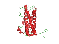
Prolactin Structure. Courtesy Boris TM at Wikipedia.
What Is Hyperprolactinemia?
H yperprolactinemia is an excess secretion of prolactin hormone causing an elevated blood level that is characterized by its effects on reproduction: in females causing amenorrhea and in males causing reduced sexual potency.
Q: What is prolactin?
A: Prolactin is a hormone produced by the the anterior pituitary gland that is transported by the bloodstream to stimulate androgen hormone production in male and estrogen hormone production in female sex glands. However, too much prolactin inhibits hormone production of estrogen in women and testosterone in men.
In pregnant females upon delivery of a baby, prolactin stimulates breast development and milk production for the purpose of feeding the infant. Sucking by the infant stimulates the release of prolactin which keeps the milk supply adequate.
Prolactin also promotes autoimmune mechanisms. Prolactin interferes specifically with B cell tolerance induction, enhances proliferative response to antigens and mitogens and increases the production of immune globulins, cytokines and autoantibodies.1
Other conditions that can stimulate prolactin release include nutritional deficiencies, thyroid stimulating hormone (TSH) produced by the pituitary gland, stress, exercise, and anterior pituitary tumors.
What Is An Elevated Prolactin Level In Celiac Disease and/or Gluten Sensitivity?
Sources:
Shelly S, Boaz M, Orbach H. Prolactin and autoimmunity. Autoimmun Rev. 2012 May;11(6-7):A465-70. doi: 10.1016/j.autrev.2011.11.009. [↩]
C heilosis is a painful feature of nutritional deficiencies, especially riboflavin and/or pyridoxine deficiency, and less frequently, from vitamin B12, folic acid, and iron. It is characterized by redness of the lips with cracking and weeping in the corner of the mouth.
Q: What is the consequence of weeping at the corners of the mouth?
A: The open sores that develop at the corners of the mouth invite infection by candida albicans, a yeast organism, and less commonly by bacteria normally present on the skin such as staphylococcus aureus.
These sores make opening the mouth very painful and so restrict eating, talking, socializing, and dental care.
What Is Cheilosis In Celiac Disease and/or Gluten Sensitivity?
Close-up Slice of a Small Intestinal Villus Showing How Enterocytes Appear Tightly Lining the Entire Outside Surface Of A Villus. Courtesy Cleo Libonati
What Is Increased Intestinal Permeability?
I ncreased intestinal permeability is characterized by dysfunctional intestinal permeability (leakiness) allowing for the penetration of harmful entities from the gut into the bloodstream such as undigested proteins and microbes. The popular name is “leaky gut.”
Q: Why does intestinal permeability increase?
A: Intestinal permeability is an essential function of the small intestinal mucosal lining by which wanted substances such as properly digested foodstuffs are allowed to permeate through the lining to enter the body via the bloodstream and lymphatics. At the same time unwanted substances are kept out.
The mucosal lining is one cell thick and makes up the surface between the digested foodstuffs inside the hollow of the intestine and the underlying tissues.
The mucosal lining is covered by millions of microscopic finger-like structures called villi that project toward the inside of the intestine giving the appearance of a shag rug.
Each one, called a villus, contains a capillary bringing blood to absorb nutrients, a vein to take away nutrients, and a lacteal to absorb and take away digested fat. Its wall is made up of a single layer of tightly connecting cells, called enterocytes.
This single layer of cells separates the contents of our small intestine from the lamina propria (underlying tissues of the small intestine) and the rest of our body. Breaching of this single layer of cells by leakiness can expose lymphocytes (immune cells) located in the lamina propria to a myriad of microorganisms and food antigens, leading to immune reactions.1
To protect the body from unwanted substances, a gatekeeping barrier system operates to regulate the passage of nutrients, or permeation, through the surface mucosal lining. This system acts to seal the inside body from the gut.
The integrity of intestinal permeability is determined by interactions among several barrier components including the unstirred water layer, mucosal surface hydrophobicity, the surface mucous coat, and cell factors (especially tight junctions).
Tight junctions hold cells tightly together side-by-side to prevent unwanted substances from passing through the lining. Tight junctions are complex structures comprising over 50 proteins, such as the claudin proteins which are considered to be the structural backbone of tight junctions.
Tight junctions include a series of special proteins forming fibrils (springy like proteins) that cross the plasma membrane and interact with proteins in the adjoining cells. Tight junctions are regulated by the protein zonulin.2
If zonulin deregulates from the action of substances such as gliadin (gluten in wheat) and bacteria, the tight junction barrier fails which results in increased intestinal permeability. Dysfunction of the barrier system allows unwanted substances to enter the body where they are damaging to many tissues.
Tight junction dysfunction has been shown to be a part of certain autoimmune diseases such as celiac disease, type I diabetes mellitus, multiple sclerosis, and rheumatoid arthritis. Other diseases associated are cancer, allergies, and infections.3
Important gastrointestinal infections that cause leaky gut include rotavirus, parasites, pathogenic bacteria (escherichia coli, clostridium difficile), and mycotoxins produced by fungi found in stored grain and dried fruit.4
Fortunately, the presence of some commensal (friendly intestinal bacteria) and probiotic strains leads to an increase in tight junctions proteins at the cell boundaries and in some cases prevents or reverses the adverse effects of pathogens, food and stress. Various dietary components are also known to regulate epithelial permeability by modifying expression and localization of tight junctions proteins.2
What Is Increased Intestinal Permeability In Celiac Disease and/or Gluten Sensitivity?
Sources:
Fahardi A, Banan A, Fields J, Keshavarzian A. Intestinal barrier: an interface between health and disease. Journal of Gastroenterology and Hepatology. 2003; 18: 479-497. [↩]
Ulluwishewa D, Anderson RC, McNabb WC, Moughan PJ, Wells JM, Roy NC. Regulation of tight junction permeability by intestinal bacteria and dietary components. J Nutr. 2011 May;141(5):769-76. doi: 10.3945/jn.110.135657. [↩] [↩]
Fasano A. Zonulin and Its Regulation of Intestinal Barrier Function: The Biological Door to Inflammation, Autoimmunity, and Cancer. Physiological Reviews. January 2011Vol. 91no. 151-175DOI: 10.1152/physrev.00003.2008 [↩]
Farhadi A, Banan A, Fields J, Keshavarzian A. Intestinal barrier: an interface between health and disease. Journal of Gastroenterology and Hepatology. 2003;18:479-91. [↩]
H ypocalcemia, or low plasma calcium, means the level of calcium in blood is too low to meet metabolic needs of the body for calcium.
Low blood calcium is characterized by bone and tooth demineralization (loss of calcium causing weak teeth and fragile bones), and these impaired functions: nerve conduction, muscle contraction, blood clotting, blood pressure regulation, glycogen to glucose conversion, many hormone actions, many enzyme activities, and acetylcholine production.
Q: Where is calcium found in the body?
A: Calcium is the most abundant mineral in the body, with 99% residing in bones and teeth where it constitutes 40% of skeletal bone weight along with 45% phosphorus. As a component of bone (hard tissue), calcium fulfills a structural role to maintain body size and act as attachments for musculoskeletal tissues. The remaining 1% of calcium is present in blood and soft tissues.
Calcium levels in the blood are maintained within very strict limits by dietary intake, hormonal regulation by the parathyroid gland and a rapidly exchangeable pool in bone tissue.
What Is Hypocalcemia In Celiac Disease and/or Gluten Sensitivity?
D ental enamel defects (DED) comprise a variety of abnormalities in the dental enamel of the second or permanent teeth and of primary or baby teeth.
Teeth may appear with pits, thin or missing enamel, non-white enamel (yellow, orange, or gray colored teeth), and demarcated opacities (very white spots) even if teeth are white.
Q: What is enamel?
A: Dental enamel is the shiny, hard, white, dense, inorganic substance covering the crowns of the teeth. The crown is the tooth portion above the gum. Under the enamel is hard dentin which surround the living pulp. In teeth with missing enamel, the areas with exposed dentin appear dull and tan colored.
What Are Dental Enamel Defects In Celiac Disease and/or Gluten Sensitivity?
Paleness and Tendon Problem: Symptoms of Hypocupremia.
What Is Hypocupremia?
H ypocupremia, or low plasma copper, means the level of copper is too low to meet metabolic needs of the body for copper and is characterized by these many features:
Impaired energy production causing weakness.
Impaired ability as part of an enzyme to oxidize vitamin C which is required to breakdown histamine.
Faulty blood cell formation and instability of blood cell membranes causing anemia, shortened life span of neutrophils causing susceptibility to infection, faulty platelet formation causing impaired blood clotting.
B leeding and/or swollen gums is an inflammatory disorder characterized by changes in gum integrity that may result in tooth loss.
Sponginess of the gum (swelling) indicates inflammation, called gingivitis.
Q: What causes gingivitis?
A: Gingivitis that is caused by inadequate dental hygiene shows as pockets of swollen gum between or along the base of teeth where sticky plaque was allowed to build up. Plaque holds bacteria that inflames the gums where it occurs.
Gingivitis that is caused by vitamin C deficiency, pregnancy, certain drugs such as the anti-epileptic drug phenytoin, and leukemia affects the whole gum irrespective of mouth care.
What Are Bleeding / Swollen Gums In Celiac Disease and/or Gluten Sensitivity?

![Aphthous_ulcer[1]](https://glutenfreeworks.com/health/wp-content/uploads/sites/10/Aphthous_ulcer1.jpg)



![Enamel_celiac[1]](https://glutenfreeworks.com/health/wp-content/uploads/sites/10/Enamel_celiac1.jpg)

