Increased appetite is the increased desire for food.
Q: What causes appetite.
A: Appetite is caused by hunger.
The brain monitors nutritional needs and employs a hunger mechanism to alert us when fresh food is needed. Hunger is mainly caused by the need for protein. Low blood sugar also triggers hunger.
The tempting sight, smell, taste and expectation of food stimulates involuntary sensory nerves. By reflex action, these sensory nerves cause muscle and sensory activity in various digestive organs. Salivary glands in the mouth begin to secrete saliva as stomach glands and muscles become active.1
What Is Increased Appetite In Celiac Disease and/or Gluten Sensitivity?
Sources:
Cleo Libonati. Recognizing Celiac Disease. Gluten Free Works Publishing, Fort Washington, PA 19034 USA [↩]
Primary biliary cirrhosis (PBC) is a biliary tract disease characterized by chronic cholestasis (build-up of bile) and gradual destruction of bile ducts within the liver, called intrahepatic bile ducts, caused by chronic inflammation.
Primary biliary cirrhosis comes under the umbrella term autoimune liver disease in which the end result is immune-mediated hepatocellular (liver cell) or hepatobiliary (bile duct) injury.1
Q: What is the end result of destruction of bile ducts?
A: The end result of destruction of bile ducts is liver damage.
Injured liver tissue from chronic inflammation and the buildup of bile leads to cirrhosis, a condition in which the liver slowly deteriorates and malfunctions.
Scar tissue replaces healthy liver tissue, partially blocking the flow of blood through the liver. Scarring also impairs the liver’s normal ability to control infections, remove bacteria and toxins from the blood, process nutrients, hormones, and drugs, make proteins that regulate blood clotting, produce bile, and effectively replace its own cells when they become damaged.2
The liver is the largest organ within the body. It lies mostly in the upper part of the abdomen on the right side just under the diaphragm. About 70% of liver tissue is made up of cube shaped cells called hepatocytes that do the main work of the liver. Other cells (epithelial) form structure and are arranged in single layers around blood vessels, sinusoids, and bile ducts.
Bile ducts carry bile, a greenish brown liquid made by the liver to the gall bladder for storage until needed to aid in the digestion and absorption of fat and fat-soluble vitamins A, D, E, and K from the small intestine. Bile emulsifies fat eaten in the diet so that the pancreatic enzyme called lypase can break it down into its fatty acid and glycerol components which can then be absorbed into the body.
Bile also carries away waste products produced by normal metabolism and toxic substances that are removed by the liver for eventual elimination in stool. Bile is continually made by the liver from phospholipids, bile acids, cholesterol, and aging blood cells it removes from circulation. As such, bile must continually flow out of the liver to prevent build-up in the liver.
There is no cure for primary biliary cirrhosis.
What Is Primary Biliary Cirrhosis In Celiac Disease and/or Gluten Sensitivity?
Sources:
Trivedi PJ, Adams DH. Mucosal immunity in liver autoimmunity: a comprehensive review. J Autoimmun. 2013 Oct;46:97-111. doi: 10.1016/j.jaut.2013.06.013. [↩]
Hypoglycemia means the level of glucose within cells is too low to meet metabolic needs of the body for this essential sugar.
Q: What are the metabolic needs for glucose?
A: Glucose is the most important simple sugar in human metabolism mainly because it is the primary source of energy for most cells of the body.
Energy contained in the glucose molecule is obtained by the body from its reaction with oxygen (oxidation). This oxidation reaction occurs in power producing mitochondria structures that are located within cells.1
Hypoglycemia is characterized by alterations in neurologic, metabolic and muscular functions:
Neurologic function because brain tissue is particularly dependent on glucose for energy,
Metabolic function of glucose-dependent tissues which include red blood cells, white blood cells, bone marrow, eye, inner heart of the kidney, and peripheral nerves because these tissues cannot metabolize fatty acids as an alternate source of energy, and
Muscle function because muscle cells continually require glucose for energy production.
Glucose is made available to cells through the regulating action of insulin, a hormone produced by specialized cells located on the surface of the pancreas.
What Is Hypoglycemia In Celiac Disease and/or Gluten Sensitivity?
Aphthous ulcers, also called canker sores, are a chronic disorder of soft mouth tissue characterized by small, painful purpuric, papular, or erosive lesions that are often surrounded by erythematous (red) margins.1
Q: What soft mouth tissues develop canker sores?
A: Canker sores can erupt on the mucosal surface of the inside lip, sides of mouth, under the tongue and along the side of the tongue. When they are forming, the area swells into a papule, or small bump. Later, the surface erodes and a crater forms with firm pus.
What Are Aphthous Ulcers In Celiac Disease and/or Gluten Sensitivity?
Sources:
Lahteenoja H, Toivanen A, Viander M, Maki M, Irjala K, Raiha I, Syrjanen S. Oral mucosal changes in coeliac patients on a gluten-free diet. European Journal of Oral Sciences. Oct 1998;106(5):899,8p. [↩]
A 3D Image From Magnetic Resonace Cholangiography showing biliary tree. NIHMS
What Is Primary Sclerosing Cholangitis?
Primary sclerosing cholangitis (PSC) is an uncommon, slowly progressive bile duct disease that results in stagnation or build-up of bile in the liver, called cholestasis.
Primary sclerosing cholangitis is characterized by sclerosis, or scarring inflammation in bile ducts both within the liver (intra-hepatic ducts), and outside the liver (extra-hepatic ducts), causing progressive narrowing and, eventually, obliteration of the bile ducts.
Primary sclerosing cholangitis comes under the umbrella term autoimune liver disease in which the end result is immune-mediated hepatocellular (liver cell) or hepatobiliary (bile duct) injury.1
Q:What happens when scarred bile ducts can no longer transport bile out of the liver?
A:Bile that cannot be removed from the liver by the biliary duct system backs up and damages the liver, causing cirrhosis.
Bile is continually made by the liver from phospholipids salt, cholesterol, and aging blood cells that it removes from circulation to be carried out of the liver. Bile also carries away waste products produced by normal metabolism and toxic substances that are removed by the liver for eventual elimination in stool. As such, bile must continually flow out of the liver to prevent build-up in the liver.
Bile is a greenish brown liquid made by the liver. Bile ducts carry it out of the liver to the gall bladder for storage until needed to aid in the digestion and absorption of fat from the small intestine. Bile emulsifies fat eaten in the diet so that the pancreatic enzyme called lypase can break it down into its fatty acid and glycerol components.
The liver is the largest organ within the body. It lies mostly in the upper part of the abdomen on the right side just under the diaphragm. About 70% of liver tissue is made up of cube shaped cells called hepatocytes that do the main work of the liver. Other cells (epithelial) form structure and are arranged in single layers around blood vessels, sinusoids, and bile ducts.
Build-up of bile in the liver is the end result of the inflammatory process in primary sclerosing cholangitis, that by swelling and scarring of bile ducts impedes and eventually prevents bile flow out of the liver, leading to liver failure. There is no curative treatment available for primary sclerosing cholangitis, besides liver transplantation.2
The appearance of the intrahepatic and extrahepatic biliary ducts can be assessed by use of cholangiography, and magnetic resonance (MR) imaging is the best way to identify patients. See image above.3
MR cholangiography offers a noninvasive method of obtaining images of the biliary system without the use of a contrast agent. There is no radiation exposure. Pulse sequences can be chosen to obtain bright bile or black bile cholangiograms. Image processing algorithms can be selected to obtain a three-dimensional representation of biliary anatomy and pathology, and those images can be rotated in any plane so that ductal anatomy and pathology can be seen to best advantage.4
There is no cure for primary sclerosing cholangitis but there are symptom treatments one of which is supplementation for low levels of vitamins A,D,E, and K. Liver transplant is the only effective option.
What Is Primary Sclerosing Cholangitis In Celiac Disease and/or Gluten Sensitivity?
Sources:
Trivedi PJ, Adams DH. Mucosal immunity in liver autoimmunity: a comprehensive review. J Autoimmun. 2013 Oct;46:97-111. doi: 10.1016/j.jaut.2013.06.013. [↩]
Kummen M, Schrumpf E, Boberg KM. Liver abnormalities in bowel diseases. Best Pract Res Clin Gastroenterol. 2013 Aug;27(4):531-42. doi: 10.1016/j.bpg.2013.06.013. [↩]
Eaton JE, Talwalkar JA, Lazaridis KN, Gores GJ, Lindor KD. Pathogenesis of primary sclerosing cholangitis and advances in diagnosis and management. Gastroenterology. 2013 Sep;145(3):521-36. doi: 10.1053/j.gastro.2013.06.052. Epub 2013 Jul 1. [↩]
Meakem TJ 3rd, Schnall MD. Magnetic resonance cholangiography. Gastroenterol Clin North Am. 1995 Jun;24(2):221-38. [↩]
Low cholesterol found in blood indicates an abnormal blood level of this essential lipid (fat) that is characterized by decreased production of steroid hormones and bile.
Cholesterol is a soft, fat-like substance found in the bloodstream and is an essential structural component in all body cells. It is required for membrane fluidity and permeability of cells. Within the cell membrane, cholesterol also functions in intracellular transport, cell signaling and nerve conduction.
Cholesterol is required for these essential body processes:
Cholesterol is converted to bile by the liver and stored in the gallbladder until needed for digestion of fats in the small intestine.
Cholesterol is used by the adrenal glands to produce the steroid hormones cortisol and aldosterone.
Cholesterol is used by the sex glands to make progesterone, estrogen, and testosterone and their derivatives.
Cholesterol is needed for the production of vitamin D.
Q: What makes cholesterol bad for you?
A: There are different forms of cholesterol that can be bad or good depending on the level. In blood vessels, low-density lipoprotein (LDL or ‘bad’) cholesterol can abnormally join with fats and other substances to build up in the inner walls of arteries. The arteries can become clogged and narrow, and blood flow is reduced. This is atherosclerosis which can lead to stroke, heart attack and thrombosis.
High-density lipoprotein (HDL or ‘good’) carries harmful cholesterol away from the arteries and helps protect from heart attack and stroke.1
What Is Low Cholesterol In Celiac Disease and/or Gluten Sensitivity?
Coagulation factors II, VII, IX, X found in blood are essential for normal blood clotting. Low coagulation factors on blood assay indicate an altered secondary coagulation disorder that is characterized by impaired clot formation.
Each coagulation factor must be present in sufficient quantity in order for normal clotting to occur, but the level required is different for each factor. Results are frequently reported as a percentage with 100% being normal. For example, a factor VIII that is 30% would be considered abnormally low.1
The production of the coagulation factors II, VII, IX, and X requires vitamin K without which the factors will be low.
Q: What happens when coagulation factors II, VII, IX, and X are low?
A: When any of the blood clotting factors are lacking or not working properly, the blood tests prothrombin (PT) and partial thromboplastin time (PTT) will be abnormally prolonged. Prothrombin and partial thromboplastin time measure the time it takes for blood to clot. When you bleed, the body launches a series of activities that help the blood clot. This is called the coagulation cascade. There are three pathways to this event. These tests looks at coagulation factors, found in two of these pathways.2
What Are Low Coagulation Factors In Celiac Disease and/or Gluten Sensitivity?
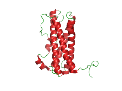
Prolactin Structure. Courtesy Boris TM at Wikipedia.
What Is Hyperprolactinemia?
Hyperprolactinemia is an excess secretion of prolactin hormone causing an elevated blood level that is characterized by its effects on reproduction: in females causing amenorrhea and in males causing reduced sexual potency.
Q: What is prolactin?
A: Prolactin is a hormone produced by the the anterior pituitary gland that is transported by the bloodstream to stimulate androgen hormone production in male and estrogen hormone production in female sex glands. However, too much prolactin inhibits hormone production of estrogen in women and testosterone in men.
In pregnant females upon delivery of a baby, prolactin stimulates breast development and milk production for the purpose of feeding the infant. Sucking by the infant stimulates the release of prolactin which keeps the milk supply adequate.
Prolactin also promotes autoimmune mechanisms. Prolactin interferes specifically with B cell tolerance induction, enhances proliferative response to antigens and mitogens and increases the production of immune globulins, cytokines and autoantibodies.1
Other conditions that can stimulate prolactin release include nutritional deficiencies, thyroid stimulating hormone (TSH) produced by the pituitary gland, stress, exercise, and anterior pituitary tumors.
What Is An Elevated Prolactin Level In Celiac Disease and/or Gluten Sensitivity?
Sources:
Shelly S, Boaz M, Orbach H. Prolactin and autoimmunity. Autoimmun Rev. 2012 May;11(6-7):A465-70. doi: 10.1016/j.autrev.2011.11.009. [↩]
H ypokalemia means the level of potassium in the bloodstream is too low to meet metabolic needs of the body for this mineral and is characterized by metabolic acidosis, altered nerve conduction and muscle contraction.
Rapid potassium loss can result in life-threatening hypokalemic rhabdomyolysis which is destruction of muscle tissue that results in kidney damage.1
Q: What are metabolic needs of the body for potassium?
A: The metabolic needs of the body for potassium are great because this mineral is crucial for life and especially for normal nerve and muscle function.
Most potassium is intracellular, meaning it is found within cells while sodium, its opposing mineral (both electrolytes), is found in the fluid surrounding cells. In muscle contraction, exchange of potassium and sodium takes place so that potassium moves out of muscle cells and sodium moves into them. With muscle relaxation, potassium moves back into the cells and sodium moves out.
What Is Hypokalemia In Celiac Disease and/or Gluten Sensitivity?
Sources:
Williams SG, Davison AG, Glynn MJ. Hypokalaemic rhabdomyolsis: an unusual presentation of celiac disease. European Journal of Gastroenterology and Hepatology. Feb 1995;7(2):183-4. [↩]
 What Is Increased Appetite?


![Aphthous_ulcer[1]](https://glutenfreeworks.com/health/wp-content/uploads/sites/10/Aphthous_ulcer1.jpg)