Erosions Resulting From Esophagitis Can Be A Cause Of Gastrointestinal Bleeding.
What Is Occult Gastrointestinal Bleeding?
O ccult gastrointestinal bleeding is characterized by unseen or minute quantities of blood in stool. The origin of bleeding is from mucosa that lines the inside of the digestive tract at a site that must be found by medical testing and procedures to look at the lining.
Q: What tests and procedures are performed to locate the bleeding?
A: The simplest test to discover blood that cannot be seen is the stool test. This consists of a card with 3 spaces for 3 separate stool collection over 3 separate days. A tiny smear of stool is placed on a test card space on each of three days. Then the card is examined for a reaction that indicates the existence of blood in the stool.
If the stool test is positive, the origin of bleeding must be located. This search requires certain procedures that look at the mucosal lining directly by a gastroenterologist, usually under light sedation.
Gastroscopy procedure can visualize the upper gastrointestinal tract, which includes the esophagus and stomach.
Endoscopy procedure can also visualize the upper small intestine. However, endoscopy procedure is limited in that it cannot visualize the middle small intestine (jejunum).
Colonoscopy can visualize the end of the small intestine and colon all the way to the rectum.
Capsule endoscopy procedure. If gastroendoscopy and colonoscopy fail to discover the source of bleeding, the physician may administer a capsule endoscopy which visualizes the entire gastrointestinal tract. In this procedure, the patient swallows a capsule size camera (encased) which takes constant pictures over the course of a day until expelled through the rectum. Of course, the patient must be able to swallow the large size capsule.
What Is Occult Gastrointestinal Bleeding In Celiac Disease and/or Gluten Sensitivity?
C ommon variable immunodeficiency (CVID) is a primary antibody deficiency disease characterized by the onset of recurrent bacterial infections resulting from markedly decreased immunoglobulin antibody production and antibody levels.
Q: What causes common variable immunodeficiency?
A: Common variable immunodeficiency is caused by a defect in any critical stage of B cell development and is characterized by impaired production of normal amounts of antigen-specific antibodies. This is a set up for infection, autoimmune disease, and cancer.
One important histologic feature is the absence or paucity of plasma cells in the biopsy that occurs in common variable immunodeficiency. The diagnosis is initially made by measuring quantitative immunoglobulins and then specific immune testing on these cell subsets in the circulation by flow cytometry.1
Early diagnosis and treatment with IgG therapy (immunoglobulin G) can decrease illness and mortality.2
Pulmonary damage is the most frequent complication and may result from recurrent infections and/or immune dysregulation. Other complications due to the underlying immune dysregulation include lymphoproliferative disease (granulomatous disease, lymphadenopathy and hepatosplenomegaly), autoimmune disease, gastrointestinal disease such as chronic inflammation and an increased risk of cancer.3
It is important to recognize that common variable immunodeficiency can occur at any age, but an early onset may be associated with an increased risk of gastric cancers and lymphoma, particularly of the intestine.3
Patients with common variable immunodeficiency should be managed by an immunologist with experience in primary immunodeficiency states, given the complications of these patients.4
What Is Common Variable Immunodeficiency In Celiac Disease and/or Gluten Sensitivity?
Sources:
Murray JA1, Rubio-Tapia A. Diarrhoea due to small bowel diseases. Best Pract Res Clin Gastroenterol. 2012 Oct;26(5):581-600. doi: 10.1016/j.bpg.2012.11.013. [↩]
Maarschalk-Ellerbroek LJ, Hoepelman AI, van Montfrans JM, Ellerbroek PM. The spectrum of disease manifestations in patients with common variable immunodeficiency disorders and partial antibody deficiency in a university hospital. JClin Immunol. 2012 Oct;32(5):907-21. doi: 10.1007/s10875-012-9671-6. [↩]
Maarschalk-Ellerbroek LJ, Hoepelman AI, van Montfrans JM, Ellerbroek PM. The spectrum of disease manifestations in patients with common variable immunodeficiency disorders and partial antibody deficiency in a university hospital. J Clin Immunol. 2012 Oct;32(5):907-21. doi: 10.1007/s10875-012-9671-6. [↩] [↩]
Murray JA1, Rubio-Tapia A. Diarrhoea due to small bowel diseases. Best Pract Res Clin Gastroenterol. 2012 Oct;26(5):581-600. doi: 10.1016/j.bpg.2012.11.013. [↩]
Z incemia means the zinc level in blood plasma is too low to meet metabolic needs of the body for this mineral.
Q: How important is a normal blood level of zinc?
A: A low blood level of zinc is characterized by widespread alterations in energy metabolism, growth, hemoglobin, carbon dioxide transport, hormone activity, insulin storage, many enzyme activities, prostaglandin function, collagen production, male fertility, protein synthesis, and vitamin A metabolism.
What Is Zincemia In Celiac Disease and/or Gluten Sensitivity?
Testing Thigh Strength. Courtesy Charlie Goldberg, M.D., UCSD School of Medicine
What Is Hypophosphatemia?
H ypophosphatemia means the level of phosphates in the bloodstream is too low to meet metabolic needs of the body for this mineral.
Q: How important is phosphorus in metabolism?
A: Phosphorus is crucial to life, being present in every cell of the body and constitutes 45% of skeletal bone weight along with 40% calcium needed to support the body as a framework.
A low blood phosphate level is characterized by alterations in blood acid-alkaline balance and serious neuromuscular, hematologic, renal, skeletal, and dental abnormalities.
Symptoms result primarily from decreased production of adenosine triphosphate (ATP), the main energy source in cells, and phosphocreatine, a secondary energy source for muscle contraction.
Acute phosphorus deficiency may precipitate rhabdomyolysis which is destruction of muscle.
Nervous system dysfunction is observed in severe hypophosphatemia.
Chronic phosphorus deficiency causes proximal myopathy (upper arms and thighs).1
Severe phosphorus deficiency has widespread and ultimately fatal consequences.
What Is Hypophosphatemia In Celiac Disease and/or Gluten Sensitivity?
Sources:
Takeda E, Ikeda S, Nakahashi O. Lack of phosphorus intake and nutrition. Clin Calcium. 2012 Oct;22(10):1487-91. [↩]
What Is Plummer-Vinson Syndrome Affecting the Esophagus?
Plummer-Vinson syndrome is a manifestation of severe, long-term, iron deficiency anemia that is characterized by post-cricoid esophageal webs and dysphagia.
Q: What are esophageal webs?
A: Esophageal webs are one or more thin horizontal membranes consisting of squamous epithelium (cells that line the surface of the esophagus) and submucosa. They usually protrude from the anterior (front) wall, extending laterally across the inside esophagus but not to the posterior (rear) wall, which means that they rarely encircle the lumen.1
Dysphagia, or difficulty swallowing, from these webs is commonly painless and intermittent or progressive and may cause obstruction.
Webs can be detected by barium swallow X-ray, but the best way for demonstration is videofluoroscopy and by upper gastrointestinal endoscopy. They appear smooth, thin, and gray with eccentric or central lumen space. The webs typically occur in the upper part of the esophagus and may be missed and accidentally ruptured unless the endoscope is introduced under direct visualization.1
Iron deficiency is believed to decrease the contraction amplitude or force of the esophageal muscle resulting in motility impairment. Slower transit times have been recorded at the proximal and middle parts of the esophagus of Plummer-Vinson syndrome patients compared to healthy volunteers.2 Transit time is how fast ingested food and fluids travel through the esophagus.
Gude et al, report that iron replacement does not necessarily reverse the dysphagia in all the cases of Plummer-Vinson syndrome and that close monitoring of the web is mandated to watch for malignant change. In fact, 3 to 15 per cent of the patients with Plummer-Vinson syndrome, mostly women between 15 and 50 years of age, have been reported to develop esophageal or pharyngeal cancer.2
What Is Plummer-Vinson Syndrome Affecting the Esophagus In Celiac Disease and/or Gluten Sensitivity?
Sources:
Novacek G. Plummer-Vinson syndrome. Orphanet J Rare Dis. 2006; 1: 36. Published online 2006 September 15. doi: 10.1186/1750-1172-1-36. [↩] [↩]
Gude D, Bansal DP, and Malu A. Revisiting Plummer Vinson Syndrome. Annals of Medical and Health Sciences Research. 2013 Jan-Mar;3(1):119-121. [↩] [↩]
A utoimmune disorders refer to those conditions that involve an abnormal attack on the body’s own tissues perpetuated by the production of autoantibodies directed against self.
Q: What happens when autoantibodies attack the body’s own tissues?
A: This abnormal immune activity by autoantibodies causes inflammation and damage to targeted body tissues.
Dermatitis herpetiformis is a skin manifestation of celiac disease characterized by extremely itchy blisters that commonly erupt on forearms and knees but may appear on the face, scalp or buttocks.
Autoimmune diseases as a group affect approximately 8.5% of people worldwide.
What Are Autoimmune Disorders In Dermatitis Herpetiformis?
Bristol Stool Chart Showing Normal and Abnormal Stool.
What Is Chronic Constipation Alternating With Diarrhea?
C hronic constipation alternating with diarrhea is an intestinal motility disorder, or irregularity, characterized by alteration in stool formation, consistency, and evacuation which results in a bowel movement that consists of some hard or balled stool along with some loose stool that can cause leakage.
Q: How do irregular movement patterns develop in the colon?
A: The colon produces irregular movements as a result of problems that originate in the colon (large intestine) itself and/or the small intestine which then affects function of the colon.
Here are listed the many types of problems or diseases that cause these abnormal bowel movements:
Disorders that adversely affect the colon, an organ which must propel stool, remove excess water, absorb electrolytes, ferment undigested food material that passes into it, and produce nutrients from the fermentation process:
Poor diet that does not contain adequate nutrition, fiber, probiotics, prebiotics, and water to form normal stool.
Diet that contains irritating, toxic or allergenic food that cause spasms.
Diseases that inflame the mucosa lining such as collagenous colitis, altering the proper absorption of water and electrolytes.
Diseases that damage and swell the colon walls, such as Crohn’s disease, ulcerative colitis, and diverticulitis.
Diseases that obstruct the lumen or passageway so that stool passes with difficulty.
Diseases that hamper normal peristalsis (muscle action), such as irritable bowel syndrome (IBS), diabetes and thyroid disease.
Disorders that adversely affect the small intestine, an organ which must digest and absorb nutrients needed by the body while passing unabsorbed food material to the colon:
Diet that conatins too much fat, sugar or artifical sweeteners, causing diarrhea.
Disorders that result in malabsorption, such as gluten enteropathy, milk enteropathy, steatorrhea (fat malabsorption), lactose intolerance, sucrose intolerance, maltose intolerance, and bacterial overgrowth, passing abnormal amounts of undigested food material to the colon where it is fermented producing excessive gas, diarrhea and spasm.
Disorders that impair peristalsis, such as active celiac disease, diabetes, scleroderma, and thyroid disease.
Tumors like cancer and lymphoma impair regular passage of material to colon.
Drugs that impair peristalsis, such as iron supplements, aluminum containing antacids, narcotics, some anti-depressants, some anti-seizure, and some diuretics.
What Is Chronic Constipation Alternating With Diarrhea In Celiac Disease and/or Gluten Sensitivity?
Figure on right shows how atherosclerosis impedes blood flow through coronary arteries while blood clots block blood flow. Courtesy Google.
What Is Coronary Artery Disease (CAD)?
Coronary artery disease (CAD), also called ischemic heart disease, is a gradual narrowing of medium and large arteries of the heart by fatty buildups, called atherosclerotic plaques.
It is characterized by slowly developing interference with blood flow to heart tissue itself, resulting in oppressive chest pain called angina and, ultimately, thrombosis (clot) causing heart attack.
The heart is a muscular organ that is working all the time, so it needs a constant supply of oxygen. Oxygen is brought to the working heart tissue by the coronary arteries with each beat of the heart. When heart muscle has to work harder, it needs more oxygen delivered to itself. Lack of oxygen causes pain.
In fact, failure of diseased coronary arteries to deliver adequate oxygen to heart tissue is the most common cause of angina pectoris – substernal pain (under breastbone) or pressure brought on by exertion and relieved by rest.
Thrombosis, or clot formation, occurs when blood cells within a narrowed artery can no longer get through. Trapped, blood cells pile up and block the artery thus triggering a cascade of events called heart attack. Coronary arteries that are narrowed by atherosclerotic plaques can rupture causing injury to the coronary blood vessel resulting in blood clotting which blocks the flow of blood to the heart muscle. Blood clots may form, partially dissolve, and later form again and angina can occur each time a clot blocks blood flow in an artery.1
Q: How does coronary artery disease develop?
A:Coronary artery disease slowly develops from this combination of events:
Dysfunction of epithelial cells that line the inside of arteries cause the vessels to stiffen, and subsequently
Accumulation of lipid (fat) in smooth muscle cells beneath the inside lining of arteries and in foam cells cause buildup of fatty deposits on the inside walls progressing to fibrous plaque formation.
Oxidized low-density lipoprotein (oxLDL), so-called bad cholesterol, and oxysterols play important roles in the development of atherosclerosis. OxLDL triggers the immune system to produce autoantibodies against oxLDL that are detectable in serum. These antibodies are called anti-oxLDL. Anti-oxLDL antibody and oxysterol concentrations are associated with coronary artery stenosis. Oxidative stress may be greatly increased in unstable angina.2 and Chronic inflammation in the general population is a major risk factor for ischemic heart disease.
The pathophysiology of atherosclerosis is, clearly, different in women when compared to the men. The women have a higher risk of blood coagulability making them at high risk for the blood clot formation. In a large number of women endothelial dysfunction, small vessel size and diffuse atherosclerosis have been identified as causes of ischemia without evidence of blockade in the coronary arteries.3
Also, atherosclerotic plaque in women is less fibrotic and contains more lipid filled foam cells, implying greater potential for reversibility but also potentially greater vulnerability for plaque rupture and thrombosis.4
Who is Affected in the General Population?
Coronary artery disease remains the leading cause of death in developed countries despite significant progress in primary prevention and treatment strategies.
It is the leading cause of death in women, as well as an important cause of disability.
Older patients are at particularly high risk of poor outcomes following acute coronary syndrome.5
What Is Coronary Artery Disease In Celiac Disease and/or Gluten Sensitivity?
Ischemic heart disease is the leading cause of death in the United States, making cardiovascular risk assessments and potential interventions or treatments imperative for patients with celiac disease.6
Yasunobu Y, Hayashi K, Shingu T, Yamagata T, Kajiyama G, Kambe M. Coronary atherosclerosis and oxidative stress as reflected by autoantibodies against oxidized low-density lipoprotein and oxysterosis. Atherosclerosis. Apr 2001;155(2):445-53. [↩]
Kunadian V, Ford GA, Bawamia B, Qiu W, Manson JE. Vitamin D deficiency and coronary artery disease: A review of the evidence. Am Heart J. 2014 Mar;167(3):283-291. doi: 10.1016/j.ahj.2013.11.012. Epub 2013 Dec 19. [↩]
Kunadian V, Ford GA, Bawamia B, Qiu W, Manson JE. Vitamin D deficiency and coronary artery disease: A review of the evidence. Am Heart J. 2014 Mar;167(3):283-291. doi: 10.1016/j.ahj.2013.11.012. Epub 2013 Dec 19. [↩]
Kunadian V, Ford GA, Bawamia B, Qiu W, Manson JE. Vitamin D deficiency and coronary artery disease: A review of the evidence. Am Heart J. 2014 Mar;167(3):283-291. doi: 10.1016/j.ahj.2013.11.012. [↩]
Robinson BL, Davis SC, Vess J, Lebel, J. Primary care management of celiac disease. Nurse Practitioner. February 2015: Vol 40 – Issue 2; 28–34. [↩]
Arrows Show Abnormal Movement of Gastric Acid in Gastroesophageal Reflux Disease.
What Is Gastroesophageal Reflux Disease (GERD)?
GERD is an upper digestive disorder that is characterized by a decrease in lower esophageal sphincter pressure (LES,) which allows the abnormal reflux or backflow of stomach contents into the esophagus. It is also called erosive esophagitis or reflux esophagitis and is the most common disorder of the esophagus.
The esophagus is a muscular tube that transports swallowed substances to the stomach. It begins at the cricoid cartilage (Adam’s apple) as a continuation of the pharynx and ends at the lower esophageal sphincter (LES).
The lower esophageal sphincter is located at the junction of the esophagus and the stomach. It functions like a circular band to tighten after food is ingested in order to prevent its going back up the esophagus.
Q: How does reflux damage the esophagus?
A: Damage to the lining of the esophagus is induced by the caustic, chemical action of acid and pepsin in gastric juice and, in severe cases, also bile salts, that back upwards from the stomach through an impaired LES. Gastric acid combined with pepsin or bile salts seems to be more harmful to the esophageal epithelial layers than gastric acid alone.1
Pepsin is normally produced by the stomach to dissolve protein in swallowed food. Unfortunately, when the esophagus is inflamed, pepsin will act on it to break down the protein in its sore wall. These sores are called erosions.
Importantly, refluxate to the esophagus in patients with acid suppression therapy is different from those in patients without. Higher levels of secondary bile acids are detected in patients with acid suppression therapy. Even if acid suppression is successful, weakly acidic reflux with bile acids can damage the esophagus.1
Damage starts at the luminal surface (inside where food passes through) of the squamous epithelium (tough surface cells) and progresses through the underlying layers into the submucosa.
One of the primary functions of the esophageal epithelium is to protect the underlying tissue from mechanical and chemical damage by acting as a barrier. The epithelial layers of the distal esophagus need to withstand reflux from the stomach and its contents. When the epithelium fails to protect the underlying tissue from this damage, it leads to erosions, esophagitis, and may lead to Barrett’s esophagus.1
Barrett’s esophagus and esophageal small cell cancer are severe complications of GERD that can be fatal.
GERD can result from too much, or more commonly, too little stomach acid.
What Is Gastroesophageal Reflux Disease (GERD) In Celiac Disease and/or Gluten Sensitivity?
Sources:
Chen X, Oshima T, Tomita T, Fukui H, Watari J, Matsumoto T, Miwa H. Acidic bile salts modulate the squamous epithelial barrier function by modulating tight junction proteins. Am J Physiol Gastrointest Liver Physiol. 2011 Aug;301(2):G203-9. doi: 10.1152/ajpgi.00096.2011. Epub 2011 May 26. [↩] [↩] [↩]
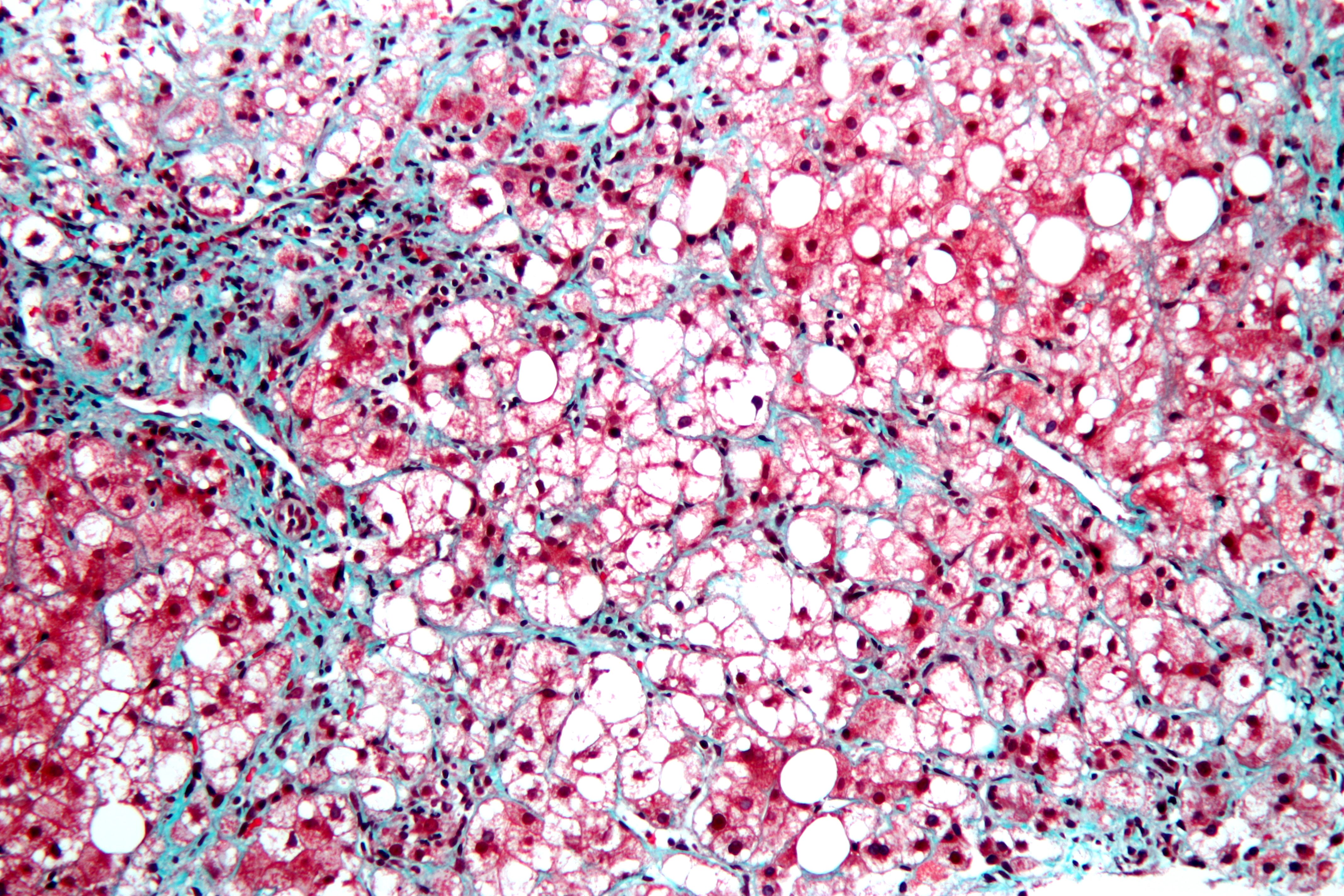
This is a stained liver biopsy sample showing advanced cellular changes in non-alcoholic fatty liver disease. Blue is fibrosis. White is fat accumulation in degenerated cells. Courtesy of Nephron’s work.
What Is Non-Alcoholic Fatty Liver Disease?
N on-alcoholic fatty liver is a non-inflammatory liver disorder characterized by degenerative changes in the liver caused by excessive accumulation of lipid (fat) in hepatocytes (liver cells) that is called free fatty acid-generated lipotoxicity.
Non-alcoholic fatty liver shows an increase in liver enzymes called transaminases.
Q: What are the enzymes that increase?
A: The transaminases that increase are ALT and AST. ALT is the abbreviation for alanine aminotransferase enzyme and AST is the abbreviation for aspartate aminotransferase enzyme. They are commonly measured in blood tests to determine liver function and when elevated indicate inflammation.
What Is Non-Alcoholic Fatty Liver In Celiac Disease and/or Gluten Sensitivity?