Skip to content

Lymphoma, Enteropathy-Associated T-Cell (EATL) 

A real photomicrograph of carcinoma of the esophagus. Panorama of 6 photos of a slide at 40x through the microscope. Some areas may appear blurry due to shallow DOF.

What Is Cancer Of The Esophagus?

C ancer of the esophagus is a malignancy arising in the stratified squamous cell lining of any part of the esophagus and having a poor prognosis.

This tumor first invades the deeper layers of the esophagus which includes the layer beneath the mucosa (submucosa) and muscle before it may invade structures close to it including the aorta and trachea. Later, it may spread to organs further away such as the liver, lungs and bones.

Q: What is the stratified squamous cell lining of the esophagus?

A: Stratified squamous cells are flat epithelial cells that are composed of several layers, called the epithelium. This type of epithelium forms the surface mucosa that lines the inside of the esophagus.

The esophagus itself is a muscular tube that transports swallowed substances to the stomach. It begins at the cricoid cartilage (Adam’s apple) as a continuation of the pharynx in the throat and ends at the lower esophageal sphincter (LES).

The LES is a circular muscle surrounding the junction of the esophagus and stomach. The LES opens to allow swallowed food and liquids to enter the stomach and closes to prevent their travelling back into the esophagus.

Who is Affected in the General Population?

Esophageal cancer is the fifth leading cause of death in men from cancer worldwide.1

What Is Cancer Of The Esophagus In Celiac Disease and/or Gluten Sensitivity?

Sources:
  1. Feldman M, Friedman LS, Brandt LJ. Sleisenger & Fordtran’s Gastrointestinal and Liver Disease. ed 9. Philadelphia: Saunders; 2010. pp. 745–767. []

Alopecia Areata

neutrophilWhat Is Neutropenia?

N eutropenia  is a blood disorder characterized by presence of an abnormally low number of neutrophils.

Neutrophils are white blood cells (leukocytes) that serves as the primary defense against infections by destroying bacteria in the blood. 

Specfically, neutrophils are a type of granulocyte that contain granules filled with potent chemicals to break down the microbes they ingest. Some of these chemicals, such as histamine, also contribute to inflammation and allergy.

The process of eating and digesting microbes is called phagocytosis. Neutrophils are phagocytes.1

Q: How do neutrophils eat microbes?

A: Segmented neutrophils  are the mature phagocytes that migrate through tissues to destroy microbes and respond to inflammatory stimuli. Segmented neutrophils comprise 40-75 % of the peripheral leukocytes. They are usually 9 to 16 µm in diameter. The nuclear lobes, normally numbering from 2 to 5, may be spread out so that the connecting filaments are clearly visible, or the lobes may overlap or twist. The chromatin pattern is coarse and clumped. The cytoplasm is abundant with a few nonspecific granules and a full complement of rose-violet specific granules.1

What Is Neutropenia In Celiac Disease and/or Gluten Sensitivity?

Sources:
  1. http://www.wadsworth.org/chemheme/heme/microscope/seg.htm [] []

Alopecia, Diffuse (Balding Hair Loss)

Courtesy of slideshow.net

What Is Transient Erythroblastopenia?

T ransient erythroblastopenia is a rare disorder of red blood cell formation characterized by brief, reversible disappearance of erythroblasts (red blood cell precursors) in the bone marrow of children.

Q: What do the red blood cells look like?

A: Circulating red blood cells appear normal so that the anemia which develops is a normocytic (normal cellular) anemia in contrast to microcytic (small cell) anemia in iron deficiency or macrocytic (large cell) anemia in vitamin B12 or folic acid deficiency.

Because new red blood cells are failing to mature, packed red blood cell transfusions may be necessary in severe anemia.

In adults, this disorder is called pure red blood cell anemia.

What Is Transient Erythroblastopenia In Celiac Disease and/or Gluten Sensitivity?

Bone Fractures 

Proptosis and lid retraction are features of  Grave's disease, or hyperthyroidism. Courtesy of Wikimedia.
Proptosis and Lid Retraction are Features of Grave’s Disease, or Hyperthyroidism.

What Is Grave’s Disease (Hyperthyroidism)?

G rave’s disease, or hyperthyroidism, is an autoimmune thyroid disease characterized by diffuse nontender goiter, elevated thyroxine hormone levels (T4, T3), suppressed thyroid stimulating hormone (TSH), and presence of thyroid receptor antibodies in the blood.

The autoantibodies involved are anti-thyroid peroxidase and anti-thyroglobulin antibodies. They bind to the thyroid stimulating hormone receptors, causing thyroid stimulation. These antibodies are detected by blood tests.

Q: What happens to the thyroid gland in Grave’s disease?

A: The thyroid gland is located in the front of the neck. This butterfly shaped gland consists of a large number of closed vesicles that contain a homogenous substance called colloid, which contains the thyroglobulin. Thyroglobulin is an iodine-containing protein secreted by the thyroid gland and stored within its colloid, from which the thyroid hormones thyroxine (T4) and triiodothyroinine (T3) are derived.1

Thyroxine molecule, chemical structure. Thyroid gland hormone that plays a role in energy metabolism regulation. It is a iodine containing derivative of thyrosine. Atoms are represented as spheres with conventional color coding: hydrogen (white), carbon (grey), oxygen (red), nitrogen (blue), iodine (purple).
Thyroxine molecule. Atoms are represented as spheres with conventional color coding: hydrogen (white), carbon (grey), oxygen (red), nitrogen (blue), iodine (purple).

T3 is the active hormone and is made from T4. Thyroid hormones are released into the bloodstream as needed to control metabolism, brain development, breathing, heart and nervous system functions, body temperature, muscle strength, skin dryness, menstrual cycles, weight, and cholesterol levels.

Thyroid hormone production is regulated by thyroid-stimulating hormone (TSH), which is made by the pituitary gland in the brain. Normally, when thyroid hormone levels in the blood are low, the pituitary releases more TSH in response to stimulation by the nearby hypothalamus which is continually monitoring levels of thyroxin. When thyroid hormone levels are high, the pituitary decreases TSH production. So in Grave’s disease, release of TSH by the pituitary gland is suppressed by the hypothalamus because thyroid hormone is elevated. Goiter develops from growth stimulation by thyroid stimulating autoantibodies.

What Is Grave’s Disease In Celiac Disease and/or Gluten Sensitivity?

Sources:
  1. Taber’s Cyclopedic Medical Dictionary. 19th ed. F.A. Davis Company. Philadelphia, PA. []

Bronchiectasis

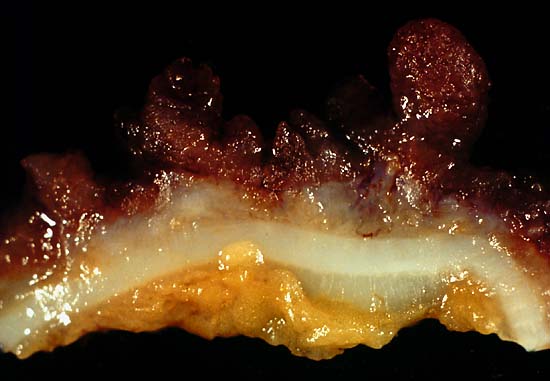
This photo was from a total colectomy done for clinically severe, intractable chronic ulcerative colitis. It shows a closer view of a longitudinal section through the colon wall. This demonstrates not only the angry red mucosa but also the tendency for the inflamed tissue to throw itself up into inflammatory pseudopolyps. Source: Ed Uthman, MD. Public domain.
This photo is from a total colectomy done for severe, intractable chronic ulcerative colitis. It shows a close view of a lengthwise section through the colon wall. This demonstrates not only the angry red mucosa, but also, the tendency for the inflamed tissue to throw itself up into inflammatory pseudopolyps.
Source: Ed Uthman, MD. Public domain.

What Is Ulcerative Colitis?

U lcerative colitis is an inflammatory disorder of the colon characterized by continuous inflammation of the mucosa and submucosa usually with small ulcers, extending from the rectum and typically involving the distal colon, rectum, and anus and producing bloody diarrhea.

While the severity of ulcerative colitis varies among patients, iron deficiency anemia often develops due to blood loss especially when there are many bloody bowel movements in a day.

The onset of ulcerative colitis is most commonly in young adulhood.

Q: Is this disease painful?

A: Yes with the passage of stool.

Psychological stress and subsequent dysbiosis exacerbate ulcerative colitis.

Psychological stress activates multiple physiological processes aimed at maintaining balance within the body. These physiological processes also have the capacity to influence the composition of microbial communities in the digestive tract, and research now indicates that exposure to stressful stimuli leads to gut microbiota dysbiosis.1

While the relative abundance of many different bacterial types can be altered during stressor exposure, findings in nonhuman primates and laboratory rodents, as well as humans, indicate that bacteria in the genus Lactobacillus are consistently reduced in the gut during stress.2

Presently, ulcerative colitis cannot be cured. This condition has a course of remissions, when symptoms subside, and flares, when symtpoms get worse. Treatment is aimed to reduce flares and promote remission. In all cases, correction of dysbiosis improves the condition.

Ulcerative colitis is associated with increased incidence of cancer of the colon.3

What Is Ulcerative Colitis In Celiac Disease and/or Gluten Sensitivity?

Sources:
  1. Galley JD, Bailey MT. Impact of stressor exposure on the interplay between commensal microbiota and host inflammation. Gut Microbes. 2014 May 1;5(3):390-396. Epub 2014 Apr 1. []
  2. Galley JD, Bailey MT. Impact of stressor exposure on the interplay between commensal microbiota and host inflammation. Gut Microbes. 2014 May 1;5(3):390-396. Epub 2014 Apr 1. []
  3. Taber’s Cyclopedic Medical Dictionary. F. A. Davis. Philadelphia, PA []

Cancer Of The Pharynx 

Areas of the Pharynx Where Cancer May Occur. Courtesy Wikimedia.

What Is Cancer Of The Pharynx?

Cancer of the pharynx is a malignant growth of stratified squamous cells that line the pharynx, or throat.

Q: What are stratified squamous cells that line the pharynx?

A: Stratified squamous cells are thin, flat epithelial cells consisting of several layers forming the surface mucosa that protects underlying layers of the pharynx. These cells lie very close together and have no blood vessels.

The pharynx is the upper passageway of the throat where swallowing food and fluids from the mouth first pass on the way to the esophagus and breathing air passes from the nose and mouth to and from the lungs.

The pharynx provides for speech production, adequate taste reception, and proper hearing through equalization of air pressure in the eustachian tubes.

The pharynx has three areas and pharyngeal cancer can occur in any of them: the nasopharynx behind the nose, the oropharynx behind the mouth, and the hypopharynx or laryngopharynx, just above the larynx. Tonsils, adenoids, and other lymph tissue lie at the back of the throat.

Unfortunately, cancers of the pharynx tend not to be detected until late in their course when symptoms become evident. This makes the prognosis poor. See symptom below.

What Is Cancer Of The Pharynx In Celiac Disease and/or Gluten Sensitivity?

Asthma

Depiction of pancreatic amylase molecule.

What Is Macroamylasemia?

M acroamylasemia is an acquired enzyme disorder that causes elevated levels of the enzyme amylase (hyperamylasaemia) in the bloodstream. It is characterized by altered amylase molecules that have become abnormally bound with plasma proteins in the bloodstream, commonly IgG (immunoglobulin G) and/or IgA (immunoglobulin A).

The resulting molecule is too large to be properly filtered by the kidneys and excreted in the urine as would be normal unbound amylase, causing sustained elevation of amylase levels in the plasma.

With normal kidney function, a hyperamylasemia without an increase in urine amylase suggests the diagnosis of macroamylasemia, and is confirmed by identifying the macromolecular components.1

Amylase is a an enzyme produced by the pancreas and the parotid glands to digest starch in the diet. Comparatively small amounts are also produced by other organs.

Q: How do the pancreas and parotid glands deliver their amylase for digestion of starch?

A: The parotid glands deliver their amylase into the mouth. The action of amylase in the mouth starts the breakdown of starch as it is chewed and moistened with saliva.

After the starchy food is liquified in the stomach, the mass empties into the duodenum. Here pancreatic amylase is delivered through the common bile duct into the duodenum where it turns starch into maltose sugar. The next step to final digestion of starch is carried out by enzymes in the small intestinal lining that split maltose into its component sugar which is glucose.

Glucose is a simple sugar that can be absorbed into the bloodstream for use in the body. Humans cannot live without adequate glucose.

Distinquishing macroamylasemia from hyperamylasemia due to pancreatic disease is necessary to prevent needless treatment and investigation for pancreatitis.2

What Is Macroamylasemia In Celiac Disease and/or Gluten Sensitivity?

Sources:
  1. Torrent Vernetta A, Segarra Cantón O, Soler Palacín P, Segura Cardona RM, Infante Pina D. Macroamylasaemia in paediatrics. An Pediatr (Barc). 2008 Nov;69(5):439-41. []
  2. Isham CA, Ridgeway NA, Hedrick R, Cate JC 4th. Screening for macroamylase in a community hospital. Clin Chem. 1984 May;30(5):741-2. []

Polymyositis

What Is Helicobacter Pylori (H. Pylori) Infection? H elicobacter pylori infection is a potentially deadly stomach disease characterized by chronic superficial inflammation and ulcerations in 100% of infected patients. This infection disrupts normal defense and… 

Adenocarcinoma Of Small Intestine (Cancer)

Section of small bowel surgically removed for adenocarcinoma that grew through the wall. By: CDC/ Dr. Edwin P. Ewing, Jr.

What Is Adenocarcinoma Of Small Intestine?

Adenocarcinomas are malignant tumors, or cancer, of the small bowel arising out of glandular tissue. They fall in the category of rare neoplasm, comprising only 3% of all gastrointestinal malignancies.

Primary adenocarcinoma is the most common histological (cell) subtype constituting 35–50% of cases.1 

Q: What does adenocarcinoma look like?

A: Adenocarcinoma may manifest as strictures, nodules, excavating masses, or annular lesions.2

What Is Adenocarcinoma Of Small Intestine In Celiac Disease and/or Gluten Sensitivity?

Sources:
  1. Benhammane H, El M’rabet FZ, Serhouchni KI, El yousfi M, Charif I, Toughray I, et al. Small Bowel Adenocarcinoma Complicating Coeliac Disease: A Report of Three Cases and the Literature Review. Case Rep Oncol Med. 2012; 2012: 935183. Published online 2012 December 1. doi: 10.1155/2012/935183 []
  2. Ramachandran I, Sinha R, Rajesh A, Verma R. Multidetector row CT of small bowel tumors.  Clinical Radiology. 2007; 62:607-614. []

Abnormal Blood Values in Childhood

canstockphoto18551453
Illustration Showing Skin Anatomy.

What Is Pruritic Skin?

P ruritic skin is a symptom of a primary disease that may involve only the skin or a systemic disorder with other features and is characterized by chronic itching.

Q: What part of skin is affected by itchiness?

A: Itching may arise from any layer of skin, depending on the cause.

In the image to the right, the outermost protective surface layer, or stratum cornem, is shown as a thin tan line. It is the top dead layer of the epidermis that normally sloughs off as new cells move up to replace it. The epidermis is shown as a dark pink layer. It is composed of five layers of keratinocytes that flatten as they move upward.

The pink layer under the epidermis is the dermis, or true skin, which is composed of collagen and elastin tissue. It contains oil glands that lubricate skin, sweat glands, blood vessels, nerve endings, lymphatics, and hair follicles. Under the dermis is subcutaneous tissue containing fat cells here colored yellow on the bottom of the image.

What Is Pruritic Skin In Celiac Disease and/or Gluten Sensitivity?