Skip to content

Early Menopause or Ovarian Failure

Hair Follicles.
Hair Follicles.

What Is Fine Hair With Rough Texture?

F ine hair (lower diameter across the width) with rough texture is an abnormal hair shaft feature altered from the normal diameter and smooth quality of hair.

Q: What is the normal diameter and smooth quality of hair?

A: Although hair may appear to be a simple structure, it is actually a complex part of the anatomy whose biology is only partially understood. Hair grows from small organs (follicles) located within the complex microenvironment of the skin which has multiple layers of tissue, three glands whose secretions bathe hair, and multiple vascular systems.1

An individual hair is a thread-like shaft made up of cornified cells. It consists of the outermost layer, or cuticle, the cortex which is a horny component, and the medulla which is the central part.

Hairs receive nourishment from capillaries via the papilla at the base of their follicles (roots). Since the number of hair follicles are determined at birth, it is important to properly nourish them so they remain healthy.

What Is Fine Hair with Rough Texture In Celiac Disease and/or Gluten Sensitivity?

Sources:
  1. Harkey MR. Anatomy and physiology of hair. Forensic Sci Int. 1993 Dec;63(1-3):9-18. []

Dry Eye, Autoimmune (Keratoconjunctivitis Sicca)

gastrointestinal complications in diabetes symptom in gluten and celiac diseaseWhat Are Gastro-Intestinal Complications Of Type 1 Diabetes?

G astrointestinal complications of type I diabetes mellitus are functional or organic changes that result from diabetes affecting every organ of the gastrointestinal tract.

Q: How do gastrointestinal tract complications affect diabetes?

A: Impaired function of individual digestive organs in diabetes can significantly influence level of diabetes compensation and vice versa. On the other side, unsatisfactory diabetes compensation can result in manifestation of digestive problems.

The most frequent (55 to 75%) and the most serious clinical complication is diabetic gastroparesis characterized by impaired evacuation and motility of the stomach and small intestine. Gastroparesis results in nausea, vomiting, early fullness after eating and constipation.

Treatment is with prokinetics. These oral medicines help motility and transport of food in the esophagus, stomach and intestines and improve sphincter function to keep food from going backwards.

Gastroesophageal reflux and heartburn are other complications in diabetes. These are treated with antacids and acid reducing medications.

Various autoimmune disease interfere with digestion. One connection between diabetes mellitus and the gastrointestinal tract can be celiac disease.1

What Are Gastro-Intestinal Complications Of Diabetes In Celiac Disease and/or Gluten Sensitivity?

Sources:
  1. Perusicova J. Gastrointestinal complications in diabetes mellitus. Vnitri Lekarstvi. May 2004;50(5):338-43. []

PMS (Premenstrual Syndrome) 

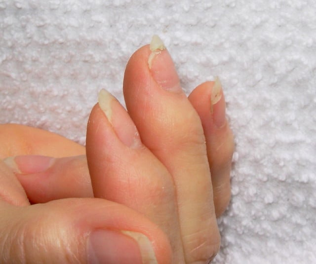
DSCN4758aWhat Are Brittle Nails?

B rittle nails are abnormalities of the nail plate that are characterized by poor nail structure affecting all fingernails and toenails seen as thin, dry nails that easily chip, split, and are difficult to maintain a clean edge. Usually longitudinal ridging occurs from the nail base to the tips.

Q: What is the nail plate?

A: The nail plate is the hard keratin cover protecting the finger tip and toe tip. The nail plate (non-living tissue) is produced by the nail matrix (living tissue) at the base of the nail plate under the lunula (moon), which is the site of brittle nail development.

Nail Anatomy. A. Nail plate; B. lunula; C. root; D. sinus; E. matrix; F. nail bed; G. hyponychium; H. free margin. Courtesy Wikipedia.org

Poor nail structure affecting all nails may be a feature of nutritional deficiency in poor diet or malabsorption such as occurs in celiac disease.

Some other causes are: idiopathic (unknown cause), the result of aging, the effects of certain drugs, or an association with systemic autoimmune disorders such as vitiligo, alopecia areata (with pitting), psoriasis (with pitting), and lichen planus. 

External (non-nutritional or disease) causes of dry, brittle nails, such as detergents and cleaners, would only affect fingernails but not toenails.

Note: It has been shown that working with your hands in water or soaking them through activities like swimming does not cause dry, brittle nails but will worsen them.

What Are Brittle Nails In Celiac Disease and/or Gluten Sensitivity?

Keratomalacia

Image From Gray's Anatomy. Courtesy Wikipedia.org
Image  of Pancreas From Gray’s Anatomy. Courtesy Wikipedia.org

What Is Pancreatic Insufficiency?

P ancreatic insufficiency is a disorder characterized by insufficient exocrine production of pancreatic enzymes for normal digestion of fats, proteins, and carbohydrates that results in maldigestion of these foodstuffs.

Pancreatic insufficiency also causes malabsorption of the fat-soluble vitamins: vitamin A, vitamin D, vitamin E, and vitamin K.

Q: What are the exocrine enzymes produced by the pancreas?

A: Exocrine enzymes produced by the pancreas include amylase for the digestion of carbohydrates, lipase for the digestion of fats, and protease for the digestion of proteins.

While lipases and amylase are secreted in the active form, proteases are secreted as pro-enzymes (need to be activated). Trypsinogen is converted to its active form trypsin in the duodenum by enterokinase, a protease secreted by the enterocytes (surface cells that line the duodenum), and trypsin in turn activates the other pancreatic proteases.1

Pancreatic enzymes are released by the pancreas into the internal pancreatic duct that empties into the common bile duct from which they are pumped through the Sphincter of Odi directly into the duodenum as needed. Tthe Sphincter of Odi is a strong circular muscle that controls the entrance of pancreatic enzymes into the duodenum.

After a meal, enzyme secretion into the duodenum increases quickly reaching peak output within the first 20 to 60 minutes, then decreasing to a stable level before reaching an interdigestive level at the end of the digestive period, that is, about 4 hours after meal intake.2

Medical treatment is with medication that contains enzymes to break down carbohydrates, fats, and protein in food at mealtime. This treatment is safe, effective, and has few side effects.

What Is Pancreatic Insufficiency In Celiac Disease and/or Gluten Sensitivity?

Sources:
  1. Fieker A, Philpott J, Armand M. Enzyme replacement therapy for pancreatic insufficiency: present and future. Clin Exp Gastroenterol. 2011;4:55-73. doi: 10.2147/CEG.S17634. []
  2. Fieker A, Philpott J, Armand M. Enzyme replacement therapy for pancreatic insufficiency: present and future. Clin Exp Gastroenterol. 2011;4:55-73. doi: 10.2147/CEG.S17634. []

Dysmenorrhea (Painful Periods)

Beaus lines in thin nails (The tiny brown streak is a splinter hemorrhage.)
Beau”s lines in a thin nail. (The tiny brown streaks are splinter hemorrhages due to vitamin C deficiency.)

What Are Horizontal Ridges In Fragile Nails?

Horizontal ridges, also called “beau’s lines,” are abnormalities of the nail plate that appear as rumpling from the base to the tips of nails and are characterized by poor nail structure of both fingernails and toenails.

The nail plate is the hard keratin cover of the finger tip and toe tip which we ordinarily call “nails.” The nail plate is produced by the nail matrix. 

Q: Why do Beau’s lines develop in nails?

A: Beau’s lines occur due to temporary cessation of proliferation (growth) of proximal nail matrix at the nail base. As the finger nail grows at the rate of 0.1 mm/day, the time course of the illness can be estimated from the position of the Beau’s line from proximal nail fold.1

Nail Anatomy. Nail Anatomy. A. Nail plate; B. lunula; C. root; D. sinus; E. matrix; F. nail bed; G. hyponychium; H. free margin. Courtesy Wikipedia.org.

A. Nail plate; B. lunula; C. root; D. sinus; E. matrix; F. nail bed; G. hyponychium; H. free margin. Courtesy Wikipedia.

Beau’s lines are frequently seen in nutritional deficiency states, bacterial illness, acute stress, and systemic disease. The conditions where Beau’s lines have been described include severe systemic illness, chemotherapy, malnutrition, zinc deficiency, trauma, paronychia, pemphigus, and Kawasaki disease.2 Beau’s lines are commonly seen in patients undergoing chemotherapy.3

 This condition of nails was named after Joseph Honoré Simon Beau (1806-1865).

What Are Horizontal Nail Ridges In Celiac Disease and/or Gluten Sensitivity?

Sources:
  1. Naik GS1, Harikrishna J. Beau’s lines. Indian J Med Res. 2013 Jan;137(1):220. []
  2. Naik GS1, Harikrishna J. Beau’s lines. Indian J Med Res. 2013 Jan;137(1):220. []
  3. Patel LM, Lambert PJ, Gagna CE, Maghari A, Lambert WC. Cutaneous signs of systemic disease. Clin Dermatol. 2011 Sep-Oct;29(5):511-22. doi: 10.1016/j.clindermatol.2011.01.019. []

Night Blindness – Nyctalopia

What Is Steatorrhea?

S teatorrhea is a condition of abnormal stool composition and consistency that is due to high fat content because fat from food is not  digested and absorbed into the body but rather passes out with stool.1

Q: How does fatty stool look?

A: The stool in steatorrhea appears pale, bulky and may float in the toilet. If stool is loose, it varies from a pudding consistency to whipped cream. If it is solid, it can vary from thin sqiggles to big turds that smear the sides of the toilet bowl. All types have a foul smell.

Fat of any kind must be digested, or broken down, by enzymes into fatty acids and glycerol and then these parts only are absorbed through the small intestinal lining into the body. The failure to digest or absorb fats deprives the body of these foodstuffs needed for many functions such as blood clotting and vital cell parts such as eye and brain structure.

Fat malabsorption produces malabsorption of vitamins A, D, E, and K because these vitamins can only be absorbed along with fatty acids.

What Is Steatorrhea In Celiac Disease and/or Gluten Sensitivity?

Sources:
  1. Murray JA, The widening spectrum of celiac disease. American Journal of Clinical Nutrition. Mar 1999;69 (3):354-365. []

Dyspareunia (Painful Intercourse), Primary

Koilonychia (Hordinsky, Sawaya, and Scher, 2000)

What Is Koilonychia?

K oilonychia is a feature of an iron deficiency state characterized by abnormally shaped nails, called spoon shaped nails, meaning that the normal downward curve is reversed.

Depending on the inadequacy of iron, nails may simply remain flat or with a slight dip without progressing to a reversed curve.

These nails develop vertical ridges from the base to the nail tip that can become deep, called washboard nails.

Q: Are all nails affected?

A: All nails are affected in varying degrees. The small finger shows the least effect while the thumb shows the greatest change which begins as flattening before scooping upward.

Nail Anatomy. Nail Anatomy. A. Nail plate; B. lunula; C. root; D. sinus; E. matrix; F. nail bed; G. hyponychium; H. free margin. Courtesy Wikipedia.org.

A. Nail plate; B. lunula; C. root; D. sinus; E. matrix; F. nail bed; G. hyponychium; H. free margin. Courtesy Wikipedia.

What Is Koilonychia In Celiac Disease and/or Gluten Sensitivity?

Ocular Myopathy

What Is Primary Hyperparathyroidism?

Tissue changes in hyperparathyroidism. Courtesy Quizlet.com
Tissue Changes in Hyperparathyroidism Causing Brown Tumor. Courtesy Quizlet.com

P rimary hyperparathyroidism is a parathyroid disorder characterized by excessive secretion of parathyroid hormone by one or more parathyroid glands for more than 6 months.

In primary hyperparathyroidism, blood calcium levels are high while phosphorus levels are decreased due to the action of parathyroid hormone.

Parathyroid hormone is produced by the four pea sized parathyroid glands that are located on the thyroid gland in the front of the neck. Partly because the thyroid and parathyroid glands share the same anatomic place in the body and partly because they have similar names, they are often confused although they have completely different actions.

Parathyroid hormone normally keeps calcium and the opposing mineral phosphorus levels in balance by drawing calcium as needed from bones to increase it in blood and releasing excess phosphorus through the kidneys to decrease blood levels.

Primary hyperparathyroidism is commonly caused by an adenoma (tumor) in a parathyroid gland (80%) or 15% due to hyperplasia of gland tissue (overgrowth). It is seldom associated with autoimmune disorders. However, cancer is a  possibility.

Q: What is a parathyroid adenoma?

A: A parathyroid adenoma is usually a solitary, well circumscribed, soft, tan reddish-brown nodule with a capsule. Gland tissues outside of the adenoma are normal or slightly shrunken (not needed anymore).1

Untreated, primary hyperparathyroidism results in cyst formations in bone marrow (osteitis fibrosa cystica) and brown tumors in bone tissue. Cysts contain large amounts of fibrous tissue with areas of hemorrhage. Brown tumors contain aggregates of osteoclasts (bone cells), hemorrhage and giant cells resembling neoplasms.2

Here is the symptomatolgy: “Painful Bones, Renal Stones, Abdominal Groans, and Mental Moans.”

What Is Primary Hyperparathyroidism In Celiac Disease and/or Gluten Sensitivity?

Sources:
  1. http://quizlet.com/32428692/pathology-of-parathyroid-and-bone-flash-cards []
  2. http://quizlet.com/32428692/pathology-of-parathyroid-and-bone-flash-cards/ []

Inflammatory Vaginitis

The tiny brown streak is a splinter hemorrhage.

What Are Splinter Hemorrhages In Nails?

S ubungual (under the nail) splinter hemorrhage is characterized by fragility of the underlying capillaries resulting in bleeding under the nail . Bleeding that results looks like a brown streak or splinter.1

Because the streak of blood is not in the nail but under the nail, it does not grow out with the nail. It can be a feature of vitamin C deficiency which resolves with adequate nutrition.

Nail Anatomy. Nail Anatomy. A. Nail plate; B. lunula; C. root; D. sinus; E. matrix; F. nail bed; G. hyponychium; H. free margin. Courtesy Wikipedia.org.

A. Nail plate; B. lunula; C. root; D. sinus; E. matrix; F. nail bed; G. hyponychium; H. free margin. Courtesy Wikipedia.

Splinter hemorrhage can be seen in a diet poor in vitamin C, disorders of food absorption, systemic disease such as psoriasis, nail lichen planus, an aggressive disease that may destroy the nail plate, and endocarditis. Some drugs, such as oral contraceptives, cause vitamin C deficiency.

Who is Affected in the General Population? Both sexes are commonly affected, though it occurs significantly more frequently in males. Where present in females, they were confined to a single digit.2

What Are Splinter Hemorrhages In Nails In Celiac Disease and/or Gluten Sensitivity?

Sources:
  1. Krause’s Food, Nutrition, & Diet Therapy. 10th Edition. Kathleen Mahan, Sylvia Escott-Stump. 2000. W.B. Saunders Company. []
  2. Monk BE. The prevalence of splinter haemorrhages. Br J Dermatol. 1980 Aug;103(2):183-5. []

Dysgeusia (Impaired Taste)

DyspareuniaWhat Is Autoimmune Thyroiditis (Hypothyroidism)?

A utoimmune thyroiditis, also called Hashimoto’s thyroiditis or Hashimoto’s Disease, is an autoimmune destruction of thyroid tissue characterized by insufficient thyroid hormone circulating in the body that causes formation of a goiter (enlarged thyroid gland) and hypothyroidism.

Hypothroidism refers to the condition of markedly reduced secretion of thyroid hormone. There are other causes of hypothyroidism besides Hashimoto’s thyroiditis.

Hashimoto’s thyroiditis is often associated with other autoimmune diseases such as celiac disease.

In Hashimoto’s thyroiditis, a profusion of antibodies are produced, which build up in the blood. Left untreated so that much of the thyroid gland is destroyed, this condition may progress to the very serious and life-threatening condition called myxedema.

Note: In myxedema, protein, electrolytes, and water abnormally accumulate in between cells which produce firm, inelastic puffy skin that is cool, dry, rough, scaly, and may appear yellow; in some people, areas such as the ankles become crusty with a look of tree bark. Many systemic changes develop shown by significant slowing of mental and physical functions. Please see below.

Q: What thyroid tissue is targeted for destruction?

A: In Hashimoto’s thyroiditis, high levels of autoantibodies target thyroglobulin and thyroid peroxidase, leading to inflammation and destruction of the thyroid gland. The resulting fibrosis or scarring of the gland results in lack of thyroid hormone production.

The thyroid gland consists of a large number of closed vesicles that contain a homogenous substance called colloid, which contains the thyroglobulin. Thyroglobulin is an iodine-containing protein secreted by the thyroid gland and stored within its colloid, from which the thyroid hormones thyroxine (T4) and triiodothyroinine (T3) are derived.1

T3 is the active hormone and is made from T4. Thyroid hormones affect metabolism, brain development, breathing, heart and nervous system functions, body temperature, muscle strength, skin dryness, menstrual cycles, weight, and cholesterol levels.

Thyroid hormone production is regulated by thyroid-stimulating hormone (TSH), which is made by the pituitary gland in the brain. Normally, when thyroid hormone levels in the blood are low, the pituitary releases more TSH. When thyroid hormone levels are high, the pituitary decreases TSH production.

Hashimoto’s disease, with or without the development of hypothyroidism, is treated with synthetic thyroxine, which is man-made T4. Health care providers prefer to use synthetic T4, such as Synthroid® (Levothyroxine), rather than synthetic T3, because T4 stays in the body longer, ensuring a steady supply of thyroid hormone throughout the day. The thyroid preparations made with animal thyroid are not considered as consistent as synthetic thyroid.2

What Is Autoimmune Thyroiditis In Celiac Disease and/or Gluten Sensitivity?

Sources:
  1. Taber’s Cyclopedic Medical Dictionary. 19th ed. F.A. Davis Company. Philadelphia, PA. []
  2. National Endocrine and Metabolic Diseases Information Service. []