Skip to content

Anti-Endomysium Antibodies (EMA) Present In Blood

Smooth muscle. Courtesy pathguy.com
Smooth Muscle. Courtesy pathguy.com

What Are Anti-Endomysium Antibodies?

Anti-endomysium antibodies (EmA) are connective tissue autoantibodies produced in persons who have inherited the genes for celiac disease, an autoimmune disease, and are reacting to gluten in their diet.

Autoantibodies are abnormal in that they attack the body’s own tissue.

Q: What is endomysium?

A: Endomysium is the delicate connective tissue that surrounds individual muscle fibers. The autoantigen, or target, that stimulates the autoimmune response is the naturally occuring enzyme in endomysium called tissue transglutaminase (tTG), or more specifically tranglutaminase-2 (TG2). Anti-tissue transglutaminase antibodies recognize the same antigen as EmA, from which they differ in terms of detection method.

Anti-endomysium antibodies (EmA) are tested by the indirect immunofluorescence method and directed against “reticulin-like” fibres in connective tissue around smooth muscle fibres in the esophagus, liver, stomach, and bladder of monkeys, in the sections of the jejunum and kidneys of rats and in sections of the human umbilical cord. In comparison, for the determination of anti-tissue transglutaminase IgA and IgG antibodies, ELISA with human extractive or recombinant transglutaminase is recommended.1

EmA‐binding patterns in serum samples from patients with celiac disease have proved to be exclusively TG2‐targeted and the correlation between EmA and TG2 antibodies is therefore good. Evidence shows that celiac autoantibodies are produced in the small‐bowel mucosa.2

What Are Anti-Endomysium Antibodies In Celiac Disease and/or Gluten Sensitivity?

Sources:
  1. Trigoni E, Tsirogianni A, Pipi E, Mantzaris G, Papasteriades C. Celiac disease in adult patients: specific autoantibodies in the diagnosis, monitoring, and screening. Autoimmune Dis. 2014;2014:623514. doi: 10.1155/2014/623514. []
  2. Salmi TT, Collin P, Korponay-Szabó IR, Laurila K, Partanen J, Huhtala H, Király R, Lorand L, Reunala T, Mäki M, Kaukinen K. Endomysial antibody‐negative coeliac disease: clinical characteristics and intestinal autoantibody deposits.Gut. 2006 Dec;55(12):1746-53. []

Progressive Myoclonic Ataxia 

biliary system primary biliary cirrhosisWhat Is Primary Biliary Cirrhosis?

P rimary biliary cirrhosis (PBC) is a biliary tract disease characterized by chronic cholestasis (build-up of bile) and gradual destruction of bile ducts within the liver, called intrahepatic bile ducts, caused by chronic inflammation.

Primary biliary cirrhosis comes under the umbrella term autoimune liver disease in which the end result is immune-mediated hepatocellular (liver cell) or hepatobiliary (bile duct) injury.1

Q: What is the end result of destruction of bile ducts?

A: The end result of destruction of bile ducts is liver damage.

Injured liver tissue from chronic inflammation and the buildup of bile leads to cirrhosis, a condition in which the liver slowly deteriorates and malfunctions.

Scar tissue replaces healthy liver tissue, partially blocking the flow of blood through the liver. Scarring also impairs the liver’s normal ability to control infections, remove bacteria and toxins from the blood, process nutrients, hormones, and drugs, make proteins that regulate blood clotting, produce bile, and effectively replace its own cells when they become damaged.2

The liver is the largest organ within the body. It lies mostly in the upper part of the abdomen on the right side just under the diaphragm. About 70% of liver tissue is made up of cube shaped cells called hepatocytes that do the main work of the liver. Other cells (epithelial) form structure and are arranged in single layers around blood vessels, sinusoids, and bile ducts.

Bile ducts carry bile, a greenish brown liquid made by the liver to the gall bladder for storage until needed to aid in the digestion and absorption of fat and fat-soluble vitamins A, D, E, and K from the small intestine. Bile emulsifies fat eaten in the diet so that the pancreatic enzyme called lypase can break it down into its fatty acid and glycerol components which can then be absorbed into the body.

Bile also carries away waste products produced by normal metabolism and toxic substances that are removed by the liver for eventual elimination in stool. Bile is continually made by the liver from phospholipids, bile acids, cholesterol, and aging blood cells it removes from circulation. As such, bile must continually flow out of the liver to prevent build-up in the liver.

There is no cure for primary biliary cirrhosis.

What Is Primary Biliary Cirrhosis In Celiac Disease and/or Gluten Sensitivity?

Sources:
  1. Trivedi PJ, Adams DH. Mucosal immunity in liver autoimmunity: a comprehensive review. J Autoimmun. 2013 Oct;46:97-111. doi: 10.1016/j.jaut.2013.06.013. []
  2. http://digestive.niddk.nih.gov/ddiseases/pubs/primarybiliarycirrhosis/ []

Anti-Gliadin Antibodies Present

This is a model of Immunoglobulin-G Antibody (IgG)

What Are Anti-Gliadin Antibodies?

Anti-gliadin antibodies (AGA) are produced by the body as an immune response against partially digested gliadin peptides (fragments) that have abnormally entered the body from the intestinal tract. Gliadin is a protein in wheat grain.

Anti-gliadin antibodies circulating in blood are measured by laboratory testing on blood that is drawn. The antibodies are both immunoglobulin A (IgA) and immunoglobulin G (IgG) types, which are termed AGA-IgA and AGA-IgG.

The presence of anti-gliadin antibodies in blood is an abnormal laboratory finding. That is, when the intestinal lining is healthy and able to keep out undigested matter from the body, there are no molecules of gliadin present to trigger the immune system for defense.

Q: Who produces anti-gliadin antibodies?

A: Persons, with or without celiac disease, can produce anti-gliadin antibodies if and when leaky gut (increased intestinal permeability) develops that then allow gliadin peptides to get into the bloodstream.

Gliadin itself causes leaky gut, but there are other causes such as various foods, intestinal infections, medications, and other diseases of the gastrointestinal tract. Whatever the cause of leaky gut, if poorly digested gliadin is present, it can slip through the leaky lining and enter the bloodstream where it can trigger an antibody response by anti-gliadin antibodies.

What Are Anti-Gliadin Antibodies In Celiac Disease and/or Gluten Sensitivity Or Non-Celiac Gluten Sensitivity?

Bone Alkaline Phosphatase (BALP), Elevated

Vertebrae. Courtesy FreePik.com

What Is Elevated Bone Alkaline Phosphatase?

Elevated bone alkaline phosphatase (BALP) is a laboratory result that indicates an abnormal blood level of this bone enzyme.

A bone alkaline phosphatase blood level is one of the most frequently used biochemical markers of bone formation.

Q: Why is the purpose of  bone alkaline phosphatase?

A: Bone alkaline phosphatase is produced by bone cells called osteoclasts in normal bone maintenance for the purpose of breaking down old or damaged bone so that other bone cells called osteoblasts can fill in the excavated areas with new bone. This process keeps bone stong and healthy.

Elevated bone alkaline phosphatase shows that more bone is being broken down than is being replaced. It can be caused by hyperparathyroidism, bone tumors from cancer, and malnutrition.

What Is Elevated Bone Alkaline Phosphatase (BALP) In Celiac Disease and/or Gluten Sensitivity?

Hypokalemia (Low Potassium Blood Level)

hypokalemia gluten celiac disease symptomWhat Is Hypokalemia?

Hypokalemia means the level of potassium in the bloodstream is too low to meet metabolic needs of the body for this mineral and is characterized by metabolic acidosis, altered nerve conduction and muscle contraction.

Rapid potassium loss can result in life-threatening hypokalemic rhabdomyolysis which is destruction of muscle tissue that results in kidney damage.1

Q: What are metabolic needs of the body for potassium?

A: The metabolic needs of the body for potassium are great because this mineral is crucial for life and especially for normal nerve and muscle function.

Most potassium is intracellular, meaning it is found within cells while sodium, its opposing mineral (both electrolytes), is found in the fluid surrounding cells. In muscle contraction, exchange of potassium and sodium takes place so that potassium moves out of muscle cells and sodium moves into them. With muscle relaxation, potassium moves back into the cells and sodium moves out.

What Is Hypokalemia In Celiac Disease and/or Gluten Sensitivity?

Sources:
  1. Williams SG, Davison AG, Glynn MJ. Hypokalaemic rhabdomyolsis: an unusual presentation of celiac disease. European Journal of Gastroenterology and Hepatology. Feb 1995;7(2):183-4. []

Vitamin B12 Deficiency Anemia

What Is Vitamin B12 Deficiency Anemia? Vitamin B12 deficiency anemia is a megaloblastic anemia that is characterized by defective DNA synthesis of red blood cells due to a lack of vitamin B12. Vitamin B12 is essential… 

Angina Pectoris

What Is Angina Pectoris?Coronary Artery Lesion

Angina pectoris, or simply angina, is a coronary syndrome characterized by an oppressive substernal pain (pain under breastbone) or pressure brought on by exertion and relieved by rest that results from failure of coronary arteries to deliver adequate oxygen to heart tissue due to ischemic heart disease.

Q: Why do coronary arteries fail to deliver adequate oxygen to heart tissue?

A: Coronary arteries are the blood vessels that serve the heart. In angina, these vessels fail to deliver adequate oxygen to heart tissue because they are narrowed or blocked by fatty buildups, called atherosclerotic plaques or by a blood clot which impair their ability to carry adequate blood that carries the oxygen. Diseased coronary arteries cannot deliver adequate oxygenated blood pumped by the heart to its own muscle cells.

The heart is a muscular organ that is working all the time without rest, so it needs a constant supply of oxygen. When heart muscle has to work harder, it needs more oxygen. Lack of oxygen causes pain which makes the affected person stop activity and rest.

Angina can be stable or unstable. Unstable angina is much more serious and can be life-threatening.

  • Stable angina produces predictable pain and responds to rest and/or medication. It is less serious than unstable angina but can be very painful or uncomfortable. Anything that makes the heart muscle need more oxygen can cause an angina attack in someone with heart disease, including: smoking, cold weather, exercise, emotional stress, obesity, and large meals. Other causes of angina include: abnormal heart rhythms (usually ones that cause the heart to beat quickly), anemia, coronary artery spasm, heart failure, heart valve disease, and hyperthyroidism (overactive thyroid).1
  • Unstable angina produces unpredictable pain that may occur at rest, lasting more than 20 minutes. It is more severe than stable angina and less responsive to medication. Atherosclerosis is by far the most common cause of unstable angina. Oxidized low-density lipoprotein, so-called bad cholesterol, and oxysterols play an important role in atherogenesis, the development of atherosclerosis. Coronary arteries that are narrowed by atherosclerotic plaques can rupture causing injury to the coronary blood vessel resulting in blood clotting which blocks the flow of blood to the heart muscle. Blood clots may form, partially dissolve, and later form again and angina can occur each time a clot blocks blood flow in an artery. People with unstable angina are at increased risk of having a heart attack.2

What Is Angina In Celiac Disease and/or Gluten Sensitivity?

Sources:
  1. http://www.ncbi.nlm.nih.gov/pubmedhealth/PMH0001247/ []
  2. http://www.heart.org/HEARTORG/Conditions/HeartAttack/SymptomsDiagnosisofHeartAttack/Unstable-Angina_UCM_437513_Article.jsp# []

Secondary Hyperparathyroidism

Hepatitis-C-Healthy-Liver-and-Inflammation-of-Liver

What Are Elevated Liver Enzymes?

H ypertransaminasemia is a chronic condition of elevated blood liver transaminase enzymes, commonly called “liver enzymes,” that signifies hepatocellular (liver) injury.

Q: What are serum transaminases?

A: Transaminases are the liver enzymes ALT and AST. ALT is the abbreviation for alanine aminotransferase enzyme and AST is the abbreviation for aspartate aminotransferase enzyme.

Transaminases are commonly measured in routine blood tests to determine liver function. Elevated ALT and AST transaminases  indicate inflammation of the liver. Other specific tests must follow to determine the cause of inflammation.

What Is Hypertransaminasemia In Celiac Disease and/or Gluten Sensitivity?

Delayed Puberty in Boys

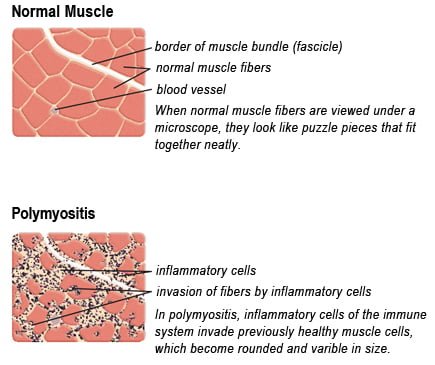
Drawing of Biopsy Showing Muscle Fibers Invaded by Immune Cells. Courtesy MDA.org

What Is Polymyositis?

P olymyositis is a body-wide connective tissue disease resulting from autoimmune attack of skeletal muscles that is characterized by inflammatory and degeneratory changes. The course is unpredictable being marked by spontaneous flare-ups and remissions.

Polymyositis can begin slowly or abruptly according to the factor that is triggering the onset such as infection, medications like phenytoin, and autoimmune disease.

Progressive muscle weakness starts in the proximal skeletal muscles (muscles closest to the trunk of the body).

Skeletal muscles, also called voluntary, are muscles that move the body as we want, such as walking and  lifting objects, as opposed to those we cannot voluntarily control, such as the muscles of digestion. 

Q: What are the degeneratory changes in skeletal muscles?

A: In polymyositis, degeneratory changes in skeletal muscles means that muscles are being destroyed (called necrosis), resulting in fibrosis, or scarring. When scar tissue takes the place of lost muscle tissue, it cannot act like muscle to contract and relax.   Muscle destruction is what causes muscle pain and weakness.

After the clinical work-up of exams and blood studies to determine muscle damage, the diagnosis of polymyositis is confirmed by muscle biopsy. See image at above left. The black dots are inflammatory cells. Edema (fluid) between cells caused by inflammation pushes muscle fibers apart.

There is no cure for polymyositis, but the symptoms can be treated.  Options include medication, physical therapy, exercise, heat therapy (including microwave and ultrasound), orthotics and assistive devices, and rest.  The standard treatment for polymyositis is a corticosteroid drug, given either in pill form or intravenously.  Immunosuppressant drugs, such as azathioprine and methotrexate, may reduce inflammation in people who do not respond well to prednisone. 

Periodic treatment using intravenous immunoglobulin can also improve recovery.  Other immunosuppressive agents used to treat the inflammation associated with polymyositis include cyclosporine A, cyclophosphamide, and tacrolimus.  Physical therapy is usually recommended to prevent muscle atrophy and to regain muscle strength and range of motion.1

Diagnosis is based on elevated muscles enzymes, increased urinary creatine level, and electromyograph abnormalities.

Polymyositis can affect people at any age. It is most common in adults between ages 50 and 70, and in children ages 5 to 15. It affects women twice as often as men and is more common in African Americans than Caucasians.2 The major causes of death from polymyositis are cancer and lung disease, including pneumonia.  The 5-year mortality rate can be as high as 1 in 5 patients.2

What Is Polymyositis In Celiac Disease and/or Gluten Sensitivity?

Sources:
  1. National Institute of Neurological Disorders and Stroke []
  2. http://www.nlm.nih.gov/medlineplus/ency/article/000428.htm [] []

Anti-tissue Transglutaminase Antibodies (tTG) Present In Blood

Image depicts tissue transglutaminase (tTg).
Image Depicts Tissue Transglutaminase (tTG).

What Are Anti-tissue Transglutaminase Antibodies?

Anti-tissue transglutaminase antibodies (anti-tTG) are connective tissue autoantibodies and can be detected in blood samples from affected persons who are reacting to gluten in the diet.

Autoantibodies are abnormal because they attack the body’s own tissue, which in the case of these antibodies is tissue transglutaminase 2 (TG2).

Q: What is tissue transglutaminase 2 (TG2)?

A: Tissue transglutaminase 2 (TG2) is an enzyme that appears in many cell locations and is particularly abundant in endothelial cells that line the small intestine. It has been implicated in a variety of cellular processes, such as differentiation, cell death, inflammation, cell migration and wound healing.

The cell appears to adapt the dynamics of this enzyme to meet specific sub-cellular needs or to respond to stress or other stimuli. Substantial evidence indicates that the location of TG2 within cells is critical for the regulation of its various biochemical activities, which subsequently trigger diverse downstream events,1

Although initially studied as an enzyme within cells, TG2 is now known to be secreted also into the extracellular space (between cells) or onto the cell surface.1

Abnormal activation of TG2 or deregulation of its function(s) is involved in a variety of human diseases, such as celiac disease, diabetes, neurodegenerative diseases, multiple sclerosis and rheumatoid arthritis. A role in inflammatory disorders and septic shock has also been shown. Moreover, multiple studies have revealed elevated TG2 expression in many types of cancer cells.1

What Are Anti-tissue Transglutaminase Antibodies In Celiac Disease and/or Gluten Sensitivity?

Sources:
  1. Piacentini M, D’Eletto M, Farrace MG, Rodolfo C, Del Nonno F, Ippolito G, Falasca L. Characterization of distinct sub-cellular location of transglutaminase type II: changes in intracellular distribution in physiological and pathological states. Cell Tissue Res. 2014 Dec;358(3):793-805. doi: 10.1007/s00441-014-1990-x. [] [] []