Skip to content

Blepharitis, Non-Infectious (Swollen Eyelids)

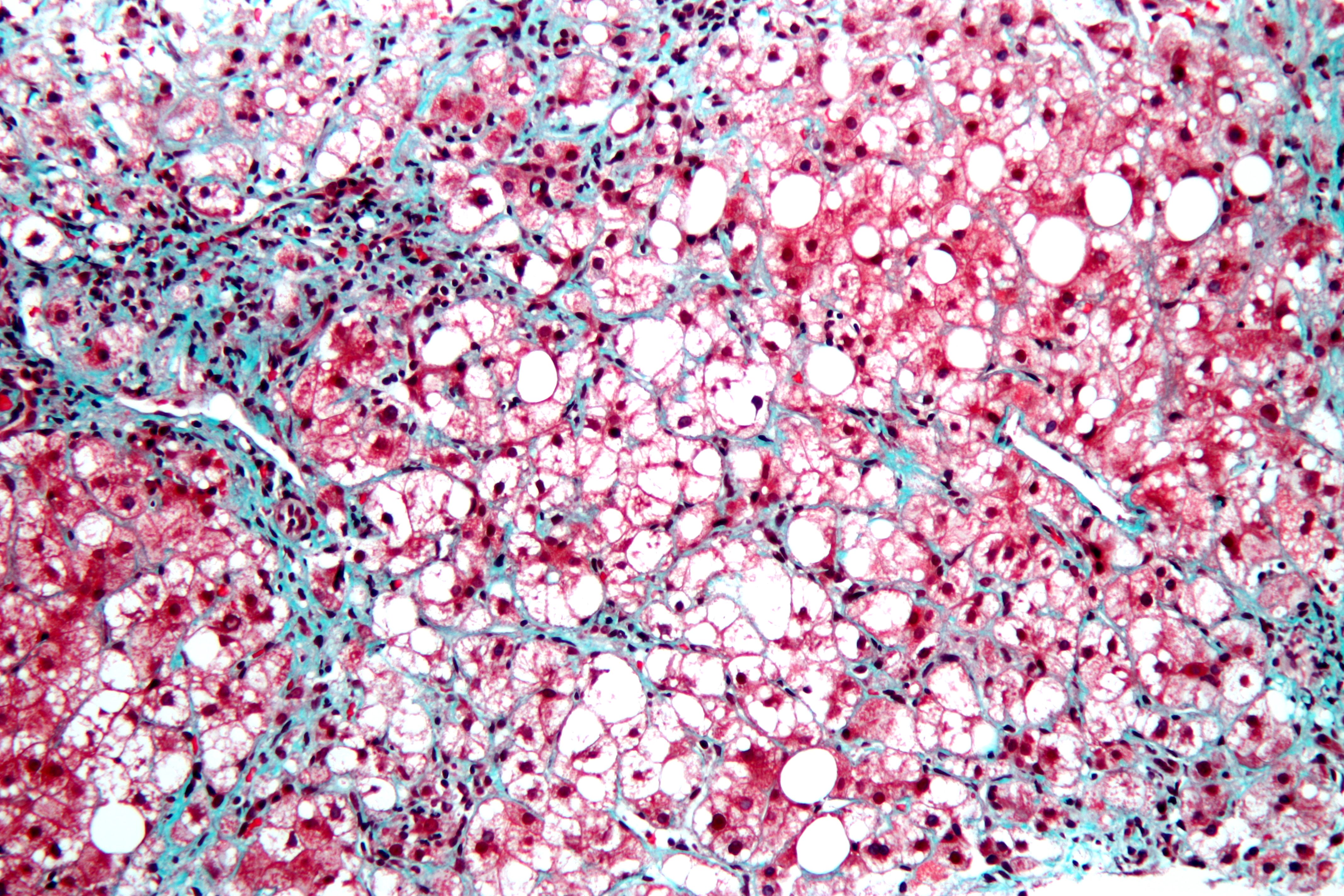
This is a stained liver biopsy sample showing advanced cellular changes in non-alcoholic fatty liver disease. Blue is fibrosis. White is fat accumulation in degenerated cells. Courtesy of Nephron's work.
This is a stained liver biopsy sample showing advanced cellular changes in non-alcoholic fatty liver disease. Blue is fibrosis. White is fat accumulation in degenerated cells. Courtesy of Nephron’s work.

What Is Non-Alcoholic Fatty Liver Disease?

N on-alcoholic fatty liver is a non-inflammatory liver disorder characterized by degenerative changes in the liver caused by excessive accumulation of lipid (fat) in hepatocytes (liver cells) that is called free fatty acid-generated lipotoxicity.

Non-alcoholic fatty liver shows an increase in liver enzymes called transaminases.

Q: What are the enzymes that increase?

A: The transaminases that increase are ALT and AST. ALT is the abbreviation for alanine aminotransferase enzyme and AST is the abbreviation for aspartate aminotransferase enzyme. They are commonly measured in blood tests to determine liver function and when elevated indicate inflammation.

What Is Non-Alcoholic Fatty Liver In Celiac Disease and/or Gluten Sensitivity?

Juvenile Autoimmune Thyroid Disease

Trouseau Testing For Tetany.
Trouseau Testing For Tetany. Courtesy Digplanet.

What Is Tetany?

T etany is a painful feature of low blood calcium or low blood magnesium causing hyperexcitability of all nerves which in turn stimulates involuntary sudden, intermittent and repetitious tonic spasms of muscles. Tonic spasms are steady rather than jerky.

Causes include low function of the parathyroid glands which regulate calcium blood levels, vitamin D deficiency which inhibits calcium absorption from the gut that leads to calcium deficiency, and alkalosis.

In alkalosis, the chemical pH (acid/alkaline measure) of blood is too high, or alkaline, which inhibits adequate free calcium ions from being available for cell use.

Q: Why do low blood levels of calcium or magnesium cause muscles to spasm?

A: Calcium controls the readiness of muscle to respond to nerve stimulation. In muscle action, calcium and magnesium are minerals that oppose each other. Calcium allows a muscle to contract while magnesium allows it to relax. Inadequate levels of these opposing minerals affect the ability of muscles to properly function.

A simple test for tetany is called Trouseau’s named after the doctor who developed it. A blood pressure cuff is placed on an upper arm as usual and pumped up to a pressure sufficient to stop blood flow.  The pressure must be held for 3 minutes. A positive result is shown in the above photo of carpopedal spasm: the wrist and hand flexes inward while the fingers extend (opposite of wrist and hand) and the thumb goes against the palm.

What Is Tetany In Celiac Disease and/or Gluten Sensitivity?

Down Syndrome

Crohnie_sores_4[1]What Is Erythema Nodosum?

E rythema nodosum is an inflammatory disorder involving the deep dermis layer of skin and subcutaneous fat septa that underlies the skin. It is characterized by eruptions of recurrent or persistent multiple painful, red nodules under the skin that leave a bruised appearance when healing and do not scar.

The lower legs are most affected, but sores can appear anywhere there is subcutaneous fat.

Q: How do the nodules develop in erythema nodosum?

A: The edges of nodules are poorly defined, and the nodules vary from 2-6 cm.

During the first week of eruption, nodules become tense, hard, and painful. During the second week, they change color from bright red to bluish or livid and may become soft, but do not ulcerate. As absorption progresses, the color gradually fades to a yellowish hue, resembling a bruise. This disappears in 1 or 2 weeks as the overlying skin sloughs off and is replaced.1

The eruptive phase of erythema nodosum begins with flulike symptoms of fever and generalized aching followed by a painful rash within 1-2 days.  Aching legs and swelling ankles may occur and precede the eruption or appear during the eruptive phase and may persist for weeks.2

Currently, the most common cause of erythema nodosum is streptococcal infection in children and streptococcal infection and sarcoidosis in adults.3 Most sores in infection-induced erythema nodosum heal within 7 weeks, but active disease may last up to 18 weeks.

In contrast, 30% of idiopathic erythema nodosum cases may last more than 6 months. Idiopathic means that the cause is not known.

What Is Erythema Nodosum In Celiac Disease and/or Gluten Sensitivity?

Sources:
  1. http://emedicine.medscape.com/article/1081633-clinical#a0217 []
  2. http://emedicine.medscape.com/article/1081633-clinical []
  3. http://emedicine.medscape.com/article/1081633-overview#a0199 []

Congenital Anomalies 

Hangnail on lowest finger. Courtesy Wikipedia.org

What Is Hangnail?

H angnail is a broken strip of epidermis (piece of skin) at root or lateral (side) edge of fingernail or toenail that causes sharp pain.

A hangnail develops because the skin around the nail is unhealthy due to inadequate nutrition. Injury from trauma including biting the skin and pushing back the cuticles or exposure to excessive detergents and water that remove protective oils promote the development of hangnail.

All ages and both sexes can be affected.

Q: Can a hangnail become infected?

A: Infection, called paronychia, may develop from invasion of sore skin by any of these pathogens: bacteria, fungus, or yeast (Candida). Infected skin is red, swollen, and painful. Topical ointment is required to treat the infection.1

What Is Hangnail In Celiac Disease and/or Gluten Sensitivity?

Sources:
  1. www.nlm.nih.gov/medlineplus/ency/article/001444.htm []

Cystic Fibrosis 

stages of melanoma
The dark brown coloring represents the progression of melanoma as it begins to grow in skin.

What Is Melanoma?

M elanoma is a cutaneous malignancy with rapid invasion and metastasis to other parts of the body.

Melanoma is characterized by growth from melanocytes appearing as a new mole or enlarging from an exisiting mole, changing shape, size or color.

Melanocytes are cells found in the lower epidermis layer that produce melanin, or pigment, that colors the skin.

What Is Melanoma In Celiac Disease and/or Gluten Sensitivity?

Spina Bifida 

pityriasis rubraWhat Is Pityriasis Rubra Pilaris?

P ityriasis rubra pilaris (PRP) is a chronic generalized exfoliative dermatitis (sloughing skin) characterized by erythema (redness), scaling, dilated plugged hair follicles, and keratoderma (thickened skin) of the hands and feet that is often associated with anemia and low serum albumin.

It may manifest either as Type I classical adult onset PRP, Type II atypical adult (onset) PRP, or Type VI PRP (HIV-associated PRP pityriasis rubra pilaris) in contrast to classical juvenile (Type III) and circumscribed juvenile (Type IV) encountered among children.1

Q: Who is affected in the general population?

A: All ages are affected. Pityriasis rubra pilaris occurs all over the world but with racial variations – it is 1 in 5,000 in Great Britain and 1 in 50,000 in India.2

What Is Pityriasis Rubra Pilaris In Celiac Disease and/or Gluten Sensitivity?

Sources:
  1. Sehgal VN, Srivastava G, Dogra S. Adult onset pityriasis rubra pilaris. Indian J Dermatol Venereol Leprol. 2008 Jul-Aug;74(4):311-21. []
  2. Sehgal VN, Srivastava G, Dogra S. Adult onset pityriasis rubra pilaris. Indian J Dermatol Venereol Leprol. 2008 Jul-Aug;74(4):311-21. []

Glycogenic Acanthosis (Esophageal Plaques)

What Is Intraepithelial Lymphocytosis In Normal Small Bowel Samples? I ntraepithelial lymphocytosis is characterized by an abnormal increase in the numbers of intraepithelial lymphocytes (IELs) that are present in the small intestinal mucosa. Q: What… 

Juvenile Diabetes (Type 1 Diabetes Mellitus)

Muscle pain in celiac disease and gluten sensitivityWhat Is Chronic Muscle Pain And Tenderness?

M uscle pain and tenderness, also called myalgia, is a disorder of muscle tissue.

Muscle pain that is ongoing or chronic can result from a variety of disorders, infections, overuse, injury, as an unwanted effect of certain medications, toxic substances, celiac disease, and certain nutritional deficiencies.

What Is Muscle Pain and Tenderness In Celiac Disease and/or Gluten Sensitivity?

Chronic Bullous Dermatosis of Childhood or Linear IgA Dermatosis

Muscle spasm and cramping in celiac disease and gluten sensitivity symptomWhat Are Muscle Spasm, Muscle Cramps?

M uscle spasm and muscle cramps are  disorders of muscle function caused by painful involuntary contractions of affected skeletal muscles characterized by limited movement.

Q: What is the difference between muscle spasm and cramps?

A: Cramps are stronger and more painful than spasms, occurring while the muscle is in its most shortened state.

Skeletal muscles are those that move the body skeleton to do work as we choose. Examples are muscles that move the mouth, arms,  hands, legs, and feet.

What Are Muscle Spasm, Muscle Cramps In Celiac Disease and/or Gluten Sensitivity?

Dermatitis Herpetiformis In Childhood

muscle wastingWhat Is Muscle Wasting?

M uscle wasting, or atrophy, is a muscle disorder resulting from the loss of muscle tissue which is characterized by thin muscles that have reduced strength and endurance.

What Is Muscle Wasting In Celiac Disease and/or Gluten Sensitivity?