Skip to content

Urticaria or Hives, Chronic

Photo of Skin Rash in ITP Courtesy Wikipedia.

What Is Immune Thrombocytopenic Purpura?

I mmune thrombocytopenia purpura (ITP), formerly called idiopathic, is an immune mediated bleeding disorder characterized by destruction of circulating platelets (thrombocytes) by autoantibodies in the presence of normal bone marrow and without other conditions that cause bleeding problems including many medications.

Q: How do autoantibodies destroy platelets?

A: Autoantibodies bind to normal platelet surface antigens (targets) which forms a complex that is cleared from blood by the reticuloendothelial system. Consequently, this condition results in an abnormal decrease in the number of platelets circulating in blood plasma.

Platelets are cells that are needed to stop bleeding. A normal platelet count is in the range of 150,000 to 450,000. With ITP, the platelet count is less than 100,000. By the time significant bleeding occurs, the patient may have a platelet count of less than 10,000. The lower the platelet count, the greater the risk of bleeding. 

Because of inability of blood to properly clot due to lack of sufficient platelets, a distinctive purplish rash develops under the skin from blood leakage and an appearance of many small red spots of varying size can cover the skin. Depending on the number of platelets, there may be minimal to extensive bleeding in various parts of the body.  Bleeding in the brain is the worst complication, causing death in approximately 1% of affected children and 5% of affected adults.

Immune thrombocytopenic purpura is common in childhood, on average 50 cases per 1,000,000 per year with same incidence in boys as girls. It may follow a viral infection or immunization and is caused by an inappropriate response of the immune system to cross react against the body’s platelets.1

The incidence of immune thrombocytopenic purpura in adults is approximately 66 cases per 1,000,000 per year.  Older persons and persons with a previous history of hemorrhage are at increased risk of severe bleeding in adult ITP.  

Spontaneous remission occurs in most children (80%).  Remission in adults usually requires treatment. 

What Is Immune Thrombocytopenic Purpura In Celiac Disease and/or Gluten Sensitivity?

Sources:
  1. Dogan M, Sal E, Akbayram S, Peker E, Cesur Y, Oner AF. Concurrent celiac disease, idiopathic thrombocytopenic purpura and autoimmune thyroiditis. Clin Appl Thromb Hemost. 2011 Nov-Dec;17(6):E13-6. doi: 10.1177/1076029610378502. Epub 2010 Aug 3. []

Tremors

chronic abdominal distention celiac disease gluten symptomWhat Is Chronic Abdominal Distention?

A bdominal distention, or bloating, is characterized by enlargement in normal size of abdomen not due to fatty tissue.

Q: Why does the abdomen enlarge?

A: The abdomen enlarges because the bowel is dysfunctional. Loops of bowel usually lack normal peristalsis (rhythmic wave-like muscle movement) trapping gas in stagnant loops of bowel, inflammation swells loops of bowel also trapping gas, and dysbiosis overproduces gas. All these factors distend the abdomen.

What Is Chronic Abdominal Distention In Celiac Disease and/or Gluten Sensitivity?

Turner’s Syndrome 

Hyperkeratosis Back of Arm. GFW Photo.
Hyperkeratosis Back of Arm. GFW Photo

What Is Follicular Hyperkeratosis?

F ollicular hyperkeratosis is an abnormal skin condition characterized by disordered overgrowth of the horny layer of the epidermis with horny plugs filling the openings of hair follicles. The plugs look like bumps in hair follicles.

Follicular hyperkeratosis with corkscrew hairs (coiled hairs in the follicles) appears in scurvy, which is advanced vitamin C deficiency.1

Typical  hyperkeratotic papules first appear on the extensor surfaces of the extremities (e.g. the back of upper arms), shoulders, and buttocks.

Phrynoderma is a distinctive form of follicular hyperkeratosis with papule formation in dilated hair follicles believed to be a manifestation of severe malnutrition.

Q: Why does hyperkeratosis develop?

A: Follicular hyperkeratosis develops from lack of necessary nutrients to support healthy skin. Although originally thought to represent only vitamin A deficiency or vitamin C in the case of scurvy, several studies have demonstrated deficiencies of B vitamins and vitamin E. General malnutrition seems to be the strongest association. The clinical picture typically improves with enhanced nutritional status.2

What Is Follicular Hyperkeratosis In Celiac Disease and/or Gluten Sensitivity?

Sources:
  1. Krause’s Food, Nutrition, & Diet Therapy. 10th Edition. Kathleen Mahan, Sylvia Escott-Stump. 2000. W.B. Saunders Company. []
  2. Maronn M, Allen DM, Esterly NB. Phrynoderma: a manifestation of vitamin A deficiency?… The rest of the story. Pediatr Dermatol. 2005 Jan-Feb;22(1):60-3. []

Psoriasis

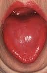
niacin deficiencyWhat Is A Fiery Red, Smooth, Burning Tongue?

A fiery red, smooth, burning tongue is an alteration in tongue tissue that is characteristic of advanced niacin deficiency.1

Q: How does niacin deficiency cause the tongue to be red and sore?

A: Niacin is an essential B vitamin that is required for a healthy tongue and by all body cells as well. Deficiency first shows in tissues with rapid cellular turnover, such as mucosal cells of the tongue.

When absorbed from the small intestines, niacin (the form in food) becomes the active form niacinamide. Niacinamide is converted by the body into co-enzymes which are present in all cells. These enzymes function in oxidation-reduction reactions essential for release of energy from carbohydrates, fats, and proteins and are needed as components for more than 200 enzymes involved in metabolism.

In addition to producing energy, niacinamide is essential for healthy skin and the mucosal lining of the digestive tract, normal functioning of the brain and nervous system, and production of steroid hormones from adrenal glands and hormones from sex glands.

What Is A Fiery Red, Smooth, Burning Tongue In Celiac Disease and/or Gluten Sensitivity?

Sources:
  1. Krause’s Food, Nutrition, & Diet Therapy. 10th Edition. Kathleen Mahan, Sylvia Escott-Stump. 2000. W.B. Saunders Company. []

Uveitis, Non-Infectious  

Courtesy Wikipedia.com
Parathyroid Glands in the Neck. Courtesy Wikipedia.com

What Is Secondary Hyperparathyroidism?

S econdary hyperparathyroidism is a parathyroid disorder resulting from hypocalcemia (low blood calcium level) that is characterized by excessive production of parathyroid hormone in the attempt to normalize the low blood calcium by releasing calcium from bone.

Parathyroid hormone is produced by the four pea sized parathyroid glands that are located on the thyroid gland in the front of the neck. In part, because the thyroid and parathyroid glands share the same anatomic place in the body and because they have similar names, they are often confused although they have completely different actions.

Parathyroid hormone regulates calcium and the opposing mineral phosphorus in the blood. In secondary hyperparathyroidism, calcium blood levels are low to normal while phosphorus levels are increased which stimulates the outpouring of parathyroid hormone.

Q: How does secondary hyperparathyroidism differ from primary hyperparathyroidism?

A: In primary hyperparathyroidism blood calcium is high and phosphorus is low, which is the opposite of secondary hyperparathyroidism.

The most common cause of secondary hyperparathyroidism is kidney disease causing failure to reabsorb calcium followed by vitamin D deficiency and malabsorption.

What Is Secondary Hyperparathyroidism In Celiac Disease and/or Gluten Sensitivity?

Attention Deficit Hyperactive Disorder (ADHD)

A small intestinal mucosa from a case of refractory coeliac disease immunostained sequentially for CD3 (alkaline phosphatase-blue) and CD8 (peroxidase-brown). Most intraepithelial lymphocytes are CD3+, CD8-.Courtesy pubcan.org
Mucosa  in refractory celiac  disease immunostained sequentially for CD3 (alkaline phosphatase-blue) and CD8 (peroxidase-brown). Most intraepithelial lymphocytes are CD3+, CD8-. Courtesy pubcan.org

What Is Refractory Celiac Disease?

R efractory celiac disease, formerly called refractory sprue, is a severe complication characterized by persistence of symptoms and intestinal inflammation despite gluten free diet after 12 months.1

Refractory celiac disease appears in two forms, ulcerative jejunitis (RCD I) and cryptic intestinal T-cell lymphoma (RCD II).

Patients with RCD I seem to profit from immunosuppressive treatment, but positive response to corticosteroid treatment does not exclude underlying enteropathy–associated T-cell lymphoma (EATL).

Patients with RCD II have a high percentage of abberant T-cells and is usually resistant to medical therapies. The presence of an aberrant clonal intraepithelial T-cell population has led to the designation of refractory celiac disease with this population as a cryptic intestinal T-cell lymphoma, characterized by frequent dissemination to the blood and the entire gastrointestinal lining.2

  • Refractory sprue may occur after an initial response to gluten free diet or without any evidence of preexisting celiac disease. All other causes of malabsorption must be excluded, such as collagenous colitis.
  • In a subgroup of patients with enteropathy-associated T-cell lymphoma (EATL) there is progressive deterioration of a refractory form of celiac disease. The prognosis is poor, although some patients respond to corticosteroids and immunosuppressive agents.3
  • A nationwide Finnish study showed that patients of male gender, older age, severe symptoms or seronegativity (negative antibody result) at the diagnosis of celiac disease are at risk of future refractory coeliac disease and should be followed up carefully.4
  • Chorea has been described as a paraneoplastic phenomenon in patients with non-Hodgkin’s lymphoma and has been described as associated with lymphoma arising from a background of refractory celiac disease. The finding of chorea in association with celiac disease should prompt a search for possible underlying intestinal T-cell lymphoma.5

How Prevalent Is Refractory Celiac Disease?

Sources:
  1. Murray JA, The widening spectrum of celiac disease. American Journal of Clinical Nutrition. Mar 1999;69 (3):354-365. []
  2. Culliford AN, Green PH. Refractory sprue. Current Gastroenterology Reports. Oct 2003;5(5):373-8. []
  3. Culliford AN, Green PH. Refractory sprue. Current Gastroenterology Reports. Oct 2003;5(5):373-8. []
  4. Ilus T, Kaukinen K, Virta LJ, Huhtala H, Mäki M, Kurppa K, Heikkinen M, Heikura M, Hirsi E, Jantunen K, Moilanen V, Nielsen C, Puhto M, Pölkki H, Vihriälä I, Collin P. Refractory coeliac disease in a country with a high prevalence of clinically-diagnosed coeliac disease. Aliment Pharmacol Ther. 2014 Feb;39(4):418-25. doi: 10.1111/apt.12606. []
  5. Kitiyakara T, Jackson M, Gorard DA. Refractory coeliac disease, small-bowel lymphoma and chorea. J R Soc Med. 2002 Mar;95(3):133-4. []

Seborrhea Dermatitis

Swollen Tongue Causing Tooth Indentations. GFW
Swollen Tongue Causing Tooth Indentations. Notice the Accompanying Denuded Area Due to Riboflavin Deficiency and Mild Candida Overgrowth. GFW

What Is A Pale, Smooth, Burning Tongue?

A pale, smooth, burning tongue is an alteration in tongue tissue characteristic of iron deficiency. The tongue is also swollen.1

Additionally, the sore tongue surface may be invaded by candida yeast which takes advantage of the sore tissue.

Iron deficiency itself increases susceptibility to infection.

Q: What is iron deficiency?

A: Iron deficiency results when the level within cells is too low to meet metabolic needs of the body for this mineral.

Deficiency is characterized by impaired red blood cell formation, free-radical disposal, oxygenation of cells, immune response to infection, enzyme activity, cognitive performance, digestion, nail structure, and fetal health.2

Iron is an essential mineral that is required for normal body function. Almost two-thirds of iron in the body is found in hemoglobin, the protein in red blood cells that carries oxygen to tissues. Smaller amounts of iron are found in myoglobin, a protein that helps supply oxygen to muscle, and in enzymes that assist biochemical reactions.

Iron is also found in proteins that store iron for future needs and that transport iron in blood. Iron stores are regulated by intestinal iron absorption.3

What Is A Pale, Smooth, Burning Tongue In Celiac Disease and/or Gluten Sensitivity?

Sources:
  1. Krause’s Food, Nutrition, & Diet Therapy. 10th Edition. Kathleen Mahan, Sylvia Escott-Stump. 2000. W.B. Saunders Company. []
  2. Kathleen Mahan and Sylvia Escott-Stump, ed. Krause’s Food, Nutrition & Diet Therapy, 10th Edition. Philadelphia, PA. USA: W.B. Saunders Company, 2000. []
  3. http://ods.od.nih.gov/factsheets/Iron-HealthProfessional accessed 12/11/12 []

Schizophrenia

Collagenous Colitis.
Microscopic Image Showing a Pink Collagen Band in Collagenous Colitis.

What Is Collagenous Colitis?

C ollagenous colitis is a disease of the large intestine (colon) that is characterized by microscopic inflammation of the surface mucosal lining and an abnormally thickened collagen band of tissue that develops wthin the lining of the colon.

The thicker than normal layer of collagen of at least 10 µm (reference value: 2–7 µm) can vary in different locations. Inflammation occurs with increased numbers of lymphocytes (white blood cells) and plasma cells and epithelial (surface cell) damage. These changes can only be seen under microscopic examination of multiple biopsied tissue samples taken during a colonoscopy procedure.

Q: What is collagen?

A: Collagen is a strong, fibrous protein found in connective tissue of the colon and many other tissues such as tendons. The normal basement membrane in the bowel consists mainly of collagen type IV, laminin, and fibronectin. The increased collagen band observed in collagenous colitis consists basically of collagen type I and III, which are the subtypes produced by repair functions, indicating a reactive origin to some irritant or drug.1

The biopsies should preferably be taken from the ascending colon, since the pathological hallmarks may be absent in the descending colon, and in the normally occurring thicker collagen layer in the rectosigmoid region.1 Inflammation of the ileum (last segment of the small intestine next to colon) is common.2

Endoscopy and radiological (x-ray) examinations are usually normal.3

Autoimmune disorders are frequently seen in adult patients with collagenous colitis.4 In the study below by Koskela et al. concomittent autoimmune diseases were present in 53% of patients with collagenous colitis.5

Importantly, the finding of collagenous colitis in patients with autoimmune diseases may reflect the treatment with NSAIDs (non-steroidal anti-inflammatory drugs), such as Ibuprofin and aspirin, PPIs (proton pump inhibitors), and other drugs. However, if secondary forms of collagenous colitis are not taken into consideration, underlying, treatable diseases may be overlooked, while only the gastrointestinal symptoms are treated symptomatically or with budesonide (a steroid).6

Treatment with budesonide steroid is efficacious irrespective of bile acid malabsorption.7

Budesonide at a mean dose of 4.5 mg/day maintained clinical remission for at least 1 year in the majority of patients with collagenous colitis and preserved health-related quality of life without safety concerns. Treatment extension with low-dose budesonide beyond 1 year may be beneficial given the high relapse rate after budesonide discontinuation.8

See below for nutritional deficiency problems caused by steroid usage and steps to be taken for correction.

What Is Collagenous Colitis In Celiac Disease and/or Gluten Sensitivity?

Sources:
  1. Ohlsson B. New insights and challenges in microscopic colitis. Therap Adv Gastroenterol. 2015 Jan;8(1):37-47. doi: 10.1177/1756283X14550134. [] []
  2. Bjørnbak C, Engel PJ, Nielsen PL, Munck LK. Microscopic colitis: clinical findings, topography and persistence of histopathological subgroups. Aliment Pharmacol Ther. 2011 Nov;34(10):1225-34. doi: 10.1111/j.1365-2036.2011.04865.x. []
  3. Abdo AA, Urbanski SJ, Beck PL. Lymphotcytic and collagenous colitis: the emerging entity of microscopic colitis. An update on pathophysiology, diagnosis and management. Canadian Journal of Gastroenterology. Jul 2003;17(7):425-32. []
  4. Leung ST, Chandan VS, Murray JA, Wu TT. Collagenous gastritis: histopathologic features and association with other gastrointestinal diseases. Am J Surg Pathol. 2009 May;33(5):788-98. doi: 10.1097/PAS.0b013e318196a67f. []
  5. Koskela RM, Niemela SE, Karttunen TJ, Lehtola JK. Clinical characteristics of collagenous and lymphocytic colitis. Scandanavian Journal of Gastroenterology. Sep 2004;39(9):837-45. []
  6. Ohlsson B. New insights and challenges in microscopic colitis. Therap Adv Gastroenterol. 2015 Jan;8(1):37-47. doi: 10.1177/1756283X14550134. []
  7. Bjørnbak C, Engel PJ, Nielsen PL, Munck LK. Microscopic colitis: clinical findings, topography and persistence of histopathological subgroups. Aliment Pharmacol Ther. 2011 Nov;34(10):1225-34. doi: 10.1111/j.1365-2036.2011.04865.x. []
  8. Münch A, Bohr J, Miehlke S, et al. Low-dose budesonide for maintenance of clinical remission in collagenous colitis: a randomised, placebo-controlled, 12-month trial. Gut. 2014 Nov 25. pii: gutjnl-2014-308363. doi: 10.1136/gutjnl-2014-308363. []

Food Allergy – IgE and Non IgE

Baby with Allergic Reaction to Peanuts. GFW
Baby with Allergic Reaction to Peanuts. GFW photo.

What Is Food Allergy?

Food allergy is an abnormal immune response to food proteins that may damage the small intestinal  lining and produce malabsorption of food. The reaction is usually delayed which makes it difficult to identify the offending food that is causing symptoms.

Q: How does food allergy develop?

A: The gastrointestinal tract serves not only to digest and absorb foodstuffs but also to protect the body from unwanted substances. When allergic food substances are eaten, the immune response that is triggered in the gut produces inflammation with symptoms such as pain, vomiting and loose bowels.

Inflammation causes swelling of the gut lining that can interfere with the passage of nutrients through it to the body which results in malabsorption. Malabsorption deprives the body of nutrients needed for normal function.

Symptoms other than digestive may involve skin rashes, hives, and respiratory difficulties that can be distressing and life-threatening.

What Is Food Allergy In Celiac Disease and/or Gluten Sensitivity?

Scleroderma (Systemic Sclerosis)

Geographic Tongue Due to Riboflavin Deficiency.

 What Is Geographic Tongue?

G eographic tongue is a painful alteration in tongue tissue that is characteristic of riboflavin (vitamin B2) deficiency.1

Q: How does the tongue surface appear?

A: Areas of inflamed tissue with flattened papillae develop among areas of normal tissue, giving a map-like appearance to the tongue surface. There are often swollen papillae that hurt or sting, such as is seen in the photo to the left.

A diet that does not provide adequate amounts of riboflavin and any condition that 1) interferes with riboflavin absorption from the gut, 2) depletes the vitamin from the body, 3) overuses the vitamin in the body, or 4) adversely affects its use in the body will result in riboflavin deficiency causing geographic tongue.

Riboflavin is essential for converting foodstuffs into energy, maintaining the normal lifespan of red blood cells and maintaining healthy skin, eyes, hair, and nervous system. Riboflavin is converted into its active forms, flavin adenine dinucleotide (FAD) and flavin mononucleotide (FMN). These enzymes are primarily involved as co-factors in oxidation reduction reactions that are essential for cellular energy production and respiration.

Riboflavin is required for the conversion of pyridoxine (vitamin B6) to its functional form and the production of niacin (vitamin B3) from the amino acid tryptophan. Riboflavin has a role in antioxidant status by activating glutathione reductase, which regenerates used glutathione, a powerful antioxidant.

Respiratory infection, exercise, certain diseases, drugs and hormones can adversely influence riboflavin status.2 Stress is a major depleter of riboflavin.

What Is Geographic Tongue In Celiac Disease and/or Gluten Sensitivity?

Sources:
  1. Krause’s Food, Nutrition, & Diet Therapy. 10th Edition. Kathleen Mahan, Sylvia Escott-Stump. 2000. W.B. Saunders Company. []
  2. Lakshmi AV. Riboflavin metabolism–relevance to human nutrition. Indian J Med Res. 1998 Nov;108:182-90. []