Skip to content

Bone Pain

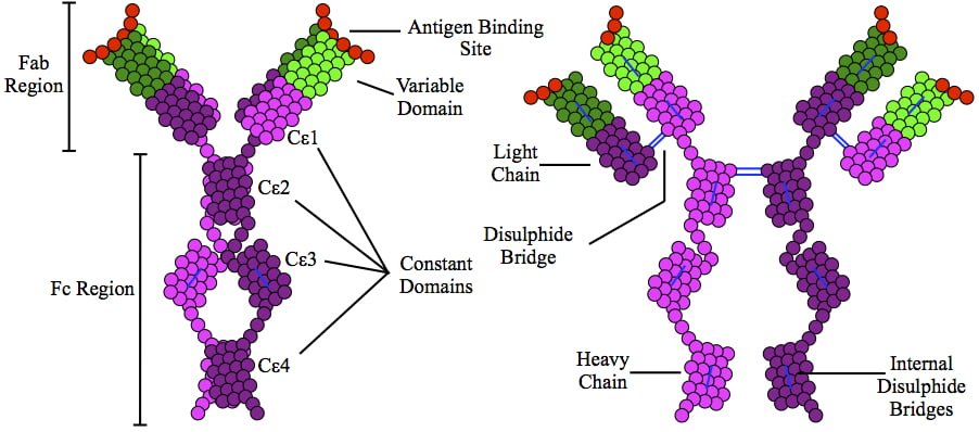
This is a depiction of immunoglobulin E (IgE) antibody which is elevated in allergic reactions.

What Is Allergic Rhinitis?

A llergic rhinitis is an immune disorder characterized by inflammation of the nasal mucosa by an IgE antibody reaction to an allergen.

An allergen is something that triggers an allergic immune response.

Q: What is the immune response?

A: Implicated in the response is an increase in T gamma-delta intraepithelial lymphocytes (IELs), which is a subset of pro-inflammatory T-cells located in the respiratory mucosa.  Lymphocytes are white blood cells.

When a person with allergic rhinitis breathes in an allergen such as pollen or dust, the body releases chemicals, including histamine that cause allergy symptoms. For example, hay fever involves an allergic reaction to pollen. A similar reaction occurs with allergy to mold, animal dander, dust, and other allergens that are breathed in.

What Is Allergic Rhinitis In Celiac Disease and/or Gluten Sensitivity?

Dysgeusia (Impaired Taste)

DyspareuniaWhat Is Autoimmune Thyroiditis (Hypothyroidism)?

A utoimmune thyroiditis, also called Hashimoto’s thyroiditis or Hashimoto’s Disease, is an autoimmune destruction of thyroid tissue characterized by insufficient thyroid hormone circulating in the body that causes formation of a goiter (enlarged thyroid gland) and hypothyroidism.

Hypothroidism refers to the condition of markedly reduced secretion of thyroid hormone. There are other causes of hypothyroidism besides Hashimoto’s thyroiditis.

Hashimoto’s thyroiditis is often associated with other autoimmune diseases such as celiac disease.

In Hashimoto’s thyroiditis, a profusion of antibodies are produced, which build up in the blood. Left untreated so that much of the thyroid gland is destroyed, this condition may progress to the very serious and life-threatening condition called myxedema.

Note: In myxedema, protein, electrolytes, and water abnormally accumulate in between cells which produce firm, inelastic puffy skin that is cool, dry, rough, scaly, and may appear yellow; in some people, areas such as the ankles become crusty with a look of tree bark. Many systemic changes develop shown by significant slowing of mental and physical functions. Please see below.

Q: What thyroid tissue is targeted for destruction?

A: In Hashimoto’s thyroiditis, high levels of autoantibodies target thyroglobulin and thyroid peroxidase, leading to inflammation and destruction of the thyroid gland. The resulting fibrosis or scarring of the gland results in lack of thyroid hormone production.

The thyroid gland consists of a large number of closed vesicles that contain a homogenous substance called colloid, which contains the thyroglobulin. Thyroglobulin is an iodine-containing protein secreted by the thyroid gland and stored within its colloid, from which the thyroid hormones thyroxine (T4) and triiodothyroinine (T3) are derived.1

T3 is the active hormone and is made from T4. Thyroid hormones affect metabolism, brain development, breathing, heart and nervous system functions, body temperature, muscle strength, skin dryness, menstrual cycles, weight, and cholesterol levels.

Thyroid hormone production is regulated by thyroid-stimulating hormone (TSH), which is made by the pituitary gland in the brain. Normally, when thyroid hormone levels in the blood are low, the pituitary releases more TSH. When thyroid hormone levels are high, the pituitary decreases TSH production.

Hashimoto’s disease, with or without the development of hypothyroidism, is treated with synthetic thyroxine, which is man-made T4. Health care providers prefer to use synthetic T4, such as Synthroid® (Levothyroxine), rather than synthetic T3, because T4 stays in the body longer, ensuring a steady supply of thyroid hormone throughout the day. The thyroid preparations made with animal thyroid are not considered as consistent as synthetic thyroid.2

What Is Autoimmune Thyroiditis In Celiac Disease and/or Gluten Sensitivity?

Sources:

  1. Taber’s Cyclopedic Medical Dictionary. 19th ed. F.A. Davis Company. Philadelphia, PA. []
  2. National Endocrine and Metabolic Diseases Information Service. []

Late Menarche (Start of Periods)

Courtesy quizlet.com

What Is Diffuse Alopecia?

D iffuse alopecia is characterized by abnormal hair loss or baldness.

Hair loss usually develops gradually and may be patchy or all over (diffuse). The average scalp contains about 100,000 hairs. Roughly 100 hairs are shed from the head every day.

Diffuse baldness not related to male pattern or heredity can be related to aging, nutritional deficiencies, some froms of dermatitis, radiation, endocrine disorders, especially thyroid hormone imbalance and diabetes, and undue stress.

Q: Can sudden stress cause hair loss?

A: A sudden physical or emotional stress may cause one-half to three-quarters of the hair throughout the scalp to shed. Other causes that need to be evaluated include use of common medications such as birth control pills, blood thinners, and anti-inflammatory pain drugs, and continued exposure to environmental chemicals such cleaning products.

What Is Diffuse Alopecia In Celiac Disease and/or Gluten Sensitivity?

Early Menopause or Ovarian Failure

Hair Follicles.
Hair Follicles.

What Is Fine Hair With Rough Texture?

F ine hair (lower diameter across the width) with rough texture is an abnormal hair shaft feature altered from the normal diameter and smooth quality of hair.

Q: What is the normal diameter and smooth quality of hair?

A: Although hair may appear to be a simple structure, it is actually a complex part of the anatomy whose biology is only partially understood. Hair grows from small organs (follicles) located within the complex microenvironment of the skin which has multiple layers of tissue, three glands whose secretions bathe hair, and multiple vascular systems.1

An individual hair is a thread-like shaft made up of cornified cells. It consists of the outermost layer, or cuticle, the cortex which is a horny component, and the medulla which is the central part.

Hairs receive nourishment from capillaries via the papilla at the base of their follicles (roots). Since the number of hair follicles are determined at birth, it is important to properly nourish them so they remain healthy.

What Is Fine Hair with Rough Texture In Celiac Disease and/or Gluten Sensitivity?

Sources:
  1. Harkey MR. Anatomy and physiology of hair. Forensic Sci Int. 1993 Dec;63(1-3):9-18. []

PMS (Premenstrual Syndrome) 

DSCN4758aWhat Are Brittle Nails?

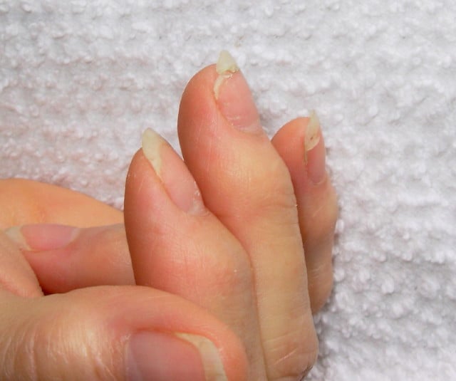
B rittle nails are abnormalities of the nail plate that are characterized by poor nail structure affecting all fingernails and toenails seen as thin, dry nails that easily chip, split, and are difficult to maintain a clean edge. Usually longitudinal ridging occurs from the nail base to the tips.

Q: What is the nail plate?

A: The nail plate is the hard keratin cover protecting the finger tip and toe tip. The nail plate (non-living tissue) is produced by the nail matrix (living tissue) at the base of the nail plate under the lunula (moon), which is the site of brittle nail development.

Nail Anatomy. A. Nail plate; B. lunula; C. root; D. sinus; E. matrix; F. nail bed; G. hyponychium; H. free margin. Courtesy Wikipedia.org

Poor nail structure affecting all nails may be a feature of nutritional deficiency in poor diet or malabsorption such as occurs in celiac disease.

Some other causes are: idiopathic (unknown cause), the result of aging, the effects of certain drugs, or an association with systemic autoimmune disorders such as vitiligo, alopecia areata (with pitting), psoriasis (with pitting), and lichen planus. 

External (non-nutritional or disease) causes of dry, brittle nails, such as detergents and cleaners, would only affect fingernails but not toenails.

Note: It has been shown that working with your hands in water or soaking them through activities like swimming does not cause dry, brittle nails but will worsen them.

What Are Brittle Nails In Celiac Disease and/or Gluten Sensitivity?

Dysmenorrhea (Painful Periods)

Beaus lines in thin nails (The tiny brown streak is a splinter hemorrhage.)
Beau”s lines in a thin nail. (The tiny brown streaks are splinter hemorrhages due to vitamin C deficiency.)

What Are Horizontal Ridges In Fragile Nails?

Horizontal ridges, also called “beau’s lines,” are abnormalities of the nail plate that appear as rumpling from the base to the tips of nails and are characterized by poor nail structure of both fingernails and toenails.

The nail plate is the hard keratin cover of the finger tip and toe tip which we ordinarily call “nails.” The nail plate is produced by the nail matrix. 

Q: Why do Beau’s lines develop in nails?

A: Beau’s lines occur due to temporary cessation of proliferation (growth) of proximal nail matrix at the nail base. As the finger nail grows at the rate of 0.1 mm/day, the time course of the illness can be estimated from the position of the Beau’s line from proximal nail fold.1

Nail Anatomy. Nail Anatomy. A. Nail plate; B. lunula; C. root; D. sinus; E. matrix; F. nail bed; G. hyponychium; H. free margin. Courtesy Wikipedia.org.

A. Nail plate; B. lunula; C. root; D. sinus; E. matrix; F. nail bed; G. hyponychium; H. free margin. Courtesy Wikipedia.

Beau’s lines are frequently seen in nutritional deficiency states, bacterial illness, acute stress, and systemic disease. The conditions where Beau’s lines have been described include severe systemic illness, chemotherapy, malnutrition, zinc deficiency, trauma, paronychia, pemphigus, and Kawasaki disease.2 Beau’s lines are commonly seen in patients undergoing chemotherapy.3

 This condition of nails was named after Joseph Honoré Simon Beau (1806-1865).

What Are Horizontal Nail Ridges In Celiac Disease and/or Gluten Sensitivity?

Sources:
  1. Naik GS1, Harikrishna J. Beau’s lines. Indian J Med Res. 2013 Jan;137(1):220. []
  2. Naik GS1, Harikrishna J. Beau’s lines. Indian J Med Res. 2013 Jan;137(1):220. []
  3. Patel LM, Lambert PJ, Gagna CE, Maghari A, Lambert WC. Cutaneous signs of systemic disease. Clin Dermatol. 2011 Sep-Oct;29(5):511-22. doi: 10.1016/j.clindermatol.2011.01.019. []

Night Blindness – Nyctalopia

What Is Steatorrhea?

S teatorrhea is a condition of abnormal stool composition and consistency that is due to high fat content because fat from food is not  digested and absorbed into the body but rather passes out with stool.1

Q: How does fatty stool look?

A: The stool in steatorrhea appears pale, bulky and may float in the toilet. If stool is loose, it varies from a pudding consistency to whipped cream. If it is solid, it can vary from thin sqiggles to big turds that smear the sides of the toilet bowl. All types have a foul smell.

Fat of any kind must be digested, or broken down, by enzymes into fatty acids and glycerol and then these parts only are absorbed through the small intestinal lining into the body. The failure to digest or absorb fats deprives the body of these foodstuffs needed for many functions such as blood clotting and vital cell parts such as eye and brain structure.

Fat malabsorption produces malabsorption of vitamins A, D, E, and K because these vitamins can only be absorbed along with fatty acids.

What Is Steatorrhea In Celiac Disease and/or Gluten Sensitivity?

Sources:
  1. Murray JA, The widening spectrum of celiac disease. American Journal of Clinical Nutrition. Mar 1999;69 (3):354-365. []

Hypokalemic Rhabdomyolysis in Celiac Disease and Dermatitis Herpetiformis

mucosal web in plummer-vinson syndromeWhat Is Plummer-Vinson Syndrome Affecting the Esophagus?

P lummer-Vinson syndrome is a manifestation of severe, long-term, iron deficiency anemia that is characterized by post-cricoid esophageal webs and dysphagia.

Q: What are esophageal webs?

A: Esophageal webs are one or more thin horizontal membranes consisting of squamous epithelium (cells that line the surface of the esophagus) and submucosa. They usually protrude from the anterior (front) wall, extending laterally across the inside esophagus but not to the posterior (rear) wall, which means that they rarely encircle the lumen.1

Dysphagia, or difficulty swallowing, from these webs is commonly painless and intermittent or progressive and may cause obstruction.

Webs can be detected by barium swallow X-ray, but the best way for demonstration is videofluoroscopy and by upper gastrointestinal endoscopy. They appear smooth, thin, and gray with eccentric or central lumen space. The webs typically occur in the upper part of the esophagus and may be missed and accidentally ruptured unless the endoscope is introduced under direct visualization.1

Iron deficiency is believed to decrease the contraction amplitude or force of the esophageal muscle resulting in motility impairment. Slower transit times have been recorded at the proximal and middle parts of the esophagus of Plummer-Vinson syndrome patients compared to healthy volunteers.2 Transit time is how fast ingested food and fluids travel through the esophagus.

Gude et al, report that iron replacement does not necessarily reverse the dysphagia in all the cases of Plummer-Vinson syndrome and that close monitoring of the web is mandated to watch for malignant change.  In fact, 3 to 15 per cent of the patients with Plummer-Vinson syndrome, mostly women between 15 and 50 years of age, have been reported to develop esophageal or pharyngeal cancer.2

What Is Plummer-Vinson Syndrome Affecting the Esophagus In Celiac Disease and/or Gluten Sensitivity?

Sources:
  1. Novacek G. Plummer-Vinson syndrome. Orphanet J Rare Dis. 2006; 1: 36. Published online 2006 September 15. doi:  10.1186/1750-1172-1-36. [] []
  2. Gude D, Bansal DP, and Malu A. Revisiting Plummer Vinson Syndrome. Annals of Medical and Health Sciences Research. 2013 Jan-Mar;3(1):119-121. [] []

Hyposplenism

Heartburn Due to Acidic Stomach Contents Re-entering the Esophagus. Courtesy kaiserpermanente.org
Heartburn Due to Acidic Stomach Contents Re-entering the Esophagus.

What Is Heartburn?

H eartburn is a functional upper digestive symptom of esophagitis that is characterized by a burning sensation behind the breastbone.

Q: Why does esophagitis cause heartburn?

A: Esophagitis is inflammation of the mucosal lining of the esophagus. The most common cause of esophagitis is the re-entering of stomach contents into the esophagus because the lower esophageal sphincter (LES) does not close tightly after food or fluids enter the stomach from the esophagus and/or the LES does not remain tightly closed.

The LES is located at the junction of the esophagus and the stomach. It functions like a circular band to tighten after food is ingested in order to prevent its going back up the esophagus when powerful stomach muscles churn ingested food.

Damage to the lining of the esophagus (esophagitis) results upon contact with hydrochloric acid and pepsin in gastric juice. Hydrochloric acid, produced by the stomach to dissolve food, inflames the esophagus, which is the opposite, or alkaline. Pepsin, produced by the stomach to dissolve protein in swallowed food, acts on the inflamed esophagus to break down the protein in its sore wall.

Heartburn can be caused by other disorders such as low stomach acid, obesity, and smoking. Relaxation of the LES can also result from dietary items such as chocolate, mint, caffeine, and alcoholic drinks.

Who is Affected in the General Population? Heartburn affects an estimated 42% of the US population.1

What Is Heartburn In Celiac Disease and/or Gluten Sensitivity?

Sources:
  1. http://informahealthcare.com/doi/abs/10.1185/03007990903553812. []

Lymphadenopathy

Beaded Barium Swallow Shows Spasms of Espophagus. Courtesy Quizlet.com

What Are Esophageal Motor Abnormalities?

E sophageal motor abnormalities are disorders of the esophagus involving altered muscular motility characterized by impaired esophageal peristalsis, or the progressive wave-like movement that occurs automatically when food or fluids are swallowed.

Q: What is altered motility?

A: Altered motility describes the lack of coordinated muscular movements or weak muscular movement needed to rapidly propel food and fluids through the highly muscular esophagus from the throat to the stomach.

The esophagus is a tube-like organ that begins at the cricoid cartilage (Adam’s apple) as a continuation of the pharynx and ends at the lower esophageal sphincter (LES).

The LES is a very strong, circular muscle surrounding the junction of the esophagus and stomach. The LES opens to allow swallowed food and liquids to enter the stomach and closes to prevent their travelling back into the esophagus.

Failure of the lower esophageal sphincter to remain tightly closed after swallowing allows stomach contents to reenter the esophagus giving symptoms of heartburn.

On the other hand, failure of the LES to relax traps the contents of the esophagus so that it cannot quickly empty into the stomach as it should. Because the esophagus is not designed to hold food/fluids, the abnormal expansion results in pain, anxiety and possibly vomiting and choking.  If the contents are aspirated into the windpipe upon choking, there will be coughing with attempts to clear it.

What Are Esophageal Motor Abnormalities In Celiac Disease and/or Gluten Sensitivity?