Skip to content

Blurred Vision, Unexplained

Microscopic view of pancreatic islet cells. Courtesy Dr. José Sánchez Gonzales

What Is Type I Diabetes Mellitus?

T ype 1 diabetes mellitus (T1DM), also termed type 1A, is an inherited autoimmune disorder in which anti-islet autoantibodies destroy the islet cells of the pancreas that secrete insulin hormone. Type 1 diabetes mellitus was formerly called juvenile diabetes because it usually afflicts persons under the age of 25 years.

Loss of insulin production results in failure to metabolize glucose. Glucose is a simple sugar that is a required source of energy for the body, especially the brain and muscles.

Type 1 diabetes mellitus is characterized by sustained fasting blood glucose levels above 126 mg/dL (hyperglycemia) with subsequent loss of glucose from the body by removal through the urine (glucosuria) as the body attempts to lower blood glucose, and cell starvation that follows.

That is, while glucose accumulates in blood, the body cannot access it. Without insulin treatment, this disorder quickly produces coma and ultimately results in death. In fact, it is 5th leading cause of death in the United States.

Q: How does insulin work?

A: Insulin moves glucose from the bloodstream into body cells where it is used or reformulated for high energy storage. For example, muscles can use glucose for immediate work or store it in the form of glygogen for later work, depending on need. Healthy insulin production keeps an 8 hour fasting blood glucose level to less than 100 mg/dL. Upon eating carbohydrate food, glucose is digested and absorbed from the small intestine into the bloodstream which then raises blood glucose levels. The elevated level is controlled by prompt action of insulin to lower it to below 140 mg/dL  within 2 hours of eating.

Insulin does not work alone. The islets of Langerhans manage glucose in the body. The islets are specialized formations located on the outer surface of the pancreas. The islets are composed of two different types of cells known as alpha and beta cells. These cells make the competing hormones that keep blood glucose within a healthy range.

Alpha cells secrete glucagon to raise blood glucose levels by triggering the body to release stored energy in the form of glycogen. In the opposite, beta cells secrete insulin to lower blood glucose by opening body cells so that glucose in blood can enter. Without insulin, glucose cannot enter cells but remains in the bloodstream where it accumulates.

Insulin is also needed to move magnesium into cells from the bloodstream. On the other side, magnesium is needed to produce insulin. Insulin has other functions such as building muscle and helping regulate cholesterol which directly impacts the sex hormones, estrogen, progesterone, and testosterone.

Onset of symptoms usually occurs over a period of days or weeks, although beta cell destruction can begin years earlier. The SEARCH for Diabetes in Youth multicenter study, funded by the Centers for Disease Control and Prevention (CDC) and the National Institutes of Health (NIH), has determined that based on data from 2002 to 2003, a total of 15,000 youth in the United States were newly diagnosed with type 1 diabetes each year. Non-Hispanic white youth had the highest rate of new cases of type 1 diabetes according to NIH.

Type 1A diabetes mellitus has become one of the most intensively studied autoimmune disorders. It is now possible to predict its development, beginning with HLA-encoded genetic susceptibility, followed by the development of a series of anti-islet autoantibodies.1

What Is Type I Diabetes Mellitus In Celiac Disease and/or Gluten Sensitivity?

Sources:
  1. Liu E, Eisenbarth GS. Type 1A diabetes mellitus-associated autoimmunity. Endocrinology and Metabolism Clinics of North America. Jun 2002;31(2):391-410, vii-viii. []

Dry Eye, Autoimmune (Keratoconjunctivitis Sicca)

gastrointestinal complications in diabetes symptom in gluten and celiac diseaseWhat Are Gastro-Intestinal Complications Of Type 1 Diabetes?

G astrointestinal complications of type I diabetes mellitus are functional or organic changes that result from diabetes affecting every organ of the gastrointestinal tract.

Q: How do gastrointestinal tract complications affect diabetes?

A: Impaired function of individual digestive organs in diabetes can significantly influence level of diabetes compensation and vice versa. On the other side, unsatisfactory diabetes compensation can result in manifestation of digestive problems.

The most frequent (55 to 75%) and the most serious clinical complication is diabetic gastroparesis characterized by impaired evacuation and motility of the stomach and small intestine. Gastroparesis results in nausea, vomiting, early fullness after eating and constipation.

Treatment is with prokinetics. These oral medicines help motility and transport of food in the esophagus, stomach and intestines and improve sphincter function to keep food from going backwards.

Gastroesophageal reflux and heartburn are other complications in diabetes. These are treated with antacids and acid reducing medications.

Various autoimmune disease interfere with digestion. One connection between diabetes mellitus and the gastrointestinal tract can be celiac disease.1

What Are Gastro-Intestinal Complications Of Diabetes In Celiac Disease and/or Gluten Sensitivity?

Sources:
  1. Perusicova J. Gastrointestinal complications in diabetes mellitus. Vnitri Lekarstvi. May 2004;50(5):338-43. []

Keratomalacia

Image From Gray's Anatomy. Courtesy Wikipedia.org
Image  of Pancreas From Gray’s Anatomy. Courtesy Wikipedia.org

What Is Pancreatic Insufficiency?

P ancreatic insufficiency is a disorder characterized by insufficient exocrine production of pancreatic enzymes for normal digestion of fats, proteins, and carbohydrates that results in maldigestion of these foodstuffs.

Pancreatic insufficiency also causes malabsorption of the fat-soluble vitamins: vitamin A, vitamin D, vitamin E, and vitamin K.

Q: What are the exocrine enzymes produced by the pancreas?

A: Exocrine enzymes produced by the pancreas include amylase for the digestion of carbohydrates, lipase for the digestion of fats, and protease for the digestion of proteins.

While lipases and amylase are secreted in the active form, proteases are secreted as pro-enzymes (need to be activated). Trypsinogen is converted to its active form trypsin in the duodenum by enterokinase, a protease secreted by the enterocytes (surface cells that line the duodenum), and trypsin in turn activates the other pancreatic proteases.1

Pancreatic enzymes are released by the pancreas into the internal pancreatic duct that empties into the common bile duct from which they are pumped through the Sphincter of Odi directly into the duodenum as needed. Tthe Sphincter of Odi is a strong circular muscle that controls the entrance of pancreatic enzymes into the duodenum.

After a meal, enzyme secretion into the duodenum increases quickly reaching peak output within the first 20 to 60 minutes, then decreasing to a stable level before reaching an interdigestive level at the end of the digestive period, that is, about 4 hours after meal intake.2

Medical treatment is with medication that contains enzymes to break down carbohydrates, fats, and protein in food at mealtime. This treatment is safe, effective, and has few side effects.

What Is Pancreatic Insufficiency In Celiac Disease and/or Gluten Sensitivity?

Sources:
  1. Fieker A, Philpott J, Armand M. Enzyme replacement therapy for pancreatic insufficiency: present and future. Clin Exp Gastroenterol. 2011;4:55-73. doi: 10.2147/CEG.S17634. []
  2. Fieker A, Philpott J, Armand M. Enzyme replacement therapy for pancreatic insufficiency: present and future. Clin Exp Gastroenterol. 2011;4:55-73. doi: 10.2147/CEG.S17634. []

Night Blindness – Nyctalopia

What Is Steatorrhea?

S teatorrhea is a condition of abnormal stool composition and consistency that is due to high fat content because fat from food is not  digested and absorbed into the body but rather passes out with stool.1

Q: How does fatty stool look?

A: The stool in steatorrhea appears pale, bulky and may float in the toilet. If stool is loose, it varies from a pudding consistency to whipped cream. If it is solid, it can vary from thin sqiggles to big turds that smear the sides of the toilet bowl. All types have a foul smell.

Fat of any kind must be digested, or broken down, by enzymes into fatty acids and glycerol and then these parts only are absorbed through the small intestinal lining into the body. The failure to digest or absorb fats deprives the body of these foodstuffs needed for many functions such as blood clotting and vital cell parts such as eye and brain structure.

Fat malabsorption produces malabsorption of vitamins A, D, E, and K because these vitamins can only be absorbed along with fatty acids.

What Is Steatorrhea In Celiac Disease and/or Gluten Sensitivity?

Sources:
  1. Murray JA, The widening spectrum of celiac disease. American Journal of Clinical Nutrition. Mar 1999;69 (3):354-365. []

Ocular Myopathy

What Is Primary Hyperparathyroidism?

Tissue changes in hyperparathyroidism. Courtesy Quizlet.com
Tissue Changes in Hyperparathyroidism Causing Brown Tumor. Courtesy Quizlet.com

P rimary hyperparathyroidism is a parathyroid disorder characterized by excessive secretion of parathyroid hormone by one or more parathyroid glands for more than 6 months.

In primary hyperparathyroidism, blood calcium levels are high while phosphorus levels are decreased due to the action of parathyroid hormone.

Parathyroid hormone is produced by the four pea sized parathyroid glands that are located on the thyroid gland in the front of the neck. Partly because the thyroid and parathyroid glands share the same anatomic place in the body and partly because they have similar names, they are often confused although they have completely different actions.

Parathyroid hormone normally keeps calcium and the opposing mineral phosphorus levels in balance by drawing calcium as needed from bones to increase it in blood and releasing excess phosphorus through the kidneys to decrease blood levels.

Primary hyperparathyroidism is commonly caused by an adenoma (tumor) in a parathyroid gland (80%) or 15% due to hyperplasia of gland tissue (overgrowth). It is seldom associated with autoimmune disorders. However, cancer is a  possibility.

Q: What is a parathyroid adenoma?

A: A parathyroid adenoma is usually a solitary, well circumscribed, soft, tan reddish-brown nodule with a capsule. Gland tissues outside of the adenoma are normal or slightly shrunken (not needed anymore).1

Untreated, primary hyperparathyroidism results in cyst formations in bone marrow (osteitis fibrosa cystica) and brown tumors in bone tissue. Cysts contain large amounts of fibrous tissue with areas of hemorrhage. Brown tumors contain aggregates of osteoclasts (bone cells), hemorrhage and giant cells resembling neoplasms.2

Here is the symptomatolgy: “Painful Bones, Renal Stones, Abdominal Groans, and Mental Moans.”

What Is Primary Hyperparathyroidism In Celiac Disease and/or Gluten Sensitivity?

Sources:
  1. http://quizlet.com/32428692/pathology-of-parathyroid-and-bone-flash-cards []
  2. http://quizlet.com/32428692/pathology-of-parathyroid-and-bone-flash-cards/ []

Dysgeusia (Impaired Taste)

DyspareuniaWhat Is Autoimmune Thyroiditis (Hypothyroidism)?

A utoimmune thyroiditis, also called Hashimoto’s thyroiditis or Hashimoto’s Disease, is an autoimmune destruction of thyroid tissue characterized by insufficient thyroid hormone circulating in the body that causes formation of a goiter (enlarged thyroid gland) and hypothyroidism.

Hypothroidism refers to the condition of markedly reduced secretion of thyroid hormone. There are other causes of hypothyroidism besides Hashimoto’s thyroiditis.

Hashimoto’s thyroiditis is often associated with other autoimmune diseases such as celiac disease.

In Hashimoto’s thyroiditis, a profusion of antibodies are produced, which build up in the blood. Left untreated so that much of the thyroid gland is destroyed, this condition may progress to the very serious and life-threatening condition called myxedema.

Note: In myxedema, protein, electrolytes, and water abnormally accumulate in between cells which produce firm, inelastic puffy skin that is cool, dry, rough, scaly, and may appear yellow; in some people, areas such as the ankles become crusty with a look of tree bark. Many systemic changes develop shown by significant slowing of mental and physical functions. Please see below.

Q: What thyroid tissue is targeted for destruction?

A: In Hashimoto’s thyroiditis, high levels of autoantibodies target thyroglobulin and thyroid peroxidase, leading to inflammation and destruction of the thyroid gland. The resulting fibrosis or scarring of the gland results in lack of thyroid hormone production.

The thyroid gland consists of a large number of closed vesicles that contain a homogenous substance called colloid, which contains the thyroglobulin. Thyroglobulin is an iodine-containing protein secreted by the thyroid gland and stored within its colloid, from which the thyroid hormones thyroxine (T4) and triiodothyroinine (T3) are derived.1

T3 is the active hormone and is made from T4. Thyroid hormones affect metabolism, brain development, breathing, heart and nervous system functions, body temperature, muscle strength, skin dryness, menstrual cycles, weight, and cholesterol levels.

Thyroid hormone production is regulated by thyroid-stimulating hormone (TSH), which is made by the pituitary gland in the brain. Normally, when thyroid hormone levels in the blood are low, the pituitary releases more TSH. When thyroid hormone levels are high, the pituitary decreases TSH production.

Hashimoto’s disease, with or without the development of hypothyroidism, is treated with synthetic thyroxine, which is man-made T4. Health care providers prefer to use synthetic T4, such as Synthroid® (Levothyroxine), rather than synthetic T3, because T4 stays in the body longer, ensuring a steady supply of thyroid hormone throughout the day. The thyroid preparations made with animal thyroid are not considered as consistent as synthetic thyroid.2

What Is Autoimmune Thyroiditis In Celiac Disease and/or Gluten Sensitivity?

Sources:
  1. Taber’s Cyclopedic Medical Dictionary. 19th ed. F.A. Davis Company. Philadelphia, PA. []
  2. National Endocrine and Metabolic Diseases Information Service. []

Bone Pain

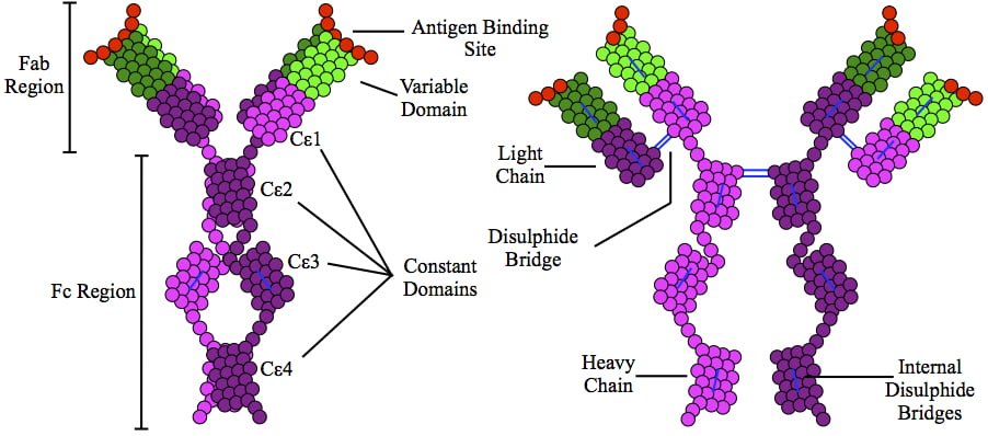
This is a depiction of immunoglobulin E (IgE) antibody which is elevated in allergic reactions.

What Is Allergic Rhinitis?

A llergic rhinitis is an immune disorder characterized by inflammation of the nasal mucosa by an IgE antibody reaction to an allergen.

An allergen is something that triggers an allergic immune response.

Q: What is the immune response?

A: Implicated in the response is an increase in T gamma-delta intraepithelial lymphocytes (IELs), which is a subset of pro-inflammatory T-cells located in the respiratory mucosa.  Lymphocytes are white blood cells.

When a person with allergic rhinitis breathes in an allergen such as pollen or dust, the body releases chemicals, including histamine that cause allergy symptoms. For example, hay fever involves an allergic reaction to pollen. A similar reaction occurs with allergy to mold, animal dander, dust, and other allergens that are breathed in.

What Is Allergic Rhinitis In Celiac Disease and/or Gluten Sensitivity?

Osteitis Fibrosa Cystica 

Deep Vein Thrombosis in the Right Leg with Swelling and Redness. Coutesy wikipedia.
Deep Vein Thrombosis in the Right Leg with Swelling and Redness. Courtesy wikipedia.

What Is Antiphospholipid Syndrome?

Antiphospholipid syndrome (APS) is an autoimmune disease and a blood clotting disorder characterized by these clinical and laboratory criteria:

Clinical criteria – recurrent vascular thrombosis (clots in veins/arteries) from hypercoagulability (abnormal excessive clotting) and/or recurrent complications of pregnancy that include loss of the fetus  (miscarriage) and pre-eclampsia or eclampsia.

Laboratory criteria – persistently elevated anticardiolipin, anti–beta-2 glycoprotein I, and/or lupus anti-coagulant antibodies in blood.

In antiphospholipid syndrome autoantibodies are produced by the body and directed against negatively charged phospholipids that are found in the outer layer of cell membranes and platelets. B2-glycoprotein-I (a protein in blood plasma) has been found as a major target antigen for antiphospholipid antibodies.

Q: Are phospholipids important in the body?

A: Yes.  Phospholipid molecules are an essential part of cell membranes. They form a barrier around cells that protect the cell, allow movement of oxygen in and carbon dioxide out of the cell, and regulate other small molecules through the cell wall. Because phospholipids are widespread in the body, this disorder can produce a large variety of symptoms and affect many organs.

One severe effect of APS is the development of a blood clot in a vein deep in the arm or leg, called deep vein thrombosis (DVT). DVT can cause pain, swelling, redness, or increased warmth in the affected limb. Deep vein clots can break off, travel to the lungs, and cause pulmonary embolism.1 Pulmonary embolism is a medical emergency.

Treatment is with anticoagulant medications and blood monitoring.

What Is Antiphospholipid Syndrome In Celiac Disease and/or Gluten Sensitivity?

Sources:
  1. http://www.nhlbi.nih.gov/health/health-topics/topics/ebc/signs.html []

Osteomalacia

Drawing shows changes in airways during asthma attack. wikipedia

What Is Asthma?

Asthma is a chronic immune respiratory condition characterized by narrowing and inflammation of the lung airways (large bronchi, bronchial tubes and small bronchioles) in response to an allergen as the trigger or stimulus. As such, asthma occurs in episodes and does not result in progressive loss of pulmonary function.

During an asthma attack, airways constrict, trapping air so lungs become overinflated.  Normally, bronchial airways bring air to millions of air sacs that are attached to the ends of bronchioles. Air sacs, called alveoli, are only one cell thick to allow for rapid exchange of gases.

That is, oxygen from air breathed into the sacs moves into the bloodsteam and carbon dioxide is released from the bloodstream to air that is breathed out of air sacs.

The outer walls of bronchioles are made up of muscles which, in the process of breathing, normally contract on expiration to help expel air and then relax. During an asthma attack, these muscles abnormally constrict, impairing airflow into and out of the alveoli. This is called bronchospasm.

Common  allergens that cause inflammation include airborne dust mite feces, mold, and pollen and foods such as wheat, cow’s milk, eggs, and peanuts. Non-allergenic triggers include exercise, air pollution, smoking, and viral respiratory infection.

Q: What effect does inflammation have on the lungs?

A: Inflammation causes local tissue edema or swelling of the bronchioles and mucus formation. Inflammation with increased mucus secretions and edema narrows the airways that connect to alveoli which makes breathing difficult.  Two things happen:

  1. Inflammation  impairs exchange of gases in alveoli, resulting in lack of sufficient oxygen (O2) for body cell functions, called hypoxia, and build-up of carbon dioxide (CO2) in blood, called CO2 retention.
  2. Inflammation narrows passageways because of swelling, which reduces the movement of air to and from the alveoli through the airways, and this puts stress on the right side of the heart.

Treatment is aimed at controlling bronchospasm and reducing inflammation. Untreated asthma can be disabling and life threatening.

What Is Asthma In Celiac Disease and/or Gluten Sensitivity?

Hypocalciuria (Low Urine Calcium)

 IgA Molecule.
Depiction of the IgA Molecule

What Is IgA Deficiency?

I gA deficiency (IgAD) is an immunodeficiency disease characterized by lack of immunoglobulin A type antibody production, called IgA antibody, with no detectable levels in blood or secretions.

Q: What is an IgA antibody?

A: IgA is an antibody of the immune system that is secreted by plasma cells (specialized white blood cells) through epithelial cell linings of mucosal surfaces into mucosa secretions to protect the lining from microbe invasion.

In fact, immunoglobulin class A is the main protein of the mucosal immune system. This includes mucosa of the eye surface, digestive tract, respiratory tract, urinary tract, and genital tract.

Both major histocompatibility complex (MHC) and non-MHC genes contribute to susceptibility to the disease. The former genes appear to be located in different parts of the MHC region depending on the HLA haplotype. The latter show a marked overlap with genes associated with a variety of autoimmune disorders including Graves’ disease, systemic lupus erythematosus, type 1 diabetes and celiac disease, suggesting common pathophysiological mechanisms. The involvement of genes associated with autoimmunity may suggest that IgAD in itself is an autoimmune disease.1

IgA deficiency may progress into a common variable immunodeficiency (CVID).2

What Is IgA Deficiency (IgAD) In Celiac Disease and/or Gluten Sensitivity?

Sources:
  1. Wang N, Hammarström L. IgA deficiency: what is new? Curr Opin Allergy Clin Immunol. 2012 Dec;12(6):602-8. doi: 10.1097/ACI.0b013e3283594219. []
  2. Binek A, Jarosz-Chobot P.Selective immunoglobulin A deficiency. Pediatr Endocrinol Diabetes Metab. 2012;18(2):76-8. []