Skip to content

Delayed Puberty in Boys
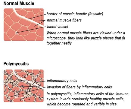
Drawing of Biopsy Showing Muscle Fibers Invaded by Immune Cells. Courtesy MDA.org

What Is Polymyositis?

[dropcap]P olymyositis is a body-wide connective tissue disease resulting from autoimmune attack of skeletal muscles that is characterized by inflammatory and degeneratory changes. The course is unpredictable being marked by spontaneous flare-ups and remissions.

Polymyositis can begin slowly or abruptly according to the factor that is triggering the onset such as infection, medications like phenytoin, and autoimmune disease.

Progressive muscle weakness starts in the proximal skeletal muscles (muscles closest to the trunk of the body).

Skeletal muscles, also called voluntary, are muscles that move the body as we want, such as walking and  lifting objects, as opposed to those we cannot voluntarily control, such as the muscles of digestion. 

Q: What are the degeneratory changes in skeletal muscles?

A: In polymyositis, degeneratory changes in skeletal muscles means that muscles are being destroyed (called necrosis), resulting in fibrosis, or scarring. When scar tissue takes the place of lost muscle tissue, it cannot act like muscle to contract and relax.   Muscle destruction is what causes muscle pain and weakness.

After the clinical work-up of exams and blood studies to determine muscle damage, the diagnosis of polymyositis is confirmed by muscle biopsy. See image at above left. The black dots are inflammatory cells. Edema (fluid) between cells caused by inflammation pushes muscle fibers apart.

There is no cure for polymyositis, but the symptoms can be treated.  Options include medication, physical therapy, exercise, heat therapy (including microwave and ultrasound), orthotics and assistive devices, and rest.  The standard treatment for polymyositis is a corticosteroid drug, given either in pill form or intravenously.  Immunosuppressant drugs, such as azathioprine and methotrexate, may reduce inflammation in people who do not respond well to prednisone. 

Periodic treatment using intravenous immunoglobulin can also improve recovery.  Other immunosuppressive agents used to treat the inflammation associated with polymyositis include cyclosporine A, cyclophosphamide, and tacrolimus.  Physical therapy is usually recommended to prevent muscle atrophy and to regain muscle strength and range of motion.1

Diagnosis is based on elevated muscles enzymes, increased urinary creatine level, and electromyograph abnormalities.

Polymyositis can affect people at any age. It is most common in adults between ages 50 and 70, and in children ages 5 to 15. It affects women twice as often as men and is more common in African Americans than Caucasians.2 The major causes of death from polymyositis are cancer and lung disease, including pneumonia.  The 5-year mortality rate can be as high as 1 in 5 patients.2

What Is Polymyositis In Celiac Disease and/or Gluten Sensitivity?

Sources:
  1. National Institute of Neurological Disorders and Stroke []
  2. http://www.nlm.nih.gov/medlineplus/ency/article/000428.htm [] []

Coronary Artery Disease

Image on left shows how atherosclerosis impedes blood flow through coronary arteries while blood clots block blood flow. Courtesy Google.
Figure on right shows how atherosclerosis impedes blood flow through coronary arteries while blood clots block blood flow. Courtesy Google.

What Is Coronary Artery Disease (CAD)?

[dropcap]C[/dropcap]oronary artery disease (CAD), also called ischemic heart disease, is a gradual narrowing of medium and large arteries of the heart by fatty buildups, called atherosclerotic plaques.

It is characterized by slowly developing interference with blood flow to heart tissue itself, resulting in oppressive chest pain called angina and, ultimately, thrombosis (clot) causing heart attack.  

The heart is a muscular organ that is working all the time, so it needs a constant supply of oxygen. Oxygen is brought to the working heart tissue by the coronary arteries with each beat of the heart. When heart muscle has to work harder, it needs more oxygen delivered to itself. Lack of oxygen causes pain.

In fact, failure of diseased coronary arteries to deliver adequate oxygen to heart tissue is the most common cause of angina pectoris – substernal pain (under breastbone) or pressure brought on by exertion and relieved by rest. 

Thrombosis, or clot formation, occurs when blood cells within a narrowed artery can no longer get through. Trapped, blood cells pile up and block the artery thus triggering a cascade of events called heart attack. Coronary arteries that are narrowed by atherosclerotic plaques can rupture causing injury to the coronary blood vessel resulting in blood clotting which blocks the flow of blood to the heart muscle. Blood clots may form, partially dissolve, and later form again and angina can occur each time a clot blocks blood flow in an artery.1

Q: How does coronary artery disease develop?

A: Coronary artery disease slowly develops from this combination of events:

  • Dysfunction of epithelial cells that line the inside of arteries cause the vessels to stiffen, and subsequently

  • Accumulation of lipid (fat) in smooth muscle cells beneath the inside lining of arteries and in foam cells cause buildup of fatty deposits on the inside walls progressing to fibrous plaque formation.

Oxidized low-density lipoprotein (oxLDL), so-called bad cholesterol, and oxysterols play important roles in the development of  atherosclerosis. OxLDL triggers the immune system to produce autoantibodies against oxLDL that are detectable in serum. These antibodies are called anti-oxLDL. Anti-oxLDL antibody and oxysterol concentrations are associated with coronary artery stenosis. Oxidative stress may be greatly increased in unstable angina.2 and Chronic inflammation in the general population is a major risk factor for ischemic heart disease.

The pathophysiology of atherosclerosis is, clearly, different in women when compared to the men. The women have a higher risk of blood coagulability making them at high risk for the blood clot formation. In a large number of women endothelial dysfunction, small vessel size and diffuse atherosclerosis have been identified as causes of ischemia without evidence of blockade in the coronary arteries.3

Also, atherosclerotic plaque in women is less fibrotic and contains more lipid filled foam cells, implying greater potential for reversibility but also potentially greater vulnerability for plaque rupture and thrombosis.4

Who is Affected in the General Population?

  • Coronary artery disease remains the leading cause of death in developed countries despite significant progress in primary prevention and treatment strategies.

  • It is the leading cause of death in women, as well as an important cause of disability.

  • Older patients are at particularly high risk of poor outcomes following acute coronary syndrome.5

What Is Coronary Artery Disease In Celiac Disease and/or Gluten Sensitivity?

Ischemic heart disease is the leading cause of death in the United States, making cardiovascular risk assessments and potential interventions or treatments imperative for patients with celiac disease.6

Sources:
  1. http://www.heart.org/HEARTORG/Conditions/HeartAttack/SymptomsDiagnosisofHeartAttack/Unstable-Angina_UCM_437513_Article.jsp# []
  2. Yasunobu Y, Hayashi K, Shingu T, Yamagata T, Kajiyama G, Kambe M. Coronary atherosclerosis and oxidative stress as reflected by autoantibodies against oxidized low-density lipoprotein and oxysterosis. Atherosclerosis. Apr 2001;155(2):445-53. []
  3. Kunadian V, Ford GA, Bawamia B, Qiu W, Manson JE. Vitamin D deficiency and coronary artery disease: A review of the evidence. Am Heart J. 2014 Mar;167(3):283-291. doi: 10.1016/j.ahj.2013.11.012. Epub 2013 Dec 19. []
  4. Kunadian V, Ford GA, Bawamia B, Qiu W, Manson JE. Vitamin D deficiency and coronary artery disease: A review of the evidence. Am Heart J. 2014 Mar;167(3):283-291. doi: 10.1016/j.ahj.2013.11.012. Epub 2013 Dec 19. []
  5. Kunadian V, Ford GA, Bawamia B, Qiu W, Manson JE. Vitamin D deficiency and coronary artery disease: A review of the evidence. Am Heart J. 2014 Mar;167(3):283-291. doi: 10.1016/j.ahj.2013.11.012. []
  6. Robinson BL, Davis SC, Vess J, Lebel, J. Primary care management of celiac disease. Nurse Practitioner. February 2015: Vol 40 – Issue 2; 28–34. []

Dysmenorrhea (Painful Periods)

Beaus lines in thin nails (The tiny brown streak is a splinter hemorrhage.)
Beau”s lines in a thin nail. (The tiny brown streaks are splinter hemorrhages due to vitamin C deficiency.)

What Are Horizontal Ridges In Fragile Nails?

Horizontal ridges, also called “beau’s lines,” are abnormalities of the nail plate that appear as rumpling from the base to the tips of nails and are characterized by poor nail structure of both fingernails and toenails.

The nail plate is the hard keratin cover of the finger tip and toe tip which we ordinarily call “nails.” The nail plate is produced by the nail matrix. 

Q: Why do Beau’s lines develop in nails?

A: Beau’s lines occur due to temporary cessation of proliferation (growth) of proximal nail matrix at the nail base. As the finger nail grows at the rate of 0.1 mm/day, the time course of the illness can be estimated from the position of the Beau’s line from proximal nail fold.1

Nail Anatomy. Nail Anatomy. A. Nail plate; B. lunula; C. root; D. sinus; E. matrix; F. nail bed; G. hyponychium; H. free margin. Courtesy Wikipedia.org.

A. Nail plate; B. lunula; C. root; D. sinus; E. matrix; F. nail bed; G. hyponychium; H. free margin. Courtesy Wikipedia.

Beau’s lines are frequently seen in nutritional deficiency states, bacterial illness, acute stress, and systemic disease. The conditions where Beau’s lines have been described include severe systemic illness, chemotherapy, malnutrition, zinc deficiency, trauma, paronychia, pemphigus, and Kawasaki disease.2 Beau’s lines are commonly seen in patients undergoing chemotherapy.3

 This condition of nails was named after Joseph Honoré Simon Beau (1806-1865).

What Are Horizontal Nail Ridges In Celiac Disease and/or Gluten Sensitivity?

Sources:
  1. Naik GS1, Harikrishna J. Beau’s lines. Indian J Med Res. 2013 Jan;137(1):220. []
  2. Naik GS1, Harikrishna J. Beau’s lines. Indian J Med Res. 2013 Jan;137(1):220. []
  3. Patel LM, Lambert PJ, Gagna CE, Maghari A, Lambert WC. Cutaneous signs of systemic disease. Clin Dermatol. 2011 Sep-Oct;29(5):511-22. doi: 10.1016/j.clindermatol.2011.01.019. []

Dyspareunia (Painful Intercourse), Primary

Koilonychia (Hordinsky, Sawaya, and Scher, 2000)

What Is Koilonychia?

[dropcap]K oilonychia is a feature of an iron deficiency state characterized by abnormally shaped nails, called spoon shaped nails, meaning that the normal downward curve is reversed.

Depending on the inadequacy of iron, nails may simply remain flat or with a slight dip without progressing to a reversed curve.

These nails develop vertical ridges from the base to the nail tip that can become deep, called washboard nails.

Q: Are all nails affected?

A: All nails are affected in varying degrees. The small finger shows the least effect while the thumb shows the greatest change which begins as flattening before scooping upward.

Nail Anatomy. Nail Anatomy. A. Nail plate; B. lunula; C. root; D. sinus; E. matrix; F. nail bed; G. hyponychium; H. free margin. Courtesy Wikipedia.org.

A. Nail plate; B. lunula; C. root; D. sinus; E. matrix; F. nail bed; G. hyponychium; H. free margin. Courtesy Wikipedia.

What Is Koilonychia In Celiac Disease and/or Gluten Sensitivity?

Hypogonadism In Adult Males That Is Unexplained  

Nail Anatomy. A. Nail plate; B. lunula; C. root; D. sinus; E. matrix; F. nail bed; G. hyponychium; H. free margin. Courtesy Wikipedia.org
Nail Anatomy. A. Nail plate; B. lunula; C. root; D. sinus; E. matrix; F. nail bed; G. hyponychium; H. free margin. Courtesy Wikimedia.org

What Are Rounded Nails With Curved Ends?

[dropcap]R ounded, curved, dark and dry nails are a well known feature of vitamin B12 deficiency.

Q: How does vitamin B12 deficiency cause the nails to grow this way?

A: Vitamin B12 deficiency affects the nail plate. The nail plate is the hard keratin cover of the finger tip and toe tip, simply called “the nail.” The nail plate is produced by the living nail matrix at its base.

What Are Rounded Nails With Curved Ends In Celiac Disease and/or Gluten Sensitivity?

Lymphomas, Extraintestinal

What Is Dysphagia? [dropcap]D ysphagia is a functional upper digestive disorder characterized by difficulty in swallowing or food getting stuck and swallowing that takes longer than 10 seconds to complete. Q: What determines the symptoms?… 

Ataxia, Gait

Normal Liver Lobule. Courtesy almostadoctor.com
Figure 1: Normal Liver Lobule. Courtesy almostadoctor.com

What Is Autoimmune Cholangitis?

[dropcap]A utoimmune cholangitis is a rare inflammatory self-targeted disease of the biliary tract produced by high levels of immunoglobulin G (IgG) antibodies and antinuclear antibodies (ANA).

Autoimmune cholangitis is characterized by progressive inflammation and scarring that leads to narrowing and destruction of the bile ducts and development of biliary cirrhosis of the liver.

Autoimmune cholangitis is also termed anti-mitochondrial antibody-negative primary biliary cirrhosis because there is no production of anti-mitochondrial antibodies.

Q: What happens when bile ducts can no longer transport bile out of the liver?

A: Bile that cannot be removed from the liver by the biliary tract backs up and damages the liver, causing cirrhosis. In Figure 1, bile back up would directly damage the hepatic (liver) cells seen here as grey “blocks.” 

Bile is a greenish brown liquid continually made by the liver cells (hepatocytes) from phospholipids salt, cholesterol, and aging blood cells that it removes from circulation. 

Bile also carries away waste products produced by normal metabolism and toxic substances that are removed by the liver for eventual elimination in stool. As such, bile must continually flow out of the liver to prevent build-up in the liver. 

Bile from the hepatic cells drains into small tubes called canniculi and then into larger bile ducts to be carried out of the liver to the gall bladder for storage until needed to aid in the digestion and absorption of fat from the small intestine.   Bile emulsifies fat eaten in the diet so that the pancreatic enzyme called lypase can break it down into its fatty acid and glycerol components. 

Unfortunately, autoimmune cholangitis is difficult to diagnose because it mimics many other diseases such as primary sclerosing cholangitis, sarcoidosis, cancer and lymphoma.

What Is Autoimmune Cholangitis In Celiac Disease and/or Gluten Sensitivity?

Ataxia, Gluten

This photo taken during laparoscopy shows the gall bladder (small white organ in middle) surrounded by yellow fat. Liver (dark red organ) is overlying.

What Is Impaired Gall Bladder Motility?

[dropcap]I mpaired gall bladder motility means the gall bladder is slow to empty or is dysfunctional.

The functional disorder of the gallbladder is caused initially either by metabolic abnormalities or by an alteration in its muscular ability to contract (motility dysfunction).

The diagnostic criteria based on symptoms of motility dysfunction of the gallbladder are episodes of moderate to severe steady pain located in the epigastrium and right upper abdominal quadrant that last at least 30 minutes.

Gallbladder motility disorder is suspected after gallstones and other structural abnormalities have been excluded.1

Q: What does the gallbladder do?

A: The gallbladder is a small pouch-like organ about the size of a pear that receives bile produced by the liver and stores it until needed during digestion. It lies just under the liver.

Bile is a complex fluid containing water, electrolytes and many organic molecules including bile acids, cholesterol, phospholipids and bilirubin. Bile  acids are critical for digestion and absorption of fats and fat-soluble vitamins in the small intestine.  Many waste products, including bilirubin, are eliminated from the body by secretion into bile and elimination in feces.2

Before a meal, the gallbladder is usually full of bile. In response to fat in the diet, the gallbladder squeezes stored bile into the small intestine through a series of ducts. When emptied after meals, the gallbladder is flat.

What Is Impaired Gall Bladder Motility In Celiac Disease and/or Gluten Sensitivity?

Sources:
  1. Behar J, Corazziari E, Guelrud M, Hogan W, Sherman S, Toouli J. Functional gallbladder and sphincter of oddi disorders. Gastroenterology. 2006 Apr;130(5):1498-509. []
  2. http://www.vivo.colostate.edu/hbooks/pathphys/digestion/liver/bile.html []

Common Variable Immunodeficiency 

Folic acid deficiency anemia gluten celiac disease symptom
Red Blood Cells.

What Is Folic Acid Or Folate Deficiency Anemia?

[dropcap]F olic acid deficiency anemia, also called folate deficiency anemia, is a macrocytic anemia characterized by defective DNA synthesis of red blood cells that results from a lack of folate in the body.

Q: How does folate deficiency cause anemia?

A: Folates are a family of B vitamins and folic acid is an active form.

Folate is required for the formation of both red and white blood cells in the bone marrow and for their maturation.

Also, folate serves as a carrier in the formation of heme, which contains iron, and is the non-protein part of the hemoglobin molecule.1

Red blood cells provide oxygen to body tissues. When there are not enough red blood cells or when they cannot properly carry oxygen, the condition is called anemia. In folic acid deficiency anemia, the red blood cells are abnormally large. Such cells are called macrocytes (macro size cells). They are also called megaloblasts (mega size cells) as seen in the bone marrow where they are produced. This is why this macrocytic anemia is also called megaloblastic anemia.2

Tests that may be done to determine folate adequacy are complete blood count (CBC), red blood cell folate level, methylmalonic acid level, and homocysteine level. Folic acid deficiency anemia shows a decrease in red blood cell folate and/or serum folate levels and normal plasma methylmalonic acid level with elevated homocysteine blood level. These levels distinguish folic acid deficiency anemia from vitamin B12 deficiency anemia.3

What Is Folate Deficiency Anemia In Celiac Disease and/or Gluten Sensitivity?

Sources:
  1. Kathleen Mahan and Sylvia Escott-Stump, ed. Krause’s Food, Nutrition & Diet Therapy, 10th Edition. Philadelphia, PA. USA: W.B. Saunders Company, 2000. []
  2. http://www.ncbi.nlm.nih.gov/pubmedhealth/PMH0001578/ []
  3. Mark Beers and Robert Berkow. The Merck Manual, 17th Edition. Whitehouse Station, N.J. USA: Merck Research Laboratories, 1999. []

Lymphoma, B-Cell Non-Hodgkin’s

e3343bf0a689b5f974a852fa2e84a718What Is Laryngospasm?

[dropcap]L aryngospasm is an acute disorder of the larynx that is characterized by life-threatening spasms of the muscles of the larynx located in the throat.

Laryngospasm is a rare feature of hypocalcemia (low blood calcium level) characterized by severe alteration in nerve conduction and muscle contraction.

What Is Laryngospasm In Celiac Disease and/or Gluten Sensitivity?