Skip to content

Tongue – Fiery Red, Smooth, Burning
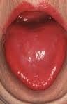
niacin deficiencyWhat Is A Fiery Red, Smooth, Burning Tongue?

[dropcap]A[/dropcap] fiery red, smooth, burning tongue is an alteration in tongue tissue that is characteristic of advanced niacin deficiency.1

Q: How does niacin deficiency cause the tongue to be red and sore?

A: Niacin is an essential B vitamin that is required for a healthy tongue and by all body cells as well. Deficiency first shows in tissues with rapid cellular turnover, such as mucosal cells of the tongue.

When absorbed from the small intestines, niacin (the form in food) becomes the active form niacinamide. Niacinamide is converted by the body into co-enzymes which are present in all cells. These enzymes function in oxidation-reduction reactions essential for release of energy from carbohydrates, fats, and proteins and are needed as components for more than 200 enzymes involved in metabolism.

In addition to producing energy, niacinamide is essential for healthy skin and the mucosal lining of the digestive tract, normal functioning of the brain and nervous system, and production of steroid hormones from adrenal glands and hormones from sex glands.

What Is A Fiery Red, Smooth, Burning Tongue In Celiac Disease and/or Gluten Sensitivity?

Sources:
  1. Krause’s Food, Nutrition, & Diet Therapy. 10th Edition. Kathleen Mahan, Sylvia Escott-Stump. 2000. W.B. Saunders Company. []

Attention Deficit Hyperactive Disorder (ADHD)

adhd-brain-maturation-dark-areasWhat Is Attention Deficit Hyperactive Disorder (ADHD)?

[dropcap]A[/dropcap]ttention deficit hyperactive disorder (ADHD) is a maladaptive behavioral disorder characterized by a persistent pattern of inattention and impulsivity, or both, that are developmentally (age) inappropriate. For these problems to be diagnosed as ADHD, they must be out of the normal range for a child’s age and development.

Inattention results from difficulty concentrating which gives the impression of not listening. The person’s speech or writing, such as when telling stories, can be disorganized.

Most children with ADHD also have at least one other developmental or behavioral problem. Depression, lack of sleep, learning disabilities, tic disorders, and behavior problems may be confused with, or appear with, ADHD. Every child suspected of having ADHD should be carefully examined by a doctor to rule out possible other conditions or reasons for the behavior.

An MRI (magnetic resonance imaging) study investigating whole-brain cortical thickness (outer layer of the brain) in male and female children, adolescents, and adults with ADHD supports the hypothesis that brain abnormalities in ADHD represent abnormal brain development rather than developmental delay.

Q: What is the abnormal development problem?

Drawing shows the 4 lobes of the human brain on the left side. The occipital lobe is pink. Courtesy Wikimedia
4 lobes of the human brain making up each hemisphere (side). In this drawing, the frontal is blue, the parietal is yellow, the temporal is green, and the occipital lobe is pink. Courtesy Wikimedia

A: Differences occur in the right side of the brain only. Reduced cortical thickness in people of all ages with ADHD was observed predominantly in the frontoparietal region (forehead and top). However, increased cortical thickness was observed predominantly in the occipital lobe (back). The brain thickness differences were correlated with severity of ADHD.1

ADHD is the most commonly diagnosed behavioral disorder of childhood. It affects about 3 – 5% of school aged children and is diagnosed much more often in boys than in girls.2

What Is ADHD In Celiac Disease and/or Gluten Sensitivity?

Sources:
  1. Almeida Montes LG, Prado Alcántara H, Martínez García RB, De La Torre LB, Avila Acosta D, Duarte MG. Brain cortical thickness in ADHD: age, sex, and clinical correlations. J Atten Disord. 2013 Nov;17(8):641-54. doi: 10.1177/1087054711434351. []
  2. http://www.ncbi.nlm.nih.gov/pubmedhealth/PMH0002518/ []

Dysgeusia (Impaired Taste)

Loss of TasteWhat Is Dysgeusia?

[dropcap]D[/dropcap]ysgeusia is impaired or altered sense of taste characterized by the inability to distinguish the flavor of a substance.

Q: What causes loss of the ability to taste?

A: Some causes of impaired taste include these health conditions: malnutrition, hypothyroidism, diabetes mellitus,  hypertension, adrenal cortical insufficiency, liver disease, and Sjogren’s syndrome.

What Is Dysgeusia In Celiac Disease and/or Gluten Sensitivity?

Ataxia, Gait

Ataxia-Thomas-fig68,69-p127-wikimediaWhat Is Gait Ataxia?

[dropcap]G[/dropcap]ait ataxia is a cerebellar (lower brain) disorder characterized by defective muscular coordination of skeletal muscles used for locomotion (walking).

Q: What is defective muscle coordination for walking?

A: The cerebellum does not initiate movements but is responsible for the coordinated execution of voluntary muscle movements by means of a complex interplay with many brainstem structures. Whatever alters this cerebellar function produces defective muscle coordination needed for walking.

What Is Gait Ataxia In Celiac Disease and/or Gluten Sensitivity?

Brain Atrophy

Comparison of Brain Atrophy. Courtesy Source: Source: csua.berkeley.edu
Comparison of Brain Atrophy. Courtesy: csua.berkeley.edu

What Is Brain Atrophy?

[dropcap]B[/dropcap]rain atrophy is a degenerative condition of the brain characterized by loss of brain tissue, causing the brain to shrink.

Q: How does loss of brain tissue affect the body?

A: Loss of brain tissue will result in dysfunction depending on the area of the brain that is affected.

For example, loss of brain tissue in the lower brain, or cerebellum, will impair muscle coordination in activities such as walking, while loss of brain tissue in the frontal lobes of the cerebrum will impair cognition such as thinking, judgment, analysis, mathematical ability, and memory.

What Is Brain Atrophy In Celiac Disease and/or Gluten Sensitivity?

Brain Blood Flow Abnormalities 

blood perfusion gluten celiac diseaseWhat Are Brain Blood Flow Abnormalities?

[dropcap]B[/dropcap]rain blood flow abnormalities, or cerebral perfusion abnormalities, such as spasm of blood vessels causing inadequate blood flow to the brain, are characterized by impaired brain function and thinking.

Q: What is cerebral perfusion?

A: Cerebral perfusion is the flow of blood in the brain. Blood supply to the brain comes from 4 major vessels: 2 carotid arteries and 2 vertebral arteries.

Cerebral perfusion abnormalities can be diagnosed using perfusion computed tomography (CT). CT scan is a technique that allows rapid qualitative and quantitative evaluation of cerebral perfusion by generating maps of cerebral blood flow (CBF), cerebral blood volume (CBV), and mean transit time (MTT). The technique is based on the central volume principle (CBF = CBV/MTT) and requires the use of commercially available software employing complex deconvolution algorithms to produce the perfusion maps.

Perfusion CT has been found to be useful for noninvasive diagnosis of cerebral ischemia (diminished blood flow) and infarction (no blood flow) and for evaluation of vasospasm (intermittent blood flow through vessels).1

What Are Brain Blood Flow Abnormalities In Celiac Disease and/or Gluten Sensitivity?

Sources:
  1. Hoeffner E, Case I, Jain R, et al. Cerebral Perfusion CT: Technique and Clinical Applications. June 2004 Radiology, 231, 632-644. []

Abdominal Distention, Chronic  (Bloating)

chronic abdominal distention celiac disease gluten symptomWhat Is Chronic Abdominal Distention?

[dropcap]A[/dropcap]bdominal distention, or bloating, is characterized by enlargement in normal size of abdomen not due to fatty tissue.

Q: Why does the abdomen enlarge?

A: The abdomen enlarges because the bowel is dysfunctional. Loops of bowel usually lack normal peristalsis (rhythmic wave-like muscle movement) trapping gas in stagnant loops of bowel, inflammation swells loops of bowel also trapping gas, and dysbiosis overproduces gas. All these factors distend the abdomen.

What Is Chronic Abdominal Distention In Celiac Disease and/or Gluten Sensitivity?

Anorexia or Loss of Appetite

anorexia nervosa celiac disease gluten symptomWhat Is Anorexia?

[dropcap]A[/dropcap]norexia or loss of appetite is a reduced desire to eat which can cause unintentional weight loss.

Anorexia is a feature of many disorders caused by malutrition such as celiac disease and aging or gastrointestinal problems such as chronic gastroenteritis, and dysgeusia (loss of taste).

Q: Are there other conditions that cause anorexia?

A: Other underlying problem include medication side effects, infection, chronic pain, depression, grief, dehydration, hormonal dysfunction such as thyroid disease, primary hyperparathyroidism, or Addison’s disease, autoimmune mechanisms such as sarcoidosis,  and cancer growth.

What Is Anorexia In Celiac Disease and/or Gluten Sensitivity?

Oral Mucosal Lesions, Chronic (Mouth Soreness)

Canker Sore Inside Mouth. Notice The White Spot on This Person's Nail Showing Zinc Deficiency.
Inflammation/Sore Inside Mouth. Notice White Spot on Fingernail Nail Showing Zinc Deficiency.

What Are Chronic Oral Mucosal Lesions?

Chronic lesions of the oral mucosa are disorders of the mouth that are characterized by soreness and sores of the soft mucosal lining of the mouth.

What Are Chronic Oral Mucosal Lesions In Celiac Disease and/or Gluten Sensitivity?

Heartburn

Heartburn Due to Acidic Stomach Contents Re-entering the Esophagus. Courtesy kaiserpermanente.org
Heartburn Due to Acidic Stomach Contents Re-entering the Esophagus.

What Is Heartburn?

[dropcap]H[/dropcap]eartburn is a functional upper digestive symptom of esophagitis that is characterized by a burning sensation behind the breastbone.

Q: Why does esophagitis cause heartburn?

A: Esophagitis is inflammation of the mucosal lining of the esophagus. The most common cause of esophagitis is the re-entering of stomach contents into the esophagus because the lower esophageal sphincter (LES) does not close tightly after food or fluids enter the stomach from the esophagus and/or the LES does not remain tightly closed.

The LES is located at the junction of the esophagus and the stomach. It functions like a circular band to tighten after food is ingested in order to prevent its going back up the esophagus when powerful stomach muscles churn ingested food.

Damage to the lining of the esophagus (esophagitis) results upon contact with hydrochloric acid and pepsin in gastric juice. Hydrochloric acid, produced by the stomach to dissolve food, inflames the esophagus, which is the opposite, or alkaline. Pepsin, produced by the stomach to dissolve protein in swallowed food, acts on the inflamed esophagus to break down the protein in its sore wall.

Heartburn can be caused by other disorders such as low stomach acid, obesity, and smoking. Relaxation of the LES can also result from dietary items such as chocolate, mint, caffeine, and alcoholic drinks.

Who is Affected in the General Population? Heartburn affects an estimated 42% of the US population.1

What Is Heartburn In Celiac Disease and/or Gluten Sensitivity?

Sources:
  1. http://informahealthcare.com/doi/abs/10.1185/03007990903553812. []